DYSKERATOSIS CONGENITA
Synonym: Zinsser–Engman–Cole syndrome
Key features
Genetically heterogeneous disorder due to defective telomere maintenance, with X-linked recessive inheritance in the most common form
Triad of reticulated hyperpigmentation, nail dystrophy (e.g.
pterygium), and leukoplakia
Bone marrow failure resulting in thrombocytopenia, anemia, or pancytopenia
Continuous lacrimation (epiphora) due to lacrimal duct atresia, pulmonary fibrosis, and liver cirrhosis may develop
Predisposition to malignancy, especially mucosal squamous cell carcinoma (e.g. mouth, anus)
Introduction
First described in 1906 by Zinsser, dyskeratosis congenita (DKC) is a genetically heterogeneous disorder caused by defective telomere maintenance. DKC is characterized by progressive bone marrow failure and the mucocutaneous triad of reticulated hyperpigmentation, nail dystrophy, and leukoplakia. In addition, affected individuals have an increased risk of developing pulmonary fibrosis, liver cirrhosis, and malignancies including squamous cell carcinoma (especially mucosal) and acute myeloid leukemia.
Epidemiology
The most common form of DKC has an X-linked recessive pattern of inheritance, but autosomal dominant and autosomal recessive forms also exist (Table 67.9). The overall male : female ratio is ~3 : 1.
Pathogenesis
DKC is caused by defective telomere maintenance, which leads to the characteristic feature of markedly shortened telomeres. Telomeres are repetitive nucleotide sequences that protect the ends of eukaryotic chromosomes. The enzyme telomerase prevents the progressive short-ening of chromosomes that would otherwise occur with DNA replication (Fig. 67.22). Tissues with high replication rates, such as the bone marrow and skin, are particularly vulnerable when telomerase is not functioning properly. When telomeres reach a critically short length, cell cycle arrest occurs and the cells undergo either senescence or apoptosis. However, telomerase dysfunction also leads to chromosome instability, which can result in tumorigenesis.
In 1998, mutations in DKC1, which encodes the dyskerin protein that interacts with telomerase, were found to underlie the X-linked recessive form of DKC. Since then, 11 additional genes have been implicated in both autosomal dominant and recessive forms of DKC that have variable clinical manifestations. These genes encode the telomerase RNA component (TERC) and reverse transcriptase (TERT) as well as other factors with roles in telomerase function and additional aspects of telomere protection (see Table 67.9 & Fig. 67.22). Advances in understanding of the pathogenesis of DKC and other telomere biology disorders may provide insights into telomere maintenance in general processes such as aging and cancer.
Clinical Features
A lacy reticulated pattern of hyperpigmentation generally develops during the first decade of life. This occurs primarily on the neck, upper chest, and upper arms and is sometimes admixed with macules of hypopigmentation (Fig. 67.23A–C). Telangiectasias and epidermal atrophy may also be seen, i.e. poikiloderma. Additional cutaneous features include wrinkled skin on the extremities, dorsal aspects of the hands, and genitalia; palmoplantar hyperhidrosis and hyperkeratosis; loss of dermatoglyphics (Fig. 67.23D); frictional bullae; acrocyanosis; and premature graying of the hair.
Nail involvement is present in the vast majority of patients, typically appearing during early childhood. Initial changes include longitudinal ridging and splitting, followed by pterygia formation and occasionally complete nail loss (Fig. 67.23E). Leukoplakia occurs in most patients, usually manifesting during early adolescence. White plaques on the oral mucosa are observed most commonly, with a predilection for the lateral portions of the tongue. Involvement of the urethra, vagina, and anus may also be seen. The teeth may be malformed, missing, or have aberrant spacing or extensive caries. Epiphora (continuous lacrimation) due to lacrimal duct atresia is common.
Bone marrow failure occurs in 50%–90% of patients and is a major cause of mortality. It presents during the second or third decade with anemia, thrombocytopenia, or pancytopenia. Malignancies tend to develop during the third or fourth decade, most notably squamous cell carcinomas of the mouth, anus, cervix, vagina, esophagus, and skin. There is also an increased risk of myelodysplastic syndrome, acute myeloid leukemia, and gastrointestinal carcinomas.
Other potential systemic manifestations include pulmonary fibrosis, liver cirrhosis, developmental delay, short stature, avascular necrosis of the femoral head, esophageal or urethral stenosis, cryptorchidism, male hypogonadism, and immunologic dysfunction leading to opportunistic infections. Of note, increased severity in successive generations (anticipation) due to progressive telomere shortening has been observed in autosomal dominant forms of DKC. Hoyeraal–Hreidarsson syndrome is a severe variant of DKC characterized by early bone marrow failure, immunodeficiency, cerebellar hypoplasia, microcephaly, enteropathy, and growth retardation (intrauterine and postnatal). Caused by mutations in several DKC genes (see Table 67.9), it typically leads to death in early childhood. Revesz syndrome is another related disorder that features progressive neurologic deterioration, intracranial calcifications, and exudative retinopathy together with the manifestations of DKC; it is caused by heterozygous mutations in TINF2.
Pathology
Histologically, the features of poikiloderma vasculare atrophicans are seen, with telangiectasias and epidermal thinning as well as foci of increased pigment within the basal layer. In the early stage, hydropic degeneration of basilar keratinocytes and a band-like inflammatory infiltrate in the upper dermis may be evident. In the late stage, the epidermis is markedly thinned and flattened and melanophages are present in the upper dermis.
Short telomere length can be detected in leukocytes from patients with DKC by flow-fluorescence in situ hybridization (FISH).
Differential Diagnosis
Fanconi anemia is also associated with pigmentary abnormalities, pancytopenia, and an increased risk of neoplasia (e.g. leukemia); this disorder is compared to DKC in Table 67.10. Naegeli–Franceschetti– Jadassohn (NFJ) syndrome and dermatopathia pigmentosa reticularis (DPR) differ by their lack of leukoplakia and bone marrow involvement (see below). Patients with chronic GVHD may develop poikilodermatous skin changes and lacy white patches of the oral mucosa. However, GVHD is usually easily distinguished by the history of a hematopoietic stem cell transplant (or, rarely, a solid organ transplant). Additional entities associated with poikiloderma are discussed in Chapter 63.
Treatment
Management of DKC patients requires multidisciplinary care. Longitudinal surveillance of all mucosal surfaces is indicated to monitor for the development of squamous cell carcinoma within areas of leukoplakia. Sun and cigarette avoidance are advisable. Bone marrow failure can be treated with supportive transfusions of blood products and the use of androgens (e.g. oxymetholone, danazol), erythropoietin, granulocyte colony-stimulating factor, and eltrombopag or romiplostim. Although hematopoietic stem cell transplantation (HSCT) can potentially cure bone marrow failure and leukemia in patients with DKC, the three-year overall survival rate following HSCT is ~65%.
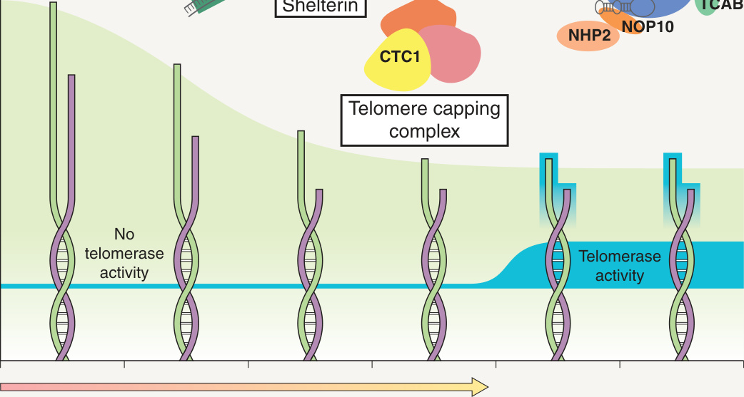
Fig. 67.22 Interactions among telomerase, dyskerin, and other telomere-associated proteins implicated in dyskeratosis congenita. Telomerase, the enzyme that protects against progressive shortening of the chromosomes with successive cell divisions, is composed of two subunits: an RNA component (TERC) and reverse transcriptase (TERT). Dyskerin interacts with telomerase and plays a role in its proper function. The NHP2 and NOP10 ribonucleoprotein homologs are also in the telomerase complex, while telomerase Cajal body protein-1 (TCAB1) controls telomerase trafficking. Poly(A)-specific ribonuclease (PARN) regulates TERC’s interaction with telomeres. TERF1-interacting nuclear factor 2 (TINF2) and TINF2-interacting protein 1 (TINT1) are components of the shelterin complex that protects the ends of telomeres. RTEL1 is a DNA helicase that regulates telomere elongation, and conserved telomere maintenance component 1 (CTC1) functions in a telomere capping complex. TERF1, telomere repeat binding factor 1.
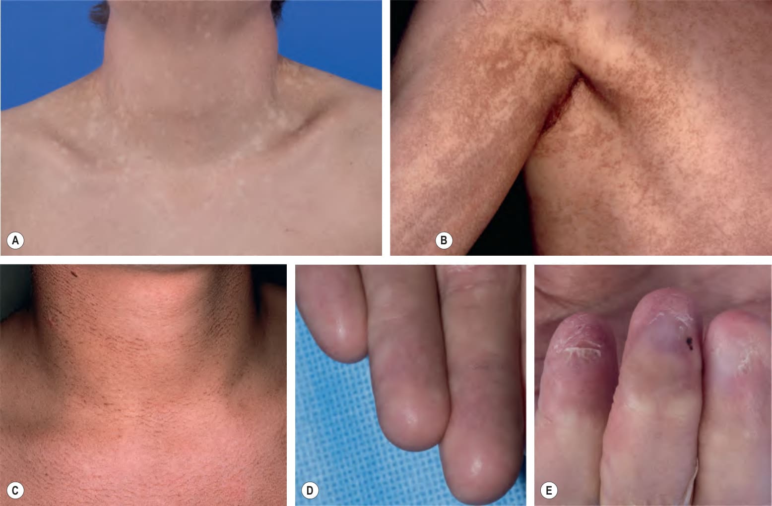
Fig. 67.23 Dyskeratosis congenita.A Diffuse and reticulated hyperpigmentation of the neck and upper chest as well as multiple hypopigmented macules. B More pronounced reticulated and confluent hyperpigmentation on the shoulder and axilla. C Reticulated hyperpigmentation on the chest and neck of a teenage boy. D Mild scaling and erythema of fingers as well as absent dermatoglyphics. E Micronychia and anonychia. A, D, E, Courtesy Edward Cowen, MD; B, Courtesy Eugene Mirrer, MD; C, Courtesy Seth Orlow, MD PhD.
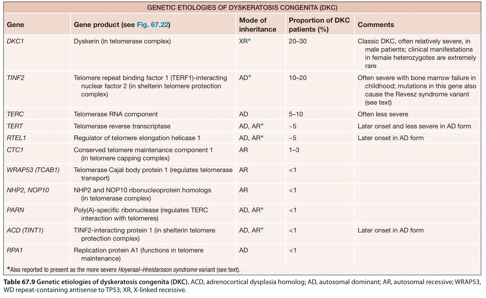
Table 67.9 Genetic etiologies of dyskeratosis congenita (DKC). ACD, adrenocortical dysplasia homolog; AD, autosomal dominant; AR, autosomal recessive; WRAP53, WD repeat-containing antisense to TP53; XR, X-linked recessive.
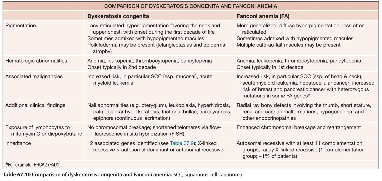
Table 67.10 Comparison of dyskeratosis congenita and Fanconi anemia. SCC, squamous cell carcinoma.