DOWLING–DEGOS DISEASE
Synonyms: Reticular pigmented anomaly of the flexures Reticulated pigmented anomaly of the flexures
Key features
Reticulated hyperpigmentation in flexural sites
Comedo-like lesions on the back and neck
Pitted facial scars
Autosomal dominant with variable penetrance
Caused by loss-of-function mutations in the gene that encodes keratin 5
Introduction
Dowling–Degos disease (DDD) is an uncommon autosomal dominant condition characterized by acquired reticular hyperpigmentation in intertriginous sites.
Epidemiology
There is no racial or sex predilection in classic DDD, and it has been reported worldwide. A founder mutation has been observed in Jewish Ashkenazi families with DDD plus hidradenitis suppurativa.
Pathogenesis
Classic DDD is often caused by loss-of-function mutations in the non-helical head domain of the keratin 5 gene (KRT5) (see Fig. 56.5) that lead to haploinsufficiency. Interestingly, a missense mutation in the same region of KRT5 underlies epidermolysis bullosa simplex with mottled pigmentation (see Ch. 32). The head domain of keratin 5 has been found to interact with Hsc70, a chaperone involved in vesicle uncoating, and it has been postulated that keratin 5 has a role in melanosome trafficking. In addition, a form of DDD with more prominent involvement of non-flexural sites can result from heterozygous loss-of-function mutations in POFUT1 and POGLUT1, which encode O-fucosyltransferase 1 and O-glucosyltransferase 1, respectively. Both of these enzymes perform post-translational modifications that regulate the Notch signaling pathway, which has roles in controlling proliferation and differentiation of melanocytes and keratinocytes as well as their interactions with one another. A variant of DDD associated with hidradenitis suppurativa is caused by mutations in the presenilin enhancer protein 2 gene (PSENEN), which encodes a component of the γ-secretase protein complex that functions in Notch signaling.
Clinical Features
Onset is typically during the third to fourth decade of life. The reticulated hyperpigmentation is admixed with and sometimes composed of lentigo-like brown macules; small brown papules with variable hyperkeratosis may also develop. These findings progressively increase over time, initially appearing in the axillae (Fig. 67.26) and groin followed by the intergluteal and inframammary folds, neck, trunk, and inner aspects of the arms and thighs. Some patients report pruritus of the affected flexural areas. Hypopigmented macules, comedo-like lesions on the back or neck, pitted perioral scars, epidermoid cysts, and hidradenitis suppurativa represent additional features in some patients.
Pathology
Histologically, there is increased pigmentation of the basal layer and finger-like elongation of the rete ridges with thinning of the suprapapillary epithelium. This results in an “antler-like” pattern arising from the under surface of the epidermis and hair follicles. Dermal melanophages and a mild perivascular lymphohistiocytic infiltrate are also observed. Galli–Galli disease represents a variant of DDD (also caused by KRT5 mutations) with prominent suprabasal non-dyskeratotic acantholysis in biopsy specimens of lesional skin in addition to the usual clinical and histologic features.
Differential Diagnosis
Acanthosis nigricans is distinguished clinically by velvety plaques and histologically by less pronounced elongation of the rete ridges. In addition, there is no follicular involvement. Patients with neurofibromatosis type 1 develop lentigines (oftentimes referred to as “freckles”) in the axillae and groin, but this disorder is usually easy to distinguish from DDD.
The spectrum of DDD-like disorders that feature reticulate pigmentation include reticulate acropigmentation of Kitamura (see below) and Haber syndrome. Patients with Haber syndrome develop a photosensitive rosacea-like facial eruption during adolescence, which is followed by the appearance of keratotic papules, comedo-like lesions, pitted scars, and reticulate hyperpigmentation on the trunk and proximal extremities as well as in the axillae.
Treatment
Topical hydroquinone, tretinoin, adapalene, and corticosteroids have been used with varying success. Improvement following treatment with the erbium:YAG or Q-switched Nd:YAG plus fractional CO lasers has also been reported.
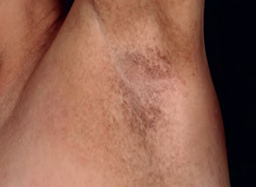
Fig. 67.26 Dowling–Degos disease. Hyperpigmented macules forming a reticulated pattern in the axilla. Courtesy Thomas Schwarz, MD.