DYSCHROMATOSIS SYMMETRICA HEREDITARIA
Synonyms: Acropigmentation of Dohi Symmetrical dyschromatosis of the extremities
Key features
Small, irregular hypopigmented and hyperpigmented macules on the dorsal aspects of the distal extremities, especially the hands and feet
Autosomal dominant disorder caused by mutations in ADAR, which encodes an RNA-specific adenosine deaminase
Introduction
Dyschromatosis symmetrica hereditaria (DSH) was first reported by Toyama in 1929. It is characterized by both hyper- and hypopigmented macules of varying size on the dorsal surfaces of the distal extremities. The vast majority of reports have come from Japan, China, and Korea, but DSH has also been described in European, Afro-Caribbean, and Indian patients. The prevalence in Japan is ~1.5 per 100 000.
Pathogenesis
DSH has an autosomal dominant inheritance pattern with high penetrance. It is caused by heterozygous mutations in ADAR (DSRAD), which encodes the RNA-specific adenosine deaminase. Of note, biallelic ADAR mutations are one cause of Aicardi–Goutières syndrome (AGS), a type I interferonopathy presenting with encephalopathy and variable pernio-like skin lesions (see Table 45.7).
Clinical Features
Hyper- and hypopigmented macules arise in infancy or early childhood. The macules often increase in size and number until adolescence, when they stabilize; darkening after sun exposure may be observed. This dyschromatosis favors the distal extremities and is usually most marked on the dorsal aspects of the hands and feet (see Fig. 67.28); it spares the palms, soles, and mucous membranes. Spontaneous clearing has not been described. A few patients with dystonia and neurologic
deterioration associated with a particular ADAR mutation have been reported, and features of both DSH and AGS have been described in individuals with biallelic ADAR mutations.
Pathology
In the hyperpigmented macules, there is increased melanin within basal keratinocytes, while the hypopigmented macules demonstrate a decrease in the density of DOPA-positive melanocytes.
Differential Diagnosis
Reticulate acropigmentation of Kitamura lacks hypopigmented macules, while vitiligo lacks hyperpigmented macules. Patients with xeroderma pigmentosum also develop hyper- and hypopigmented macules during childhood, but the lesions are in a photodistribution that includes the face and upper trunk. In dyschromatosis universalis hereditaria (see below), the dyschromic lesions have a similar clinical appearance, but they may be present at birth in some patients and, more importantly, the distribution is primarily truncal (see Table 67.11).
Treatment
Current treatments are unsatisfactory. Strict sun avoidance may decrease the contrast between the hypo- and hyperpigmented macules.
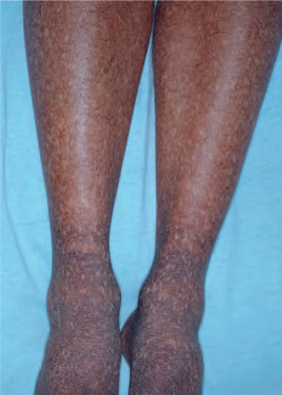
Fig. 67.27 Dyschromic amyloidosis cutis. An admixture of reticulated hyperpigmentation and guttate hypopigmentation are evident. Courtesy Seth J. Orlow, MD PhD.
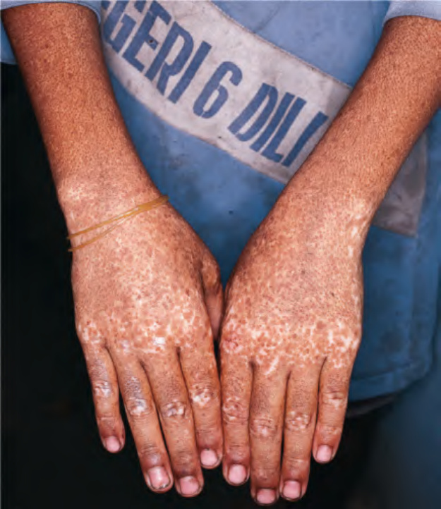
Fig. 67.28 Dyschromatosis symmetrica hereditaria. An admixture of multiple hypo- and hyperpigmented macules on the hands more so than on the forearms. Courtesy Peter Ehrnstrom, MD.
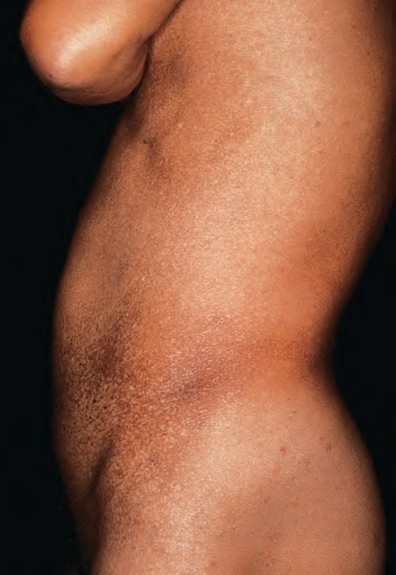
Fig. 67.29 Dyschromatosis univer- salis hereditaria. Both hypo- and hyperpigmentation were present in a widespread distribution. With permission from Urabe K, Hori Y. Dyschromatosis. Semin Cutan Med Surg 1997;16:81–5.
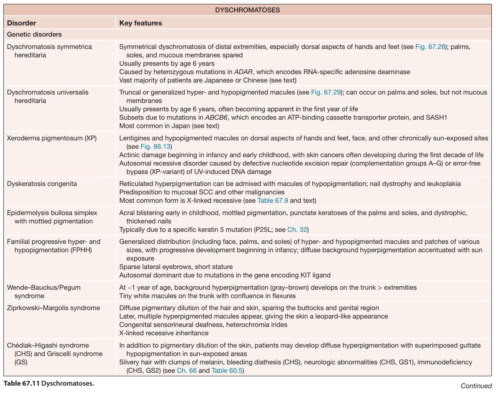
Table 67.11 Dyschromatoses. Continued