Androgenetic Alopecia
Synonyms: Male pattern and female pattern hair loss (MPHL and FPHL) Female androgenetic alopecia (FAGA)
Key features
Dihydrotestosterone (DHT) is a key hormone in the pathogenesis of androgenetic alopecia
There is a conversion of terminal scalp hairs into miniaturized vellus hairs
Treatment options include oral 5α-reductase inhibitors, topical minoxidil, and low-dose oral minoxidil
Introduction
Androgenetic alopecia (AGA) is an androgen-dependent disorder characterized by the conversion of terminal scalp hairs into miniaturized vellus hairs in a characteristic pattern. Both prevalence and severity increase with age, with up to 80% of men and 50% of women having evidence of AGA during their lifetime. Female androgenetic alopecia (FAGA) and male androgenetic alopecia share several pathogenetic, histopathologic, and trichoscopic features, but differ in their typical distribution patterns. Onset can occur during adolescence, but pediatric androgenetic alopecia is rarely observed.
Pathogenesis
In men, a polygenic basis for AGA is now generally accepted and is supported by the results of genome-wide association studies. Polymorphisms in the androgen receptor gene are thought to play a significant pathogenic role in men, but not in women. A second gene with potential importance in both men and women is WNT10A whose protein product is expressed in the follicular bulge during anagen. The WNT signaling pathway is known to play a critical role in hair follicle development (see Ch. 68), and loss-of-function mutations in WNT10A lead to several types of ectodermal dysplasia. Of note, polymorphisms
Alopecias 69
Lidia Rudnicka and Catherine M. Stefanato
in APCDD1, which encodes a WNT signaling inhibitor, have also been implicated in AGA. Lastly, FAGA has been associated with polymorphisms in CYP19A1 and ESR2, whose protein products are an aromatase that converts C19 androgens to C18 estrogens and estrogen receptor 2, respectively.
Dihydrotestosterone (DHT) plays a central role in the pathogenesis of AGA, serving as a trigger for hair follicle miniaturization (see Fig. 157.1). The enzyme 5α-reductase, of which there are three isoenzymes (types I, II, and III), converts testosterone to DHT (see Fig. 69.10). The latter androgen may not directly miniaturize hair follicles, but rather serve as a co-mediator of hair sheath thickening, perifollicular fibrosis, and
sometimes even calcification. These changes could then result in miniaturization via restriction of the follicle’s growth space as well as its oxygen and nutrient supply.
In patients with AGA, 5α-reductase activity and DHT levels are increased in balding scalp skin when compared to non-balding scalp skin. Decreasing DHT levels in both the skin and the blood have been associated with a reversal of miniaturization in men with AGA. In women, a similar androgen-related pathophysiology has been postulated. However, most patients, both men and women, with AGA have normal circulating testosterone levels and signs and symptoms of systemic hyperandrogenism are unusual. Thus, AGA represents a local androgen-driven process.
Clinical features
In both men and women, the most common presentation is progressive non-synchronized hair miniaturization. The end stage of hair miniaturization is the development of vellus hairs. Because vellus hairs are short, thin, and hypopigmented, they are nearly invisible on clinical examination. Therefore, the “hair loss” in AGA represents “terminal hair loss” with retention of vellus hairs. However, prolonged disease eventually leads to follicular dropout.
The areas with the most pronounced hair miniaturization differ between men and women. In men, progressive recession of the frontotemporal hairline and diffuse thinning of the crown constitute the predominant sites of involvement (see Ch. 157). In women, the frontal hairline is usually preserved and hair loss favors the mid scalp, especially the frontal region; this can lead to a “Christmas tree” pattern of hair thinning (Fig. 69.3A). The degree to which there is widening of the central part line is also indicative of the severity of FAGA (Fig. 69.4). Occasionally, men may develop a female pattern of hair loss and vice versa. A history of increased hair shedding may precede the clinical appearance of hair thinning.
The Hamilton and Norwood scales are most commonly used to grade and classify male AGA. They are depicted in Fig. 157.2. In female patients with AGA, the Ludwig grading system is often employed and consists of three major grades (see Fig. 157.3). The Sinclair scale represents another grading system for FAGA and it divides patients into five stages (Fig. 69.5; Table 69.2).
Diagnosis
In most men with AGA, the diagnosis is based upon clinical features and is relatively straightforward. However, additional evaluation can be helpful in women and this includes trichoscopy, i.e. dermoscopy of the hair and scalp (see Fig. 69.2). In AGA, there is heterogeneity of hair shaft thickness (Fig. 69.6) as well as a predominance of follicular units with only one terminal hair. Both findings are due to non-synchronized hair miniaturization (including within a single follicular unit). Sparse yellow dots, which represent empty hair follicles containing sebum and keratotic material, are also seen (Fig. 69.7).
In the absence of signs or symptoms of hyperandrogenism, the authors do not perform screening laboratory tests. However, some experts routinely order screening blood tests (e.g. serum DHEA-S, total and free testosterone; see Ch. 70) in women with AGA while others recommend detailed testing for possible androgen excess. As there may be an increased risk of metabolic syndrome, cardiovascular disease, and prostate cancer in men with AGA, assessment and longitudinal evaluation for these disorders should be considered in the appropriate clinical setting.
In some patients, especially those with confusing clinical presentations, scalp biopsies may provide helpful information. Interpretation of the histopathologic findings requires an appreciation of the characteristic
structures at different levels within the follicular unit (Fig. 69.8). With the movement towards horizontal sections, this has become even more essential. The characteristic histopathologic findings in AGA are outlined in Table 69.3 and illustrated in Fig. 69.9A,C, & D.
Differential diagnosis
The diagnosis of AGA in men is relatively straightforward unless it is coincidentally associated with other non-scarring hair diseases such
Additional investigations include fungal culture, Wood’s lamp examination, scalp biopsy, polarized light microscopy of scalp hairs, and blood tests.
as alopecia areata or telogen effluvium. Initial recognition of FAGA may coincide with a superimposed telogen effluvium. Early-onset or severe patterned hair loss in women should suggest the possibility of pathologic hyperandrogenism, and appropriate screening laboratory tests (e.g. total and free testosterone, DHEA-S, and 17 hydroxyprogesterone) should be performed (see Ch. 70). Senile (senescent) alopecia is age-related and is due to more synchronized hair follicle miniaturization than in AGA. While the two disorders share some
clinical and trichoscopic features, senile alopecia does not respond to antiandrogen therapies.
Treatment
Topical formulations of minoxidil (2% and 5%) have been approved by the US Food and Drug Administration (FDA) for the treatment of AGA in both men and women. Minoxidil-induced hair growth is commonly associated with shedding of telogen hairs and a paradoxical worsening of hair loss ~3 to 6 weeks following initiation of treatment. However, this resolves with continued treatment. Low-dose oral minoxidil is an off-label, but well documented treatment, for AGA. The optimal dose is currently being investigated, with recommended regimens ranging from 0.625 to 2.5 mg/day in women and 0.625 to 5 mg/day in men. Side effects, including facial hypertrichosis, periorbital edema, light-headness, and other potential cardiovascular effects, are reviewed in Chapter 157.
Oral finasteride, a type II 5α-reductase inhibitor (Fig. 69.10), is approved for AGA in men at a dose of 1 mg/day. It halts hair loss in 90% of men and partial hair regrowth occurs in ~65%. Continued use of the product is necessary to sustain these effects. Oral finasteride is not FDA-approved for women with AGA, but its efficacy and safety is well documented at a daily dose of 2.5–5 mg. As there is some indirect evidence that finasteride may be teratogenic and DHT is required for development of male genitalia, adequate contraception is required for women of child-bearing age.
Possible finasteride side effects include reversible loss of libido, reduced volume of ejaculate fluid, and erectile dysfunction, which occurs in ~2% of men. There are reports of long-lasting sexual dysfunction (post-finasteride syndrome), but its incidence is unknown. While the overall incidence of prostatic carcinoma is reduced among elderly men who take finasteride 5 mg daily for benign prostatic hypertrophy, patients should be informed that this higher dose has been associated with an increased risk of the diagnosis of high-grade prostatic carcinoma. It is uncertain if the latter represents overdiagnosis due to enhanced screening or whether it is predictive of an increased risk of metastasis and a higher mortality rate. With regard to the effects of finasteride (1 mg/day) on lowering serum concentrations of prostatespecific antigen (PSA), the recommendation is to adjust the measured serum PSA concentrations upwards by 40%–50% for the purposes of prostate cancer screening.
Additional scoring systems include: Ebling and Rook 5-stage classification, Savin’s pictorial grading (9), and Olsen’s classification (3) for female androgenetic alopecia and the alopecia barbae severity score (ALBAS; 0–100) for alopecia areata.
Oral dutasteride is more potent than finasteride due to its ability to inhibit 5α-reductase types I and II. It is not FDA-approved for AGA, but its efficacy is well documented at a dose of 0.5 mg/week to 0.5 mg/day. In both men and women, the effectiveness of finasteride and dutasteride decreases with age. In a recent meta-analysis of oral therapies for male AGA, dutasteride (0.5 mg/day) was found to be most effective, followed by finasteride (5 mg/day) and then minoxidil (5 mg/day). Use of intralesional or topical dutasteride is currently under investigation, with topical 0.01% dutasteride solution plus microneedling leading to improvement. In a recent randomized controlled trial, topical 0.5% finasteride spray solution was shown to lead to a significant improvement in hair counts.
There are additional therapies that are based upon less rigorous evidence. For FAGA, these include spironolactone, cyproterone acetate plus ethinyl estradiol, and alfatradiol (17α-estradiol) (Fig. 69.3B; see Fig. 69.10). Use of low-level light therapy and platelet-rich plasma are reviewed in Chapter 157.
Hair transplantation remains a mainstay in the treatment of AGA and is combined with medical therapy for optimal results (see Ch. 157).
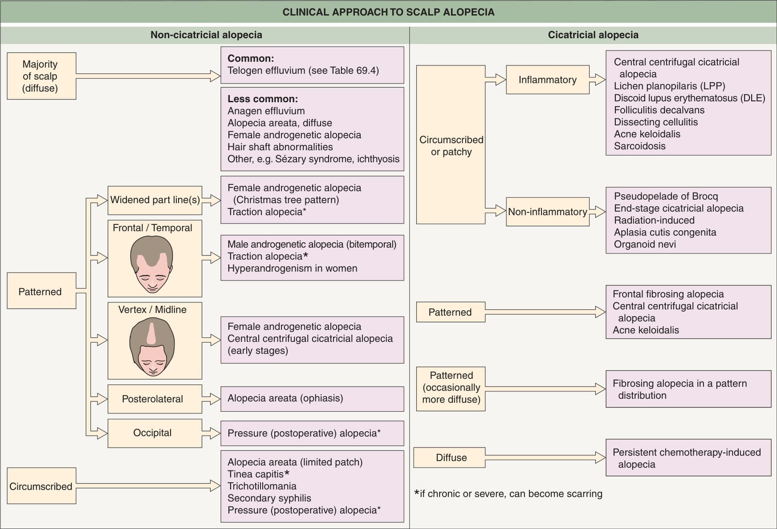
Fig. 69.1 Clinical approach to more common causes of scalp alopecia.
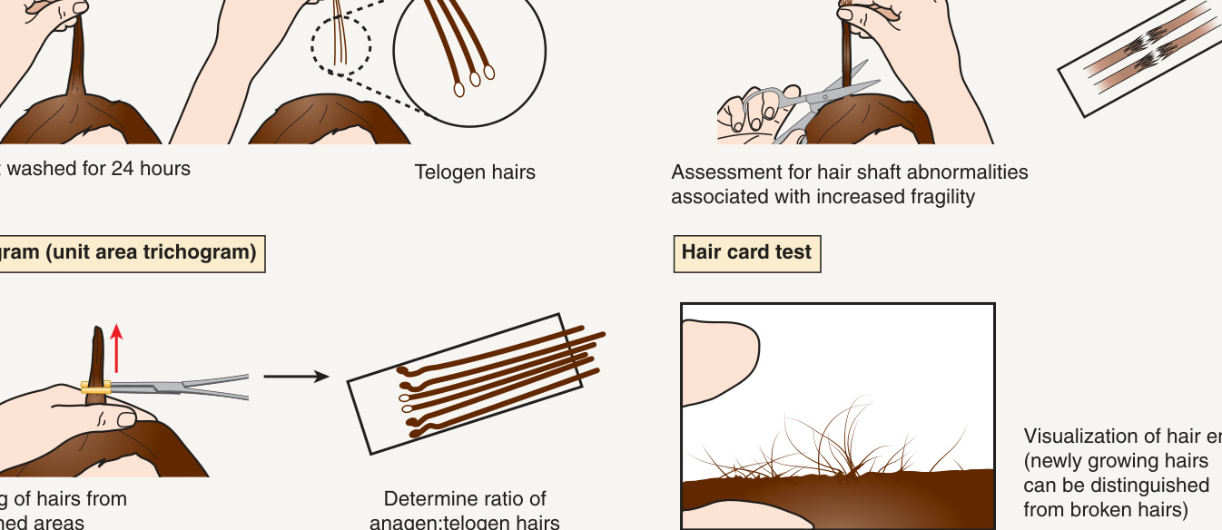
Fig. 69.2 Diagnostic tests for the assessment of alopecia.
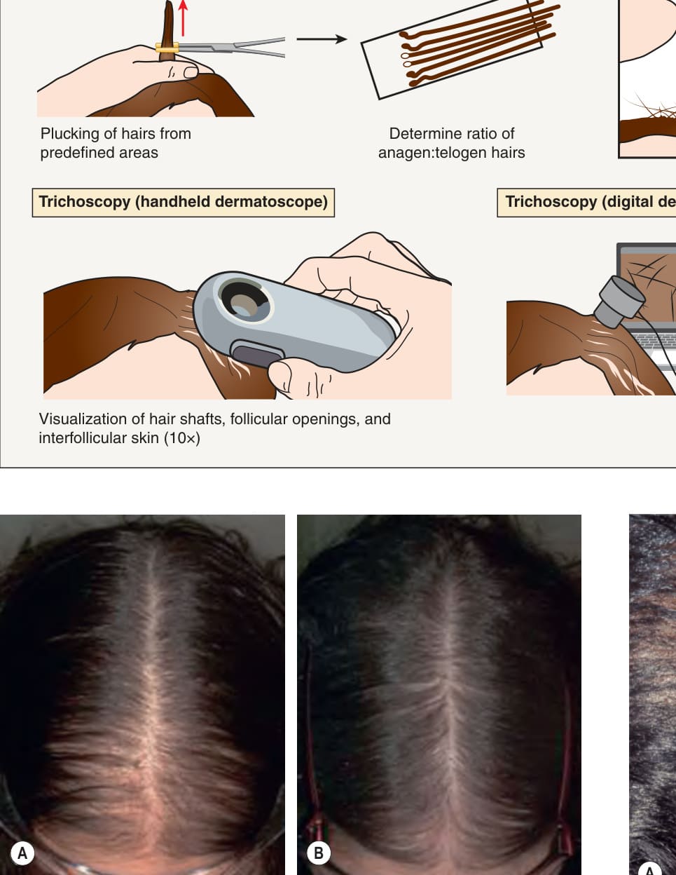
Fig. 69.3 Female androgenetic alopecia.A Prior to treatment. Note the greater hair loss anteriorly. B Obvious improvement following 5 years of therapy. Androgenetic alopecia may be at least partially reversible, when treated early. Courtesy Rodney D. Sinclair, MD.
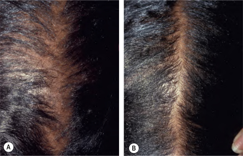
Fig. 69.4 Female androgenetic alopecia. Comparison of the part line on the top of the scalp (A) and the occiput (B). Courtesy Leonard C. Sperling, MD.
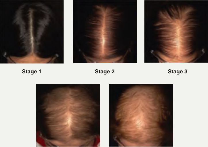
Fig. 69.5 The Sinclair scale for female androgenetic alopecia. Stage 1 – normal; Stage 2 – widening of the central part line; Stage 3 – widening of the part line with translucency of the hairs at its border; Stage 4 – development of a bald area anteriorly along the part line; and Stage 5 – advanced hair loss. Courtesy Rodney D. Sinclair, MD.
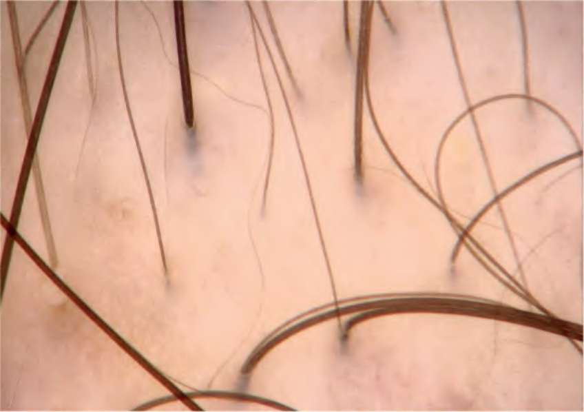
Fig. 69.6 Androgenetic alopecia – trichoscopic features. Full-thickness terminal hairs, intermediate hairs, thin hairs, and very thin (vellus) hairs are all observed simultaneously. This heterogeneity of hair shaft thickness is due to non-synchronized hair miniaturization.
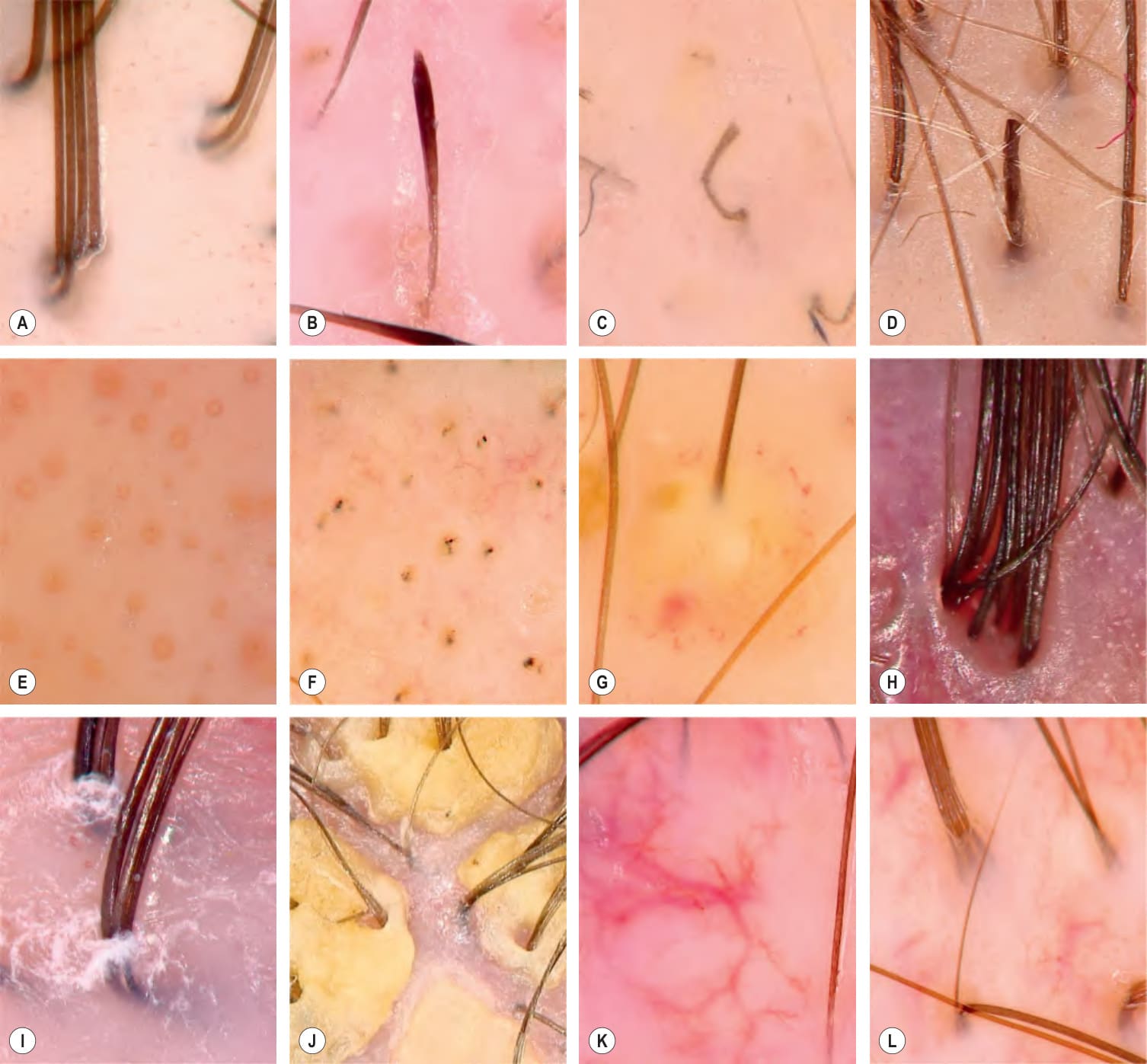
Fig. 69.7 Common trichoscopy findings.A Healthy hairs are uniform in color and shape throughout their length. The follicular units contain 1 to 4 hairs (average, 2.3). B Exclamation point hair – observed primarily in alopecia areata, trichotillomania, and chemotherapy-induced alopecia. C Comma hairs – tinea capitis. D Broken hair – trichotillomania, tinea capitis, and alopecia areata. E Yellow dots – alopecia areata and androgenetic alopecia. F Black dots – alopecia areata, tinea capitis, trichotillomania, and anagen effluvium. G Perifollicular pustule – folliculitis decalvans. H Hair tufts consisting of more than 5 hairs per follicular unit (“tufted folliculitis”) – folliculitis decalvans. I Perifollicular scaling – lichen planopilaris, folliculitis decalvans, and frontal fibrosing alopecia. J Seropurulent exudate – dissecting cellulitis and tinea capitis. K Thick arborizing vessels and tortuous vessels – systemic lupus erythematosus. L Upright regrowing hair (healthy regrowing). Zigzag hairs can be seen in tinea capitis.
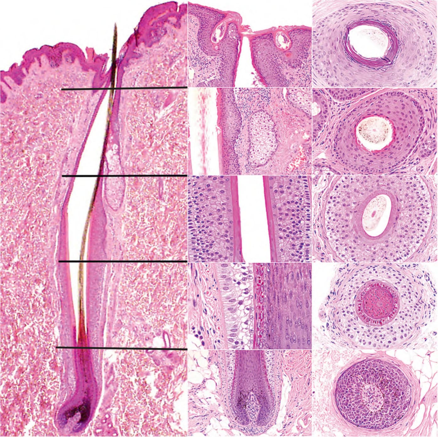
Fig. 69.8 Vertical and horizontal sections of a hair follicle cut at different levels
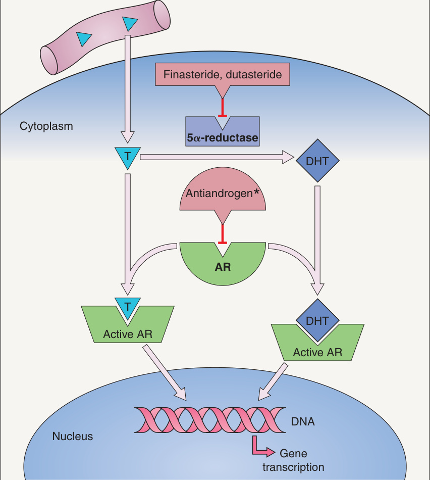
Fig. 69.10 Mechanism of action of antiandrogens and 5α-reductase inhibitors in androgenetic alopecia.
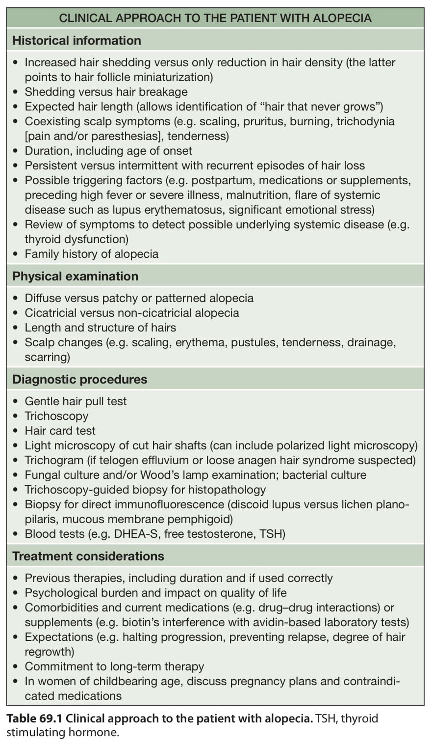
Table 69.1 Clinical approach to the patient with alopecia. TSH, thyroid stimulating hormone.
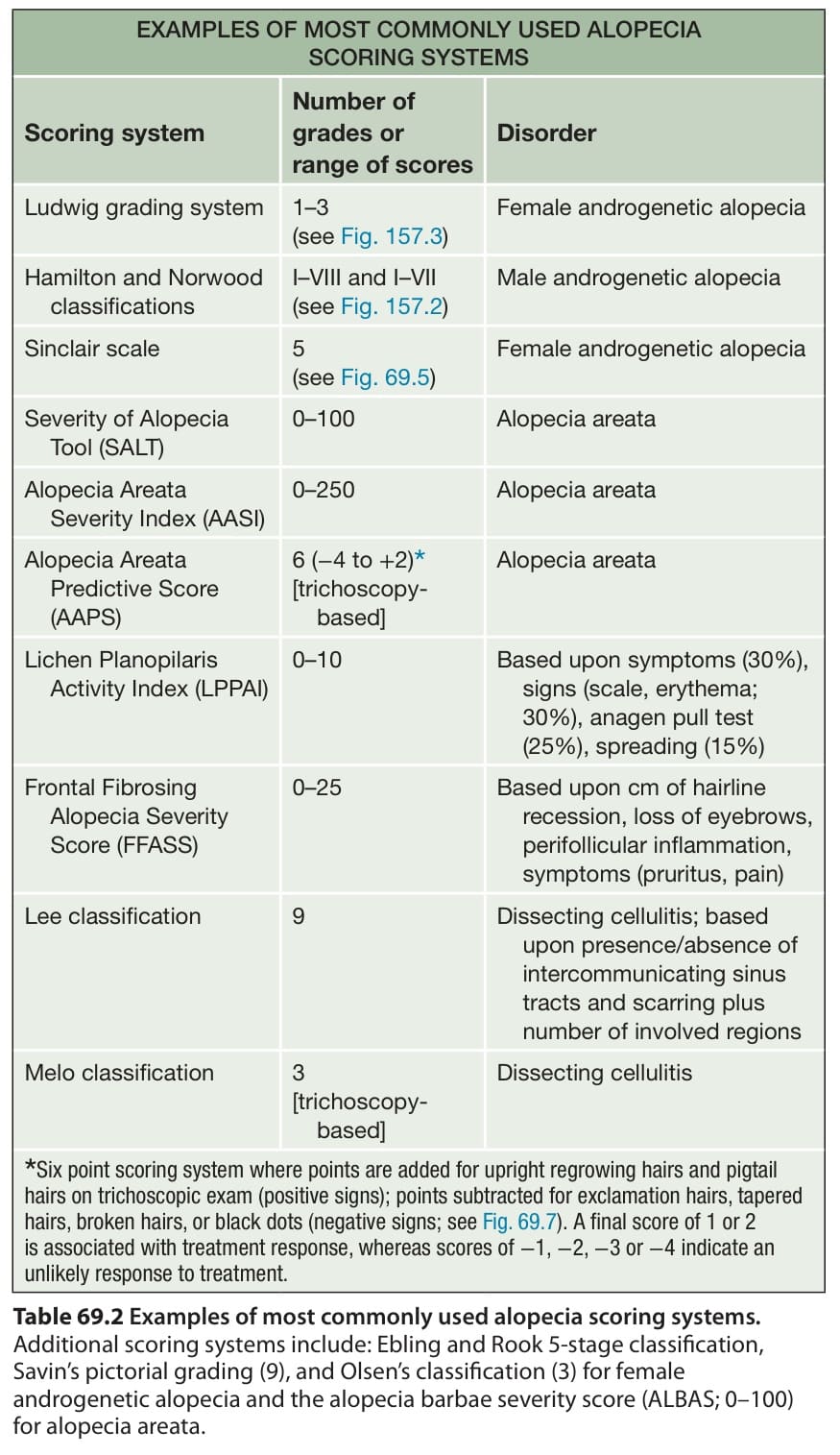
Table 69.2 Examples of most commonly used alopecia scoring systems.
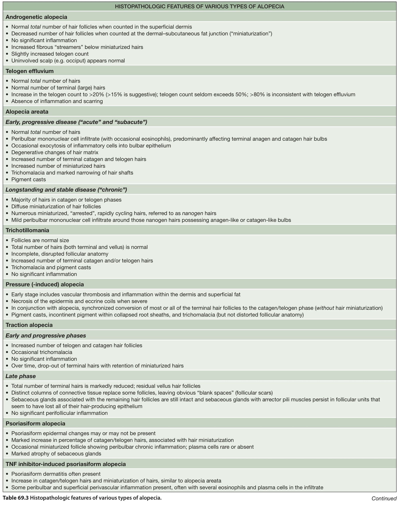
Table 69.3 Histopathologic features of various types of alopecia. Continued