Telogen Effluvium
Key features
Increased shedding of otherwise normal telogen hairs in response to an external or internal trigger (e.g. medication, high fever, malnutrition, systemic disease, significant emotional stress)
A chronic form with no identifiable precipitating factor is observed in some women
Laboratory evaluation is directed by history, physical exami- nation, trichoscopy, and microscopic evaluation of plucked hairs (trichogram)
Introduction
Scalp hair grows longer than hair on other parts of the body because its anagen phase is longer. Unlike mammals that molt, hair cycling in humans is asynchronous such that shedding of each follicle is independent of surrounding follicles. Consequently, a fairly uniform density of hair is maintained at all times.
In the normal scalp, there are ~100 000 hair follicles of which ~90% are in the anagen phase of the hair growth cycle; this phase typically lasts for 2–4 years. Hairs then enter the catagen phase (0.5%–1% of hair follicles), followed by a telogen phase (10% of hair follicles). The latter phase usually lasts for three months after which the hairs undergo conversion to exogen and are shed; daily ~50–200 hairs are shed (see Ch. 68). Internal or external factors that interfere with this normal hair cycle can trigger telogen effluvium. The most common disturbance is a shortened anagen phase.
Pathogenesis
Various external and internal factors may induce a simultaneous transition of a large number of hair follicles from anagen phase into telogen phase. This transition begins the hair shedding process which eventuates in clinically apparent hair loss (telogen effluvium) at the conclusion of the telogen phase, thus explaining its usual occurrence three months after the inciting cause. Potential triggers of telogen effluvium include pregnancy, medications, high fever, malnutrition, systemic diseases such as lupus erythematosus, and significant emotional stress as well as inflammatory scalp disorders (Table 69.4). Systemic infections represent an additional cause; for example, a quarter of individuals infected with SARS-CoV-2 experienced telogen effluvium within the following two months.
(from bottom to top).Bulb: the hair bulb embraces the dermal papilla and is composed of matrical cells admixed with pigmented melanocytes; the nucleated hair shaft is surrounded by: (1) the early inner root sheath (IRS); (2) a paler outer root sheath (ORS); and (3) a dermal fibrous sheath in continuity with the dermal papilla. Suprabulbar area: the hair shaft is nucleated and is surrounded by (moving outwards): (1) the hair cuticle – a single layer of overlapping elongated cells; (2) the Huxley layer of the IRS – 3–4 layers of cuboidals cells with keratohyaline granules; (3) the Henle layer of the IRS – single layer of elongated cells; and (4) the pale ORS. Subisthmus: the IRS is completely keratinized, the hair shaft is no longer nucleated, and the ORS is less pale and more pink, resembling epidermis. Isthmus: upon entering the isthmus, abrupt disintegration of the keratinized IRS occurs and is replaced by tricholemmal keratinization by the ORS. Infundibulum: the epithelium has reached maturation and is identical to, and in continuity with, the epidermis.
Although a wide range of medications have been reported to induce telogen effluvium, only a few have been proven. The severity of hair loss depends on the drug (see Table 69.4), its dosage, and patient susceptibility. Lastly, a significant number of women have a chronic form of telogen effluvium that lacks any identifiable trigger.
Clinical features and diagnosis
Thinning and loss of terminal hairs involves the entire scalp and may also be noted on other hairy regions of the body. A trichogram will show an increased percentage of telogen hairs, with >20% diagnostic of telogen effluvium (Fig. 69.11). By trichoscopy, telogen effluvium is often a diagnosis of exclusion. An increased percentage of follicular units with only one hair is the most characteristic feature. During the regrowth phase, multiple short upright regrowing hairs can be seen.
When due to past events such as a severe illness or pregnancy, complete hair regrowth is expected. If the underlying etiology is an ongoing issue such as hypothyroidism, then the latter needs to be addressed first (see Table 69.4). While chronic telogen effluvium can continue for years or decades, it usually does not lead to baldness.
After a history (including medications and supplements) and physical examination have been performed and the triggering factor is still unclear, then thyroid function, complete blood count, metabolic panel, ESR, and serum ferritin level can be assessed. Serum ferritin levels should be at least 40 ng/dl, but the relationship of reduced iron stores to the hair loss still needs to be determined.
For persistent cases of telogen effluvium, i.e. those lasting beyond 6 months, trichoscopy should be repeated and a trichoscopyguided biopsy from the vertex area may be considered to exclude early AGA. The histopathologic findings in telogen effluvium are outlined in Table 69.3, with the ratio of terminal to vellus hairs at the level of the isthmus allowing for distinction from AGA. That said, the coexistence of telogen effluvium and AGA is a common occurrence.
Treatment
With the exception of correcting underlying disorders or discontinuing (or substituting) medications outlined in Table 69.4, there is no well-documented treatment for telogen effluvium. Iron replacement in those with a serum ferritin level <40 ng/dl is recommended by many hair experts following determination of its etiology. Topical or low-dose oral minoxidil may be considered as an off-label, nonspecific hair growthpromoting agent as can platelet-rich plasma injections.
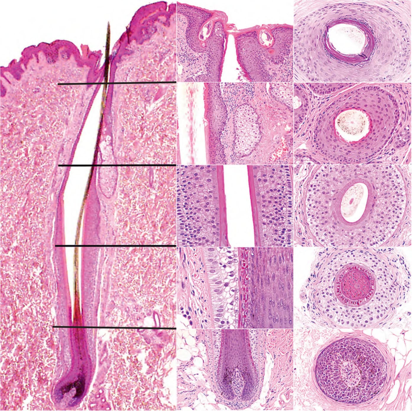
Fig. 69.8 Vertical and horizontal sections of a hair follicle cut at different levels
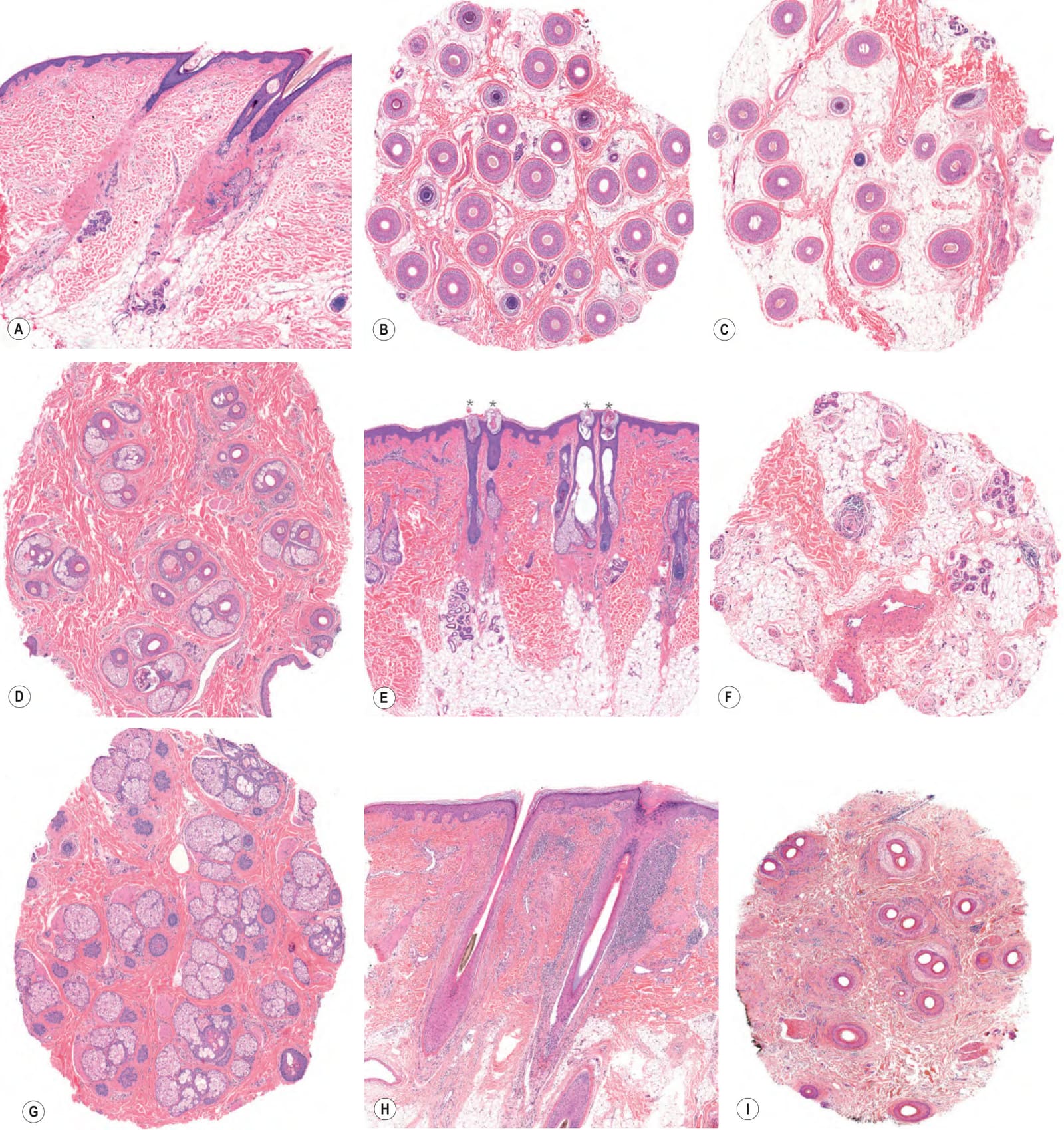
Fig. 69.9 Histopathologic features of androgenetic alopecia, alopecia areata, and lichen planopilaris.AAndrogenetic alopecia with the characteristic miniaturization of hair follicles and fibrous streamers (vertical section). B Normal scalp (horizontal section). CAndrogenetic alopecia with variation in hair follicle size (horizontal section; compare to B). DAndrogenetic alopecia at the level of the isthmus with miniaturized vellus-like hair follicles and no significant inflammation (horizontal section). EAlopecia areata with miniaturized, vellus-like hair follicles, one of which has a peribulbar lymphocytic infiltrate (vertical section). A clue to the diagnosis is the dilatation of the follicular infundibula which corresponds to the yellow dots by trichoscopy (asterisks). FAlopecia areata with the characteristic peribulbar lymphocytic infiltrates (horizontal section). GAlopecia areata at the level of the isthmus demonstrating exclusively telogen follicles (horizontal section). HLichen planopilaris. Focal hypergranulosis at the acrotrichium and a dense perifollicular lymphocytic infiltrate (vertical section). Note the Max-Joseph spaces between the hair follicle and the perifollicular sheath. ILichen planopilaris at the level of the isthmus showing complete loss of sebaceous glands, perifollicular fibrosis, perifollicular lymphocytic infiltrate, and follicular tufting (horizontal section). A–H, Courtesy Laila El Shabrawi-Caelen, MD.
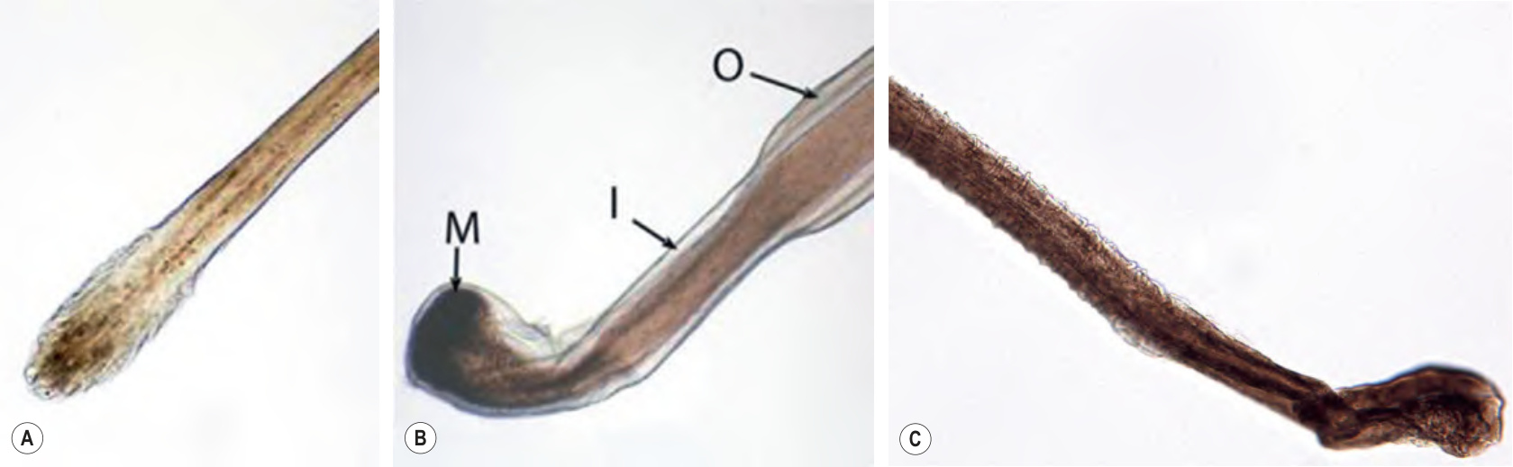
Fig. 69.11 Microscopic features of anagen and telogen hairs.A Telogen shaft showing a club-shaped bulb. B Anagen shaft with attached root sheaths, demonstrating a pigmented and distorted bulb. M, matrix; I, inner root sheath; O, outer root sheath. C Ruffled cuticle of loose anagen hair. C, Courtesy Julie V. Schaffer, MD.
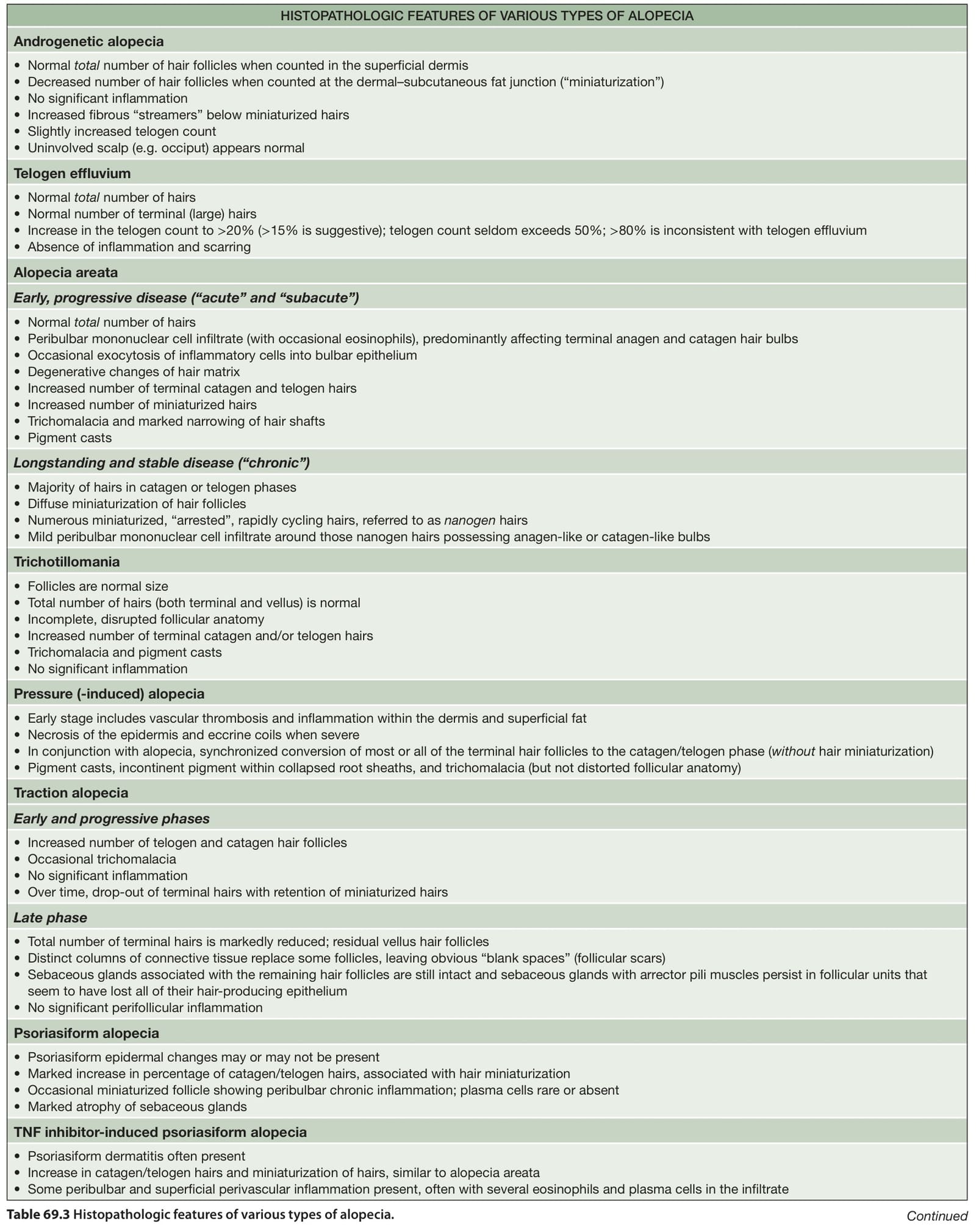
Table 69.3 Histopathologic features of various types of alopecia. Continued
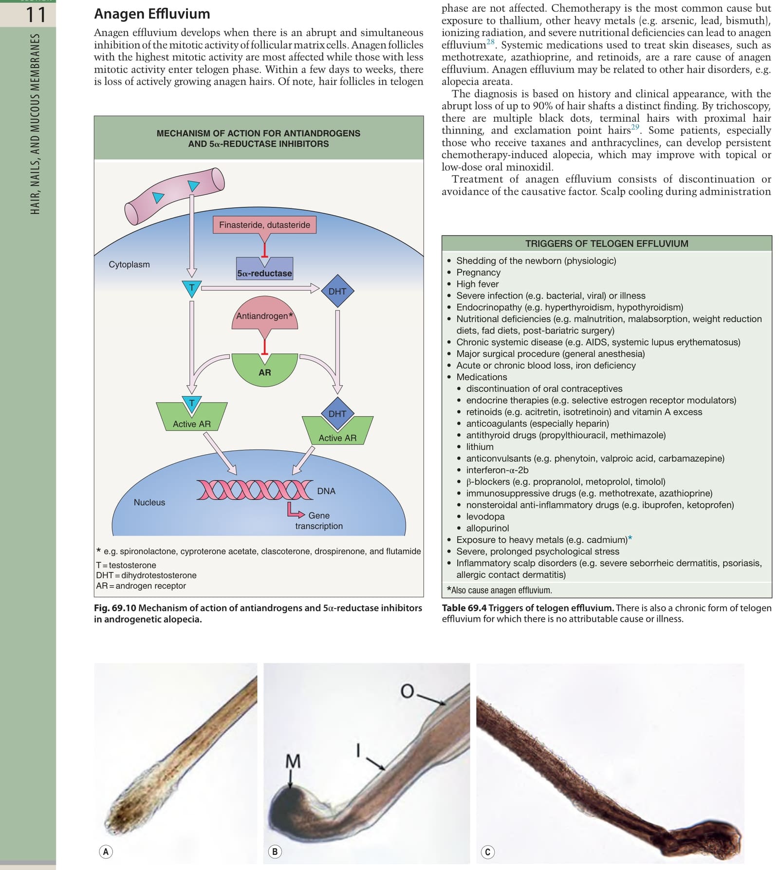
Table 69.4 Triggers of telogen effluvium. There is also a chronic form of telogen effluvium for which there is no attributable cause or illness.