Alopecia Areata
Key features
Autoimmune disease characterized by chronic or episodic non-cicatricial hair loss
Presents with hairless patches, most commonly on the scalp; may result in total hair loss
Typical trichoscopic features are exclamation point hairs, black dots, and yellow dots
Long-standing alopecia areata is associated with follicular dropout and may become irreversible
Introduction
Alopecia areata is an autoimmune disease, with genetic factors playing a role in disease susceptibility. It has a chronic relapsing course and although varying hair-bearing areas can be affected, the scalp and beard are the most common sites of involvement. The prevalence of alopecia areata in the population is ~0.1% to 0.2%, and the lifetime risk of developing alopecia areata is estimated to be ~2%.
Pathogenesis
The pathogenesis of alopecia areata is complex, with interactions involving immune, genetic, and environmental factors. It is an autoimmune T-cell mediated disease with a predominant Th1/IFN-γ response that leads to stimulation of CD8+NKG2D+ effector T cells. The latter infiltrate affected hair follicles, leading to upregulation of major histocompatibility complexes and subsequent loss of hair follicle immune privilege. IL-15 is also upregulated in diseased hair follicles, activating IFN-γ-producing CD8+NKG2D+ cytotoxic T cells which then leads to a positive feedback loop that promotes additional IL-15 production. As a result, there is a continued autoimmune attack on hair follicles and a transition from anagen to catagen. IFN-γ and IL-15 are dependent on the JAK-STAT signaling pathway for the activation and proliferation of autoreactive T cells (see Fig. 128.11), providing an explanation for the therapeutic response to JAK inhibitors. Of note, follicular stem cells are not usually targeted, allowing for future hair regrowth via spontaneous remission or effective treatment.
For some patients, environmental factors such as viral infections, vaccines, medications, and stress have been implicated as possible triggers30a. From a genetic standpoint, alopecia areata is an example of a complex polygenic disease30b,30c. Genome-wide association studies have confirmed an association with particular HLA genes, with the HLA-DQB1*03 allele identified as an important susceptibility marker30d,30e. There is also a strong association with genes in the ULBP gene cluster (chromosome 6q25) which code for activating ligands of the NKG2D receptor30f. Patients with alopecia areata have an increased risk of other autoimmune diseases (e.g. thyroid disease, rheumatoid arthritis) and this may be due to a shared inherited autoimmune pathway. Indeed, several susceptibility loci linked to other autoimmune diseases have been identified in patients with alopecia areata, e.g. CTLA-4, IL-2/IL-21, IL-2RA30g. Lastly, both PTPN22, which encodes a lymphoid protein tyrosine phosphatase that normally suppresses T cell proliferation, and IL-1 cluster genes (IL-1RA) have been associated with severe forms of alopecia areata30h.
Clinical features
Alopecia areata commonly presents as round or oval patches of non-scarring hair loss (patchy alopecia areata; Fig. 69.12A,B). Other presentations include alopecia totalis (loss of all scalp hair), alopecia universalis (loss of all scalp and body hair), ophiasis (band-like pattern of hair loss along the periphery of the temporal and occipital scalp), and alopecia involving the beard, eyebrows, or other hair-bearing sites (Fig. 69.12C–F). The reticular variant is characterized by recurrent patchy disease, in which the patient experiences hair loss in one area while at the same time undergoing spontaneous regrowth in other sites. An unusual variant is sisaipho (ophiasis inversus) in which the hair loss spares temporal and occipital areas. The rare diffuse variant of alopecia areata can present with widespread thinning or may primarily affect the vertex. Non-pigmented hairs may remain unaffected such that some patients with diffuse scalp involvement appear to develop rapid graying of the hair.
The hair loss in alopecia areata is generally reversible, but late-stage disease is characterized by progressive follicular drop-out. Prognosis worsens when alopecia areata persists beyond 5 years and hair loss can become irreversible when the alopecia persists for >10 years. However, irreversibility should not be assumed nor serve as a contraindication for a therapeutic trial. The Alopecia Areata Predictive Score (AAPS) can be used to estimate the probability of hair regrowth based upon trichoscopy findings (see below).
Alopecia areata, especially when severe, may be associated with nail involvement. The most common abnormality is nail pitting, often in a grid-like pattern (see Fig. 71.2). Other changes include trachyonychia (sandpaper-like roughness due to excessive longitudinal ridging; Fig. 69.12G), brittle nails, onycholysis, koilonychia, and rarely, onychomadesis.
In alopecia areata, the three most common trichoscopic findings are: (1) exclamation point hairs – proximal hair shaft thinning due to episodes of arrested mitotic activity within the germinal matrix; (2) black dots – when the exclamation point hairs break off, the residual dark hair shaft is visible in the follicular opening; and (3) yellow dots – empty hair follicles filled with keratotic material and sebum (Fig. 69.13; see Fig. 69.7B,E,F). The latter may be more subtle in prepubescent children. Tapered hairs represent very long exclamation point hairs and they indicate moderate disease activity. Pigtail (circle) and upright regrowing hairs indicate hair regrowth (see Fig. 69.7L).
Thyroid dysfunction, atopy, and systemic lupus erythematosus are the most common immune-mediated disorders associated with alopecia areata, with odds ratios of 4.4, 2.4, and 3.5, respectively. Reported comorbidities include metabolic syndrome, depression, and anxiety. Patients with Down syndrome have an increased incidence of alopecia areata and the latter occurs in a third of individuals with the genodermatosis autoimmune polyendocrinopathy–candidiasis–ectodermal dystrophy (APECED) syndrome. Some families with vitamin D-dependent rickets type II also develop alopecia areata.
Satoyoshi syndrome is a multisystem disorder in which patients can develop severe alopecia areata. Hair loss represents the syndrome’s major criterion while intermittent painful muscle spasms, diarrhea, and anti-nuclear antibodies are the minor criteria (2 of 3 required for diagnosis. Lastly, in a small study of patients with alopecia areata, gut dysbiosis was observed.
Differential diagnosis
Tinea capitis, trichotillomania, temporal triangular alopecia, traction alopecia, secondary syphilis, and loose anagen syndrome need to be considered in the differential diagnosis of alopecia areata as well as pressure-related alopecia, aplasia cutis, and “burnt-out” cicatricial alopecia. The diffuse variant may initially be confused with telogen
effluvium and AGA. Usually, the history and clinical examination (including trichoscopy) is sufficient to distinguish between these conditions, but performing a scalp biopsy may prove useful for less distinctive presentations. The histopathologic features of alopecia areata are illustrated in Fig. 69.9E,F,G and are outlined in Table 69.3.
Treatment
Multiple off-label, variably successful topical, intralesional, and systemic therapies are available for the treatment of alopecia areata, with the therapeutic approach depending upon several factors such as age, extent of disease, and patient expectations. The FDA recently approved two oral JAK inhibitors for severe alopecia areata in adults (baricitinib) and for alopecia areata with extensive hair loss in individuals age 12 years and older (ritlecitinib). Most experts view a Severity of Alopecia Tool (SALT) score of >20 as an indication for systemic therapy.
First-line topical therapy usually consists of an ultrapotent (class I) corticosteroid. Additional topical options include calcineurin inhibitors, minoxidil, anthralin, and especially for eyebrows, prostaglandin analogues (e.g. bimatoprost, latanoprost) and topical JAK inhibitors. Children with severe alopecia areata can be offered contact immunotherapy (e.g. diphenylcyclopropenone [diphencyprone], squaric acid dibutyl ester) before systemic therapy is considered. In all age groups, concurrent systemic immunosuppressants should be avoided as they can blunt the desired inflammatory response from contactants.
A solitary patch on the scalp is best treated with intralesional triamcinolone acetonide (2.5 to 5 mg/ml) every 3–4 weeks. Approximately 0.1 ml is injected into the mid to deep dermis, with injection sites at intervals of 1 cm apart. In an average adult, the recommended maximum dose administered during a single session is 10 to 20 mg.
In adults and adolescents with severe disease, options include prednisone or prednisolone, Janus kinase (JAK) inhibitors, and cyclosporine; pulse corticosteroids (e.g. dexamethasone 0.1 mg/kg twice weekly, prednisolone 200 mg weekly, prednisolone 5 mg/kg [up to 300 mg] monthly) can also be considered in children as well as adults. When daily corticosteroids are administered for rapidly progressive disease, an initial dose of prednisolone is typically 0.4 to 0.6 mg/kg/day with a gradual taper over at least 12 weeks and a possible transition to a pulse regimen. Either cyclosporine (3 to 5 mg/kg/day) or methotrexate (15 to 20 mg weekly) may be used as a steroid-sparing agent or as monotherapy. In younger patients, the methotrexate target dose is ~0.4 mg/kg/week. For other steroid-sparing agents, including mycophenolate mofetil, azathioprine, dapsone, sulfasalazine, IL-17 inhibitors, apremilast and dupilumab, there are limited data regarding efficacy. The latter has been successful in patients who had concomitant atopic dermatitis.
Multiple more recent studies have demonstrated the effectiveness of oral JAK inhibitors (see Ch. 128), in particular baricitinib and ritlecitinib41a, which are now FDA-approved for the treatment of alopecia areata (see above), as well as tofacitinib and deuruxolitinib. However, maintenance therapy is necessary to avoid relapse. Topical JAK inhibitors (e.g. 2% tofacitinib, 1.5% ruxolitinib) are less effective than their oral counterparts, and use of JAK inhibitors in children (maximum daily dose of 0.625 mg [<20 kg] to 1.25 mg [20–40 kg]) is currently off-label. Irrespective of the preferred treatment, low-dose oral minoxidil can be employed as supplemental therapy.
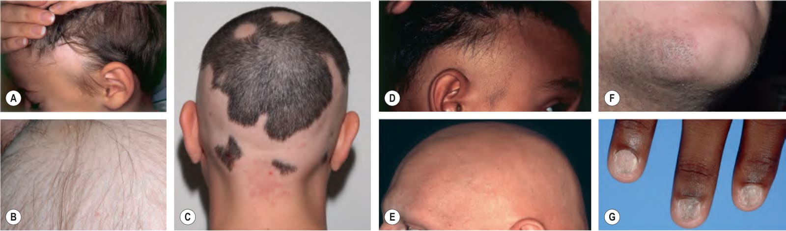
Fig. 69.12 Alopecia areata.A A circular patch of alopecia areata in a child. B Within a patch of alopecia areata, hair loss may be incomplete as telogen hairs remain; note the multiple exclamation point hairs. C Circular and confluent areas of alopecia, leading to an ophiasis pattern. There is an incidental nevus simplex. D Ophiasis with a band-like pattern of hair loss along the periphery of the temporal and occipital scalp. E Total alopecia of the scalp, eyebrows, and eyelashes in a patient with alopecia universalis. F Alopecia areata of the beard. G Trachyonychia, which is usually associated with severe alopecia areata. B, G, Courtesy Julie V. Schaffer, MD; C, Courtesy Edward Cowen, MD; E, Courtesy Leonard C. Sperling, MD; F, Courtesy Rodney D. Sinclair, MD.
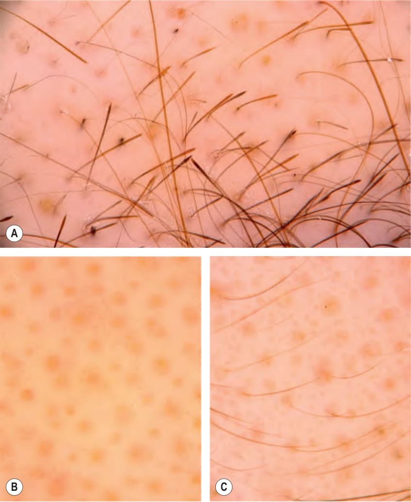
Fig. 69.13 Trichoscopy of alopecia areata.A Active disease with multiple exclamation point hairs and black dots; some yellow dots are also seen. B Chronic phase with only yellow dots. C Regrowth phase with thin hair shafts emerging from some of the yellow dots (eyebrow).
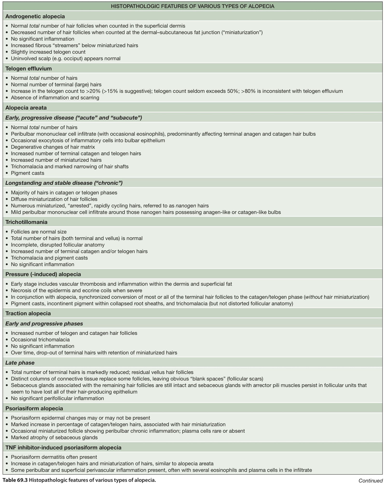
Table 69.3 Histopathologic features of various types of alopecia. Continued