CICATRICIAL (SCARRING) ALOPECIAS
Introduction
“Cicatricial” (or “scarring”) alopecia implies that the follicular epithelium has been replaced by fibrotic tissue. The lack of hair follicles, in particular replacement of the follicular bulge region by collagen and other extracellular matrix proteins, results in irreversible hair loss. Cicatricial alopecia may be primary or secondary. In primary cicatricial alopecia, the follicle is destroyed by an inflammatory process which targets the follicular unit. In secondary cicatricial alopecia, the follicle is merely an “innocent bystander” in the disease process. Examples of secondary cicatricial alopecia include deep burns, radiation dermatitis, cutaneous malignancies, cutaneous sarcoidosis, morphea, necrobiosis lipoidica, and certain chronic infections such as cutaneous tuberculosis. The various forms of secondary cicatricial alopecia should have distinctive clinical and histologic features typical of the underlying disease.
Performing a scalp biopsy may be helpful not only in establishing a diagnosis but also in assessing the degree of inflammation and injury to the stem cell region. Specimens should be obtained at the hair-bearing margin (guided by trichoscopy), be at least 4 mm in diameter, and extend into the fat. If the central portion is sampled, only end-stage scarring with fibrosis will be seen. Ideally, two biopsies should be performed – one for vertical sectioning, the other for horizontal sectioning. The North American Hair Research Society proposed a classification for primary cicatricial alopecias and, despite some controversies, it remains the best available working classification.
Lichen Planopilaris
Several distinct entities are considered to be within the broader lichen planopilaris spectrum: (1) classic lichen planopilaris; (2) frontal fibrosing alopecia; and (3) Graham Little–Piccardi–Lasseur syndrome. Some authors also consider fibrosing alopecia in a pattern distribution (FAPD) a distinct entity within this spectrum.
Classic Lichen Planopilaris
Key features
Chronic, progressive, inflammatory cicatricial alopecia
Trichoscopy shows perifollicular scaling and areas with no hair follicle openings
Histologically, there is a lymphocytic infiltrate involving the isthmus and infundibulum
Treatment consists primarily of systemic immunosuppressive drugs
Pathophysiology
In lichen planopilaris, the infundibulo-isthmic (bulge) region of the hair follicle is targeted by an immune-mediated lymphocytic infiltrate. This folliculocentric inflammatory process leads to localized fibrosis and replacement of hair follicles by fibrous tracts. The factors which trigger this disease process remain unknown.
Clinical features and diagnosis
Lichen planopilaris occurs predominantly in White women with an onset of disease between 40 and 60 years of age. Lichen planopilaris in children is rare and is usually associated with a positive family history.
Hyperesthesia of the scalp may be a premonitory symptom. A burning sensation, pain, or pruritus is usually associated with active hair loss. During the early phase, one or more small (<1 cm) patches of cicatricial alopecia appear on the vertex or in the occipital region (Fig. 69.17A). In the hair-bearing margin, there is perifollicular erythema and hyperkeratosis (Fig. 69.17B). The patches of alopecia tend to increase in size
and become confluent over time. Occasionally “lonely hairs” may be present in the central portion of the lesions.
Up to 15% of patients have coexisting mucosal lichen planus. Vulvovaginal–gingival–pilar lichen planus is a distinct entity that combines the vulvovaginal–gingival syndrome (see Ch. 11) with extensive and recalcitrant lichen planopilaris and circulating stratified epithelium-specific anti-nuclear antibodies (SES-ANA).
Trichoscopy of lichen planopilaris shows perifollicular erythema and scaling, with the scales entangling hair shafts up to 2–3 mm above the scalp surface in a tubular fashion (Fig. 69.17B inset). Hair casts and elongated linear blood vessels may also be present. Late-stage disease is characterized by milky-red areas with no follicular openings. The histopathologic features of lichen planopilaris are outlined in Table 69.3 and illustrated in Fig. 69.9H,I. Of note, perifollicular inflammation can be observed in areas with no clinically evident disease, raising the possibility that lichen planopilaris may be a generalized process that affects the entire scalp.
Treatment
The goal of treatment is to reduce pruritus and halt any further scarring. Recommended first-line treatments include hydroxychloroquine, doxycycline, and corticosteroids (intralesional or oral). Topically, potent corticosteroids and calcineurin inhibitors may be used as adjuvant therapy. Second-line therapeutic options include retinoids (acitretin and isotretinoin), mycophenolate mofetil, methotrexate, and cyclosporine as well as JAK inhibitors, apremilast, and pioglitazone. Topical or low-dose oral minoxidil and 5α-reductase inhibitors may also be used as adjuvant therapy. While lichen planopilaris can burn out spontaneously, many patients have chronic progressive disease.
Frontal Fibrosing Alopecia
Key features
Cicatricial alopecia with selective involvement of the hairline, most commonly frontotemporal
Additional findings are a loss of eyebrows and facial papules
Trichoscopy shows perifollicular scaling and “lonely hairs”
Systemic immunosuppressants and 5α-reductase inhibitors may halt or slow progression of the disease
Pathogenesis
In frontal fibrosing alopecia (FFA), as in classic lichen planopilaris, the infundibulo-isthmic (bulge) region of the hair follicle is attacked by an immune-mediated inflammatory infiltrate; the latter has a predominance of CD8+ T lymphocytes. The inciting factors that lead to this inflammation are unknown. Environmental exposures such as leave-on cosmetics, facial sunscreens, fragrances, haircare products, contact allergens, and diet have all been implicated as potential triggers in genetically predisposed patients, but to date there are no definitive associations.
Clinical features
Frontal fibrosing alopecia was first described by Kossard and colleagues in 1994. It occurs primarily in perimenopausal women, but younger women and men may also be affected. A band-like recession of the frontotemporal hairline is typically seen, which usually progresses at a rate of ~0.3–1.5 mm per month. The occipital and/or retro-auricular hairlines can also be involved.
Three clinical patterns of hair loss have been described: (1) linear – a clearly demarcated hairline is seen; (2) diffuse – there is a decrease in hair density in the frontal hairline; and (3) pseudo-fringe – a “fringe” of frontal hairs is isolated from the hairline by a hairless band (Fig. 69.18). In all three patterns, only full-thickness terminal hair shafts are visible within the involved hairline. Vellus hairs, which would be present in the “fringe” associated with traction alopecia, are not present because they are the first to disappear during the disease process. The skin is smooth, slightly atrophic, devoid of follicular openings, and lighter in color than the remaining skin of the forehead.
Additional clinical features of FFA include “lonely” hairs (Fig. 69.18A inset), lateral or complete eyebrow loss, partial loss of eyelashes, prominent or depressed forehead veins, multiple facial papules (see Fig. 69.18C), and glabellar follicular red dots. The latter two findings are due to involvement of facial vellus hairs. In late-stage FFA, there may also be thinning of axillary, pubic, limb, and truncal hair. Of note, classic lichen planus or lichen planus pigmentosus (especially in patients with darker skin phototypes) may accompany FFA. There are also rare reports of FFA coexisting with cutaneous lupus.
Treatment
Treatment options for FFA are similar to those described previously for lichen planopilaris. Of note, oral finasteride or dutasteride may be beneficial. Possible explanations for the efficacy of 5α-reductase inhibitors include an effect on coexisting androgenetic alopecia or inhibition of the conversion of terminal to vellus hair follicles, which would then lead to a reduction in the proportion of vellus hair follicles, the primary target in FFA.
Graham Little–Piccardi–Lasseur Syndrome
The Graham Little–Piccardi–Lasseur syndrome (Graham Little syndrome) was first described by Piccardi in 1913. The description by Ernst Graham Little (in a mutual patient with Lasseur) appeared in 1915 and was the inspiration for the eponym “Graham Little syndrome”. The syndrome consists of a triad of lichen planopilaris of the scalp, non-cicatricial alopecia of the axilla and groin, and follicular lichen planus of the body (see Ch. 11).
Fibrosing Alopecia in a Pattern Distribution
Fibrosing alopecia in a pattern distribution (FAPD) is a more recently recognized type of cicatricial alopecia that has characteristics of both lichen planopilaris and AGA. It is considered by many to be a subtype of lichen planopilaris. Although the etiology is not well understood, FAPD is thought to be due to an exaggerated inflammatory response to damaged hair follicles, perhaps triggered by AGA. It occurs primarily in androgen-dependent areas of the scalp in which there has been hair follicle miniaturization. Clinically and by trichoscopy, there is both perifollicular inflammation (e.g. erythema, scaling) and fibrosis with loss of follicular ostia. Sometimes FAPD is misdiagnosed as AGA
A Progressive hair loss along the anterior hairline with absence of solar lentigines (diffuse pattern). There are scattered “lonely” hairs (inset) and an eyebrow pencil has been used because of loss of eyebrow hairs. B Hypopigmented, atrophic band of cicatricial alopecia with a clearly demarcated, receded hairline (linear pattern) in addition to a “pseudofringe”; the latter is a fringe of hairs that marks the previous hair line. C Facial papules and loss of eyebrows; the patient also has rosacea. D By trichoscopy, discrete perifollicular scaling around remaining hairs and beige areas with no follicular openings (dots). A, Courtesy Kalman Watsky, MD.
plus seborrheic dermatitis, but histologic examination of early-stage sites of scalp inflammation can help confirm the diagnosis. Although there are no well-documented treatments, a combination of an anti-inflammatory drug (see Lichen Planopilaris, Treatment), antiandrogen medication (see AGA, Treatment), and hair growth-promoting agent (e.g. minoxidil) has been recommended.
Classic Pseudopelade (Pseudopelade of Brocq)
Classic pseudopelade is a non-inflammatory or minimally inflammatory form of cicatricial alopecia that most commonly affects middle-aged and older women. It most likely represents a distinct clinical entity that falls within the spectrum of lichen planopilaris. The hair loss usually begins on the vertex and is characterized by multiple, small (<2 cm), smooth, slightly depressed, asymptomatic hairless patches described as appearing like “footprints in the snow” (Fig. 69.19). Over time, these lesions can become confluent and form larger areas of alopecia. While pseudopelade is classified as a cicatricial alopecia (Table 69.5), there is debate as to whether the hair follicles undergo atrophy rather than being replaced by fibrotic tissue.
Pseudopelade is usually slowly progressive but in some patients may be self-limiting. Treatment options are similar to those described previously for lichen planopilaris, with the caveat that immunosuppressive drugs are less likely to be effective in the absence of significant inflammatory infiltrates.
Central Centrifugal Cicatricial Alopecia
Key features
Slowly progressive, symmetric cicatricial alopecia centered on the mid scalp or vertex
Most often develops in Black women; in some patients has been associated with mutations in PADI3
Treatment includes topical or intralesional corticosteroids, oral tetracyclines, and adjuvant immunosuppressive drugs
Brocq). Irregularly shaped areas of scarring alopecia along the midline. Despite no visible features of inflammation, the disease is usually progressive. Trichoscopy shows milky-red areas of cicatricial alopecia, sparse terminal hairs, and no features of inflammation (inset). Courtesy Kalman Watsky, MD.
Pathophysiology
Recent insights into the underlying pathophysiology of central centrifugal cicatricial alopecia (CCCA) include the discovery of mutations in PADI3 in some patients. The latter encodes peptidyl arginine deiminase type III, which modulates hair structural proteins including filaggrin and trichohyalin via deimination. Perifollicular lymphocytic infiltrates also play a significant pathogenic role in that they induce premature desquamation of the inner root sheath which facilitates hair loss. In addition, hair styling practices that cause traction have been implicated as potential triggers as have infectious etiologies and autoimmunity, but they remain unproven.
Clinical features
The disease begins and remains most severe on the vertex and/or mid-scalp, expanding outward in a centrifugal manner (Fig. 69.20). Most patients note mild, episodic pruritus or tenderness in the involved areas. A few isolated hairs or tufts of hair may be stranded in the otherwise denuded central zone. Perifollicular pustules may also be present. By trichoscopy, there is loss of follicular openings, mild perifollicular scaling, small hair tufts, and in darkly pigmented skin, fibrotic white dots.
Differential diagnosis
All conditions that can result in a progressive pattern of alopecia affecting the crown can mimic CCCA. These include AGA, lichen planopilaris, folliculitis decalvans, and even unusual cases of discoid lupus erythematosus. Other entities in the differential diagnosis are tinea capitis (kerion) and true bacterial infections of the scalp.
Treatment
Most authors suggest that the management should begin with reduction in chemical straightening and traction-producing hair styles as well as instituting healthy hair practices (Table 69.6). Corticosteroids (potent topical or intralesional triamcinolone) are usually employed as firstline treatment. Once stabilization is achieved, maintenance therapy can consist of less frequent application of a potent topical corticosteroid. Oral doxycycline or minocycline are frequently prescribed while other possible therapeutic options include low-dose oral minoxidil as well as hydroxychloroquine, thalidomide, cyclosporine, or mycophenolate mofetil. These systemic medications can be supplemented by topical minoxidil and calcineurin inhibitors. There are anecdotal reports of the use of oral JAK inhibitors.
Discoid Lupus Erythematosus
Synonym: Chronic cutaneous lupus erythematosus, discoid form
Key features
Early lesions are characterized by erythema, scaling, and dilated, plugged follicular ostia
Inflammatory lesions are gradually replaced by cicatricial alopecia
The diagnosis is based on clinical evaluation, trichoscopy, histo pathology, and, if needed, direct immunofluorescence (DIF) of lesional skin
Antimalarial drugs and intralesional corticosteroids represent first- line therapies
Introduction
Discoid lupus erythematosus (DLE) can be an isolated disease or it can occur in the setting of systemic lupus erythematosus (SLE). Among patients with skin disease only, the majority will have lesions limited to the head and neck region and most do not develop systemic disease (see Ch. 41). There is, however, an increased risk of SLE in those individuals who have lesions beyond the head and neck, i.e. disseminated DLE.
Clinical features
DLE is one of the four entities classified as chronic cutaneous LE (see Fig. 41.4), and the vast majority of patients are women. Early lesions are characterized by erythema, scaling, and dilated, plugged follicular ostia. Central hypopigmentation and peripheral hyperpigmentation are commonly seen in individuals with darker skin phototypes. The inflammatory lesions are gradually replaced by cicatricial alopecia, which develops in the central portion of the lesion and progresses outwards (Fig. 69.21).
Diagnosis
By trichoscopy, there are large yellow dots, thick arborizing vessels, and fine scaling (Fig. 69.21 inset). Scattered dark-brown to bluish discoloration is seen in patients with darker skin phototypes. In late-stage disease, features are similar to those observed in other cicatricial alopecias, e.g. milky-red or white areas with no follicular openings (dots) (see Fig. 69.19 inset).
The clinical diagnosis of DLE should be confirmed by histologic examination (see Table 69.3). If distinction from lichen planopilaris or other disorders in the differential diagnosis is not possible, DIF of the active peripheral edge of a lesion can be performed. It is recommended that application of topical corticosteroids be suspended for at least several days to proposed biopsy sites.
Treatment
Photoprotection and avoidance of sun exposure are important preventive measures. Active, inflammatory discoid lesions usually respond to oral antimalarial drugs (e.g. hydroxychloroquine) and corticosteroids (topical, intralesional). Second-line therapies for antimalarial-resistant cutaneous disease, such as systemic retinoids, methotrexate, dapsone, mycophenolate mofetil, thalidomide, lenalidomide, and IVIg, are outlined in Table 41.11. Monoclonal antibodies, e.g. anifrolumab, litifilimab, daxdilimab, that inhibit production or actions of type I inter-ferons and are usually prescribed for SLE can also improve associated mucocutaneous disease. There are scattered reports of JAK inhibitors and ustekinumab being effective in severe cases of recalcitrant DLE.
Follicular mucinosis
Synonym: Alopecia mucinosa (clinical term)
Alopecia mucinosa is characterized histologically by the accumulation of mucin within the pilosebaceous unit. Patches, plaques, and papules commonly develop in the head and neck region. When the scalp is involved, well-defined areas of hair loss, usually 2–6 cm in diameter, can be seen; they are often accompanied by inflammation. Grouped erythematous follicular papules can also be observed within the hairless patches (see Fig. 46.15).
With the exception of urticaria-like follicular mucinosis in which there is no associated alopecia, patients with alopecia mucinosa are subdivided into primary and secondary forms (see Ch. 46). Primary disease favors children and adolescents and often resolves within 1–2 years, including hair regrowth. In adults, a greater number of lesions that are more persistent may be seen. However, excluding a secondary cause, in particular mycosis fungoides, is important in this latter group. In addition to primary alopecia mucinosa and an association with cutaneous lymphomas, deposition of mucin within hair follicles (follicular mucinosis) can be observed as an incidental finding in a number of disorders including eczematous dermatoses, SLE, and medications (e.g. imatinib). Trichoscopy shows multiple enlarged follicular openings filled with keratotic material.
In secondary follicular mucinosis, treatment focuses on the under-lying disease, including phototherapy. Topical, intralesional, or oral corticosteroids may be used for primary disease, realizing that, especially in children and adolescents, the disease may resolve spontaneously. Other treatment options include hydroxychloroquine, minocycline, dapsone, indomethacin, and isotretinoin.
Keratosis Follicularis Spinulosa Decalvans
Key features
Rare X-linked recessive disorder within the spectrum of keratosis pilaris atrophicans
Keratosis pilaris-like papules favor the scalp and face
Follicular atrophy and scarring alopecia can follow resolution of papules
Keratosis follicularis spinulosa decalvans is a rare genodermatosis with an X-linked recessive (XLR) pattern of inheritance when due to mutations in MBTPS2 (see Table 38.2). It is allelic with the XLR form of ichthyosis follicularis with atrichia and photophobia (IFAP) syndrome. The disease usually becomes apparent during infancy or early childhood and remains active through adolescence. Keratosis pilaris-like papules favor the face and scalp, but are sometimes more widespread. Additional findings include facial erythema, palmoplantar keratoderma, and keratitis (see Table 38.2). Hypotrichosis is often noted early on and progression of lesions leads to cicatricial alopecia of the scalp, eyebrows, and eyelashes.
Histologically, there is follicular hyperkeratosis and follicular destruction with a mixed lymphohistiocytic infiltrate as well as perifollicular and interfollicular fibrosis. In the one case described to date, trichoscopic findings (e.g. perifollicular scaling) were similar to those seen in lichen planopilaris. Oral medications, including isotretinoin, dapsone, and antibiotics, have been tried with varied results.
Folliculitis Decalvans
Key features
Neutrophilic cicatricial alopecia
It may possibly represent an altered immune response to the follicular microbiome
When active, follicular pustules and hair tufts are the most charac- teristic findings
The most common treatment is long-term oral antibiotics
Pathophysiology
Folliculitis decalvans is a neutrophilic primary cicatricial alopecia. The presence of Staphylococcus aureus has been observed in 20%–75% of patients. It has been suggested that folliculitis decalvans may result from an abnormal immune response to the follicular microbiome, with the inflammation leading to permanent follicular damage.
Clinical features
This disease typically affects young and middle-aged adults, predominantly men. The vertex and occipital area of the scalp are most often involved. During its early phase, there are recurrent follicular pustules; perifollicular erythema, scaling, erosions, and hemorrhagic crusts may also be seen. A hallmark of folliculitis decalvans is the presence
of hair tufts (5–20 or more hairs in one follicular unit; Fig. 69.22), sometimes referred to as “tufted folliculitis”. Although this is a characteristic finding in folliculitis decalvans, hair tufts also develop following repeated follicular injury in other inflammatory cicatricial alopecias (e.g. acne keloidalis, dissecting cellulitis). With progression, hairless patches develop that can coalesce to form large areas of cicatricial alopecia. In addition to hair tufts, active inflammation (e.g. erythema, follicular pustules, scaling, crusting) may be present at the margins of the alopecic lesions.
By trichoscopy, tufted hairs surrounded by perifollicular hyperplasia in a starburst pattern are seen. Previously described changes associated with inflammation may also be observed. The typical histopathologic features are outlined in Table 69.3. Occasionally, patients will have clinical features of both folliculitis decalvans and lichen planopilaris, either simultaneously or sequentially. Of note, EGFR inhibitorinduced scarring alopecia can resemble folliculitis decalvans.
Treatment
In a recent analysis, a 10-week course of clindamycin plus rifampin led to the longest period of disease remission while doxycycline or azithromycin were associated with shorter remission times. Such courses of antibiotics may be repeated, if needed. Some authors prefer continuous treatment with antibiotics, either as monotherapy or in combination with immunosuppressive drugs. Isotretinoin may sometimes be effective, but its use in folliculitis decalvans remains controversial. There is a low level of evidence for other treatment options, including TNF inhibitors (e.g. adalimumab, infliximab), secukinumab, cyclosporine, apremilast, trimethoprim–sulfamethoxazole, JAK inhibitors, and photodynamic therapy. Topical corticosteroids and calcineurin inhibitors may be used as supplemental therapy.
Acne Keloidalis
Synonyms: Acne keloidalis nuchae Folliculitis keloidalis
Key features
Persistent folliculitis on the nape of the neck with formation of keloid-like scars and cicatricial alopecia
Most common in Black men, but can occur in women and White individuals
Treatment options include intralesional triamcinolone, oral antibiotics
(e.g. doxycycline), low-dose isotretinoin, and surgical excision
Pathophysiology
The etiology of acne keloidalis is still under investigation. It may be triggered by chronic follicular occlusion or trauma due to friction (e.g. rubbing from shirt collars or helmets), heat, humidity, or hair cutting practices. Other possible contributory factors include autoimmunity, infection, androgens, and medications (e.g. cyclosporine). Hypothetically, acute perifollicular inflammation leads to weakening of the follicular wall. Naked hair shafts then act as foreign bodies, with development of granulomatous inflammation and ultimately fibrosis.
Clinical features
Acne keloidalis typically affects young Black men and less often young Black women. It is significantly less common in White individuals. Papules, admixed with occasional pustules, appear in a band-like distribution on the posterior neck and lower occipital scalp (see Fig. 38.12). The initial papules are small (2–4 mm), smooth, and firm. Over time, they can coalesce to form larger, keloid-like, hairless plaques (see Fig. 38.13). Descriptions of trichoscopic features are limited, but include white halos around normal and dilated hair follicle openings, follicular ostia filled with keratin, and tufted hairs. The histopathologic findings are outlined in Table 69.3 and illustrated in Figure 38.14.
Treatment
Treatment options are discussed in detail in Chapter 38.
Dissecting Cellulitis
Synonym: Perifolliculitis capitis abscedens et suffodiens
Key features
Neutrophilic cicatricial alopecia
May coexist with hidradenitis suppurativa, acne conglobata, and pilonidal sinus/cyst (“follicular occlusion tetrad”)
Scalp nodules appear first then develop into interconnecting, boggy, fluctuant, oval and linear ridges
Isotretinoin and oral antibiotics are first-line therapies
Clinical features and diagnosis
Dissecting cellulitis is part of the “follicular occlusion triad” that includes hidradenitis suppurativa and acne conglobata (see Ch. 38) or “tetrad” that also includes pilonidal sinus/cyst. However, isolated scalp disease is often seen. Dissecting cellulitis most commonly affects young men, especially Black men. It occurs less commonly in White individuals and women.
Lesions begin as multiple, firm scalp nodules, most commonly on the mid and posterior vertex and the upper occiput (Fig. 69.23A). The hairless nodules rapidly develop into interconnecting, boggy, fluctuant, oval and linear ridges that eventually discharge purulent material (Fig. 69.23B). Over time the nodules become fibrotic, resulting in cicatricial alopecia. By trichoscopy, yellow structureless areas, 3D-shaped black dots, yellow dots, and dystrophic hair shafts are seen. The histopathologic findings are listed in Table 69.3.
Treatment
Isotretinoin (0.5–1.5 mg/kg daily until 4 months after achieving a clinical remission) may be the most effective therapy, but this disease can prove difficult to treat. Additional therapeutic options include: antimicrobials (e.g. clindamycin, rifampin, ciprofloxacin, trimethoprim– sulfamethoxazole, metronidazole); TNF inhibitors (e.g. adalimumab, infliximab); other systemic retinoids (acitretin, alitretinoin); dapsone and colchicine. Corticosteroids (oral or intralesional) may be used to halt the inflammatory process. Some patients have been treated with laser therapy (e.g. 1064 nm Nd:YAG), photodynamic therapy, and external beam radiation therapy. Surgical therapy may be helpful in managing disfiguring, scarring, and recalcitrant disease.
Acne Necrotica
Synonyms: Necrotizing lymphocytic folliculitis Acne necrotica varioliformis
The etiology of this disease is unknown, but an altered immune reaction to microorganisms within the skin has been suggested. It primarily affects adults, with the face and scalp the most common sites of involvement followed by the neck and upper trunk. Acne necrotica can have a chronic or recurrent course lasting decades.
Lesions begin as umbilicated follicular vesicles or papulovesicles that then become pustular. Over time, hemorrhagic crusts develop and eventually varioliform scars. During a flare, the affected areas may be quite inflamed with significant erythema and numerous follicular lesions. Based upon case reports and expert recommendations, long-term treatment with antibiotics (e.g. doxycycline) or isotretinoin is recommended combined with oral corticosteroids when needed.
Erosive Pustular Dermatosis of the Scalp
Erosive pustular dermatosis of the scalp is an inflammatory disease, and in some patients, it can lead to cicatricial alopecia. The disease occurs primarily on the central scalp in older adults within sites of significant photodamage (see Ch. 25). Less often lesions develop on the lower extremities in those with venous insufficiency or at sites of injury, including in neonates (see Table 34.1).
Clinically, there are erosions and they can be covered with thick keratotic crusts (see Fig. 87.24I). When the crusts are removed, purulent discharge may be present. On the scalp, the background skin is often atrophic and fragile with evidence of AGA, which represents a risk factor for the associated photodamage. Thus, unlike with other types of cicatricial alopecia, the inflammatory lesions are not limited to hair-bearing margins, but may involve the entire area of alopecia.
Trichoscopy shows follicular pustules, serous exudates, black crusts, follicular hyperkeratosis, and areas with an absence of follicular openings. In areas of skin atrophy, hair bulbs may be visible. Histologic findings during the early stage include laminated orthokeratosis and psoriasiform hyperplasia of the epidermis with mixed inflammatory infiltrates in the dermis. Late-stage disease is characterized by extensive fibrosis with atrophy, absence of sebaceous glands, and moderate mixed inflammatory infiltrates.
Multiple treatment options are outlined in Chapter 25.
Nonspecific (End-Stage) Cicatricial Alopecia
In patients with progressive cicatricial alopecia, “burnt-out” or “end-stage” disease presents with scalp fibrosis, which may be limited to one or a few hairless patches or may involve the entire scalp. Clinically, no inflammation is apparent at this stage, and it is not possible to identify the preceding disease which led to the fibrosis. This clinical presentation should not be confused with the histopathologic pattern of “end-stage” disease; the latter can be seen when an older, inactive area is sampled even when there is active disease elsewhere on the scalp.
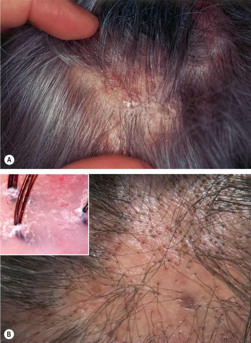
Fig. 69.17 Lichen planopilaris.A Patchy areas of alopecia with perifollicular erythema and scale at the active margins. B Later stage showing scarring with loss of follicular openings. By trichoscopy, there is perifollicular scaling and areas with no follicular openings (dots) due to loss of hair follicles (inset). A, Courtesy Jean L. Bolognia, MD.
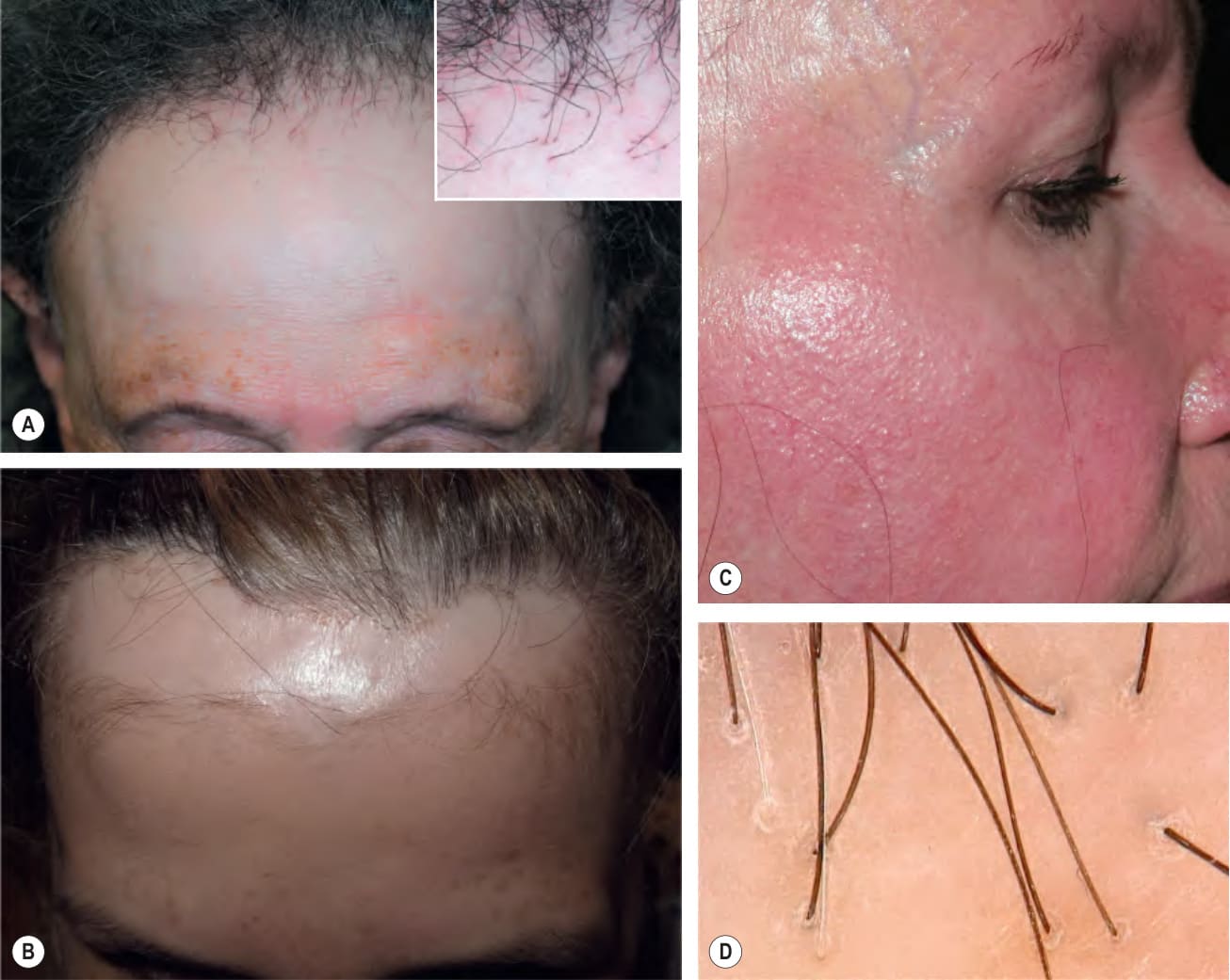
Fig. 69.18 Frontal fibrosing alopecia.
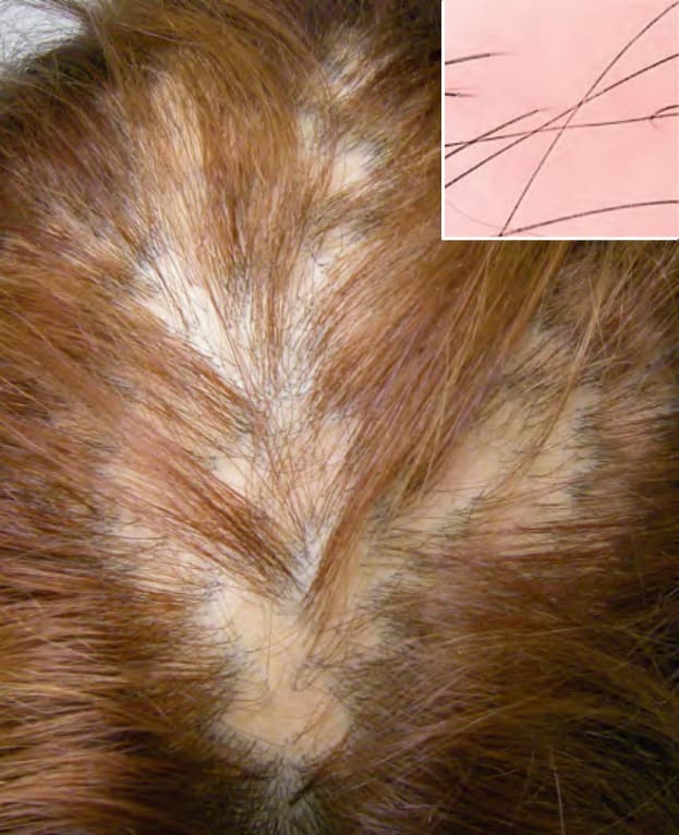
Fig. 69.19 Classic pseudopelade (pseudopelade of
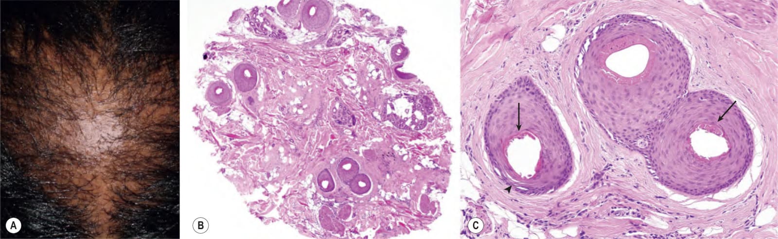
Fig. 69.20 Central centrifugal cicatricial alopecia in an African-American woman – clinical and histopathologic features.A Alopecia is most prominent on the crown and/or vertex. B, C Subisthmus horizontal section demonstrating scarring alopecia with reduction of terminal hair follicles and scant inflammatory cell infiltrate as well as premature desquamation of the inner root sheath (arrows) and eccentric thinning of the follicular epithelium (arrowhead) on higher magnification. A, Courtesy Leonard C. Sperling, MD and Rodney D. Sinclair, MD.
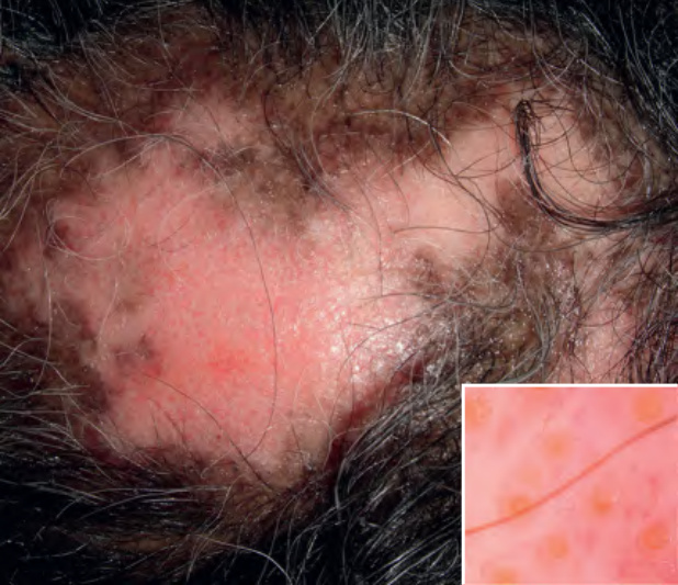
Fig. 69.21 Discoid lupus erythema- tosus. Coalescing areas of cicatricial alopecia in which there is obvious erythema. Central hypopigmentation is accompanied by peripheral hyperpigmentation. Trichoscopy shows follicular plugs (large yellow dots) and arborizing vessels (inset). Courtesy Leonard C. Sperling, MD and Rodney D. Sinclair, MD.
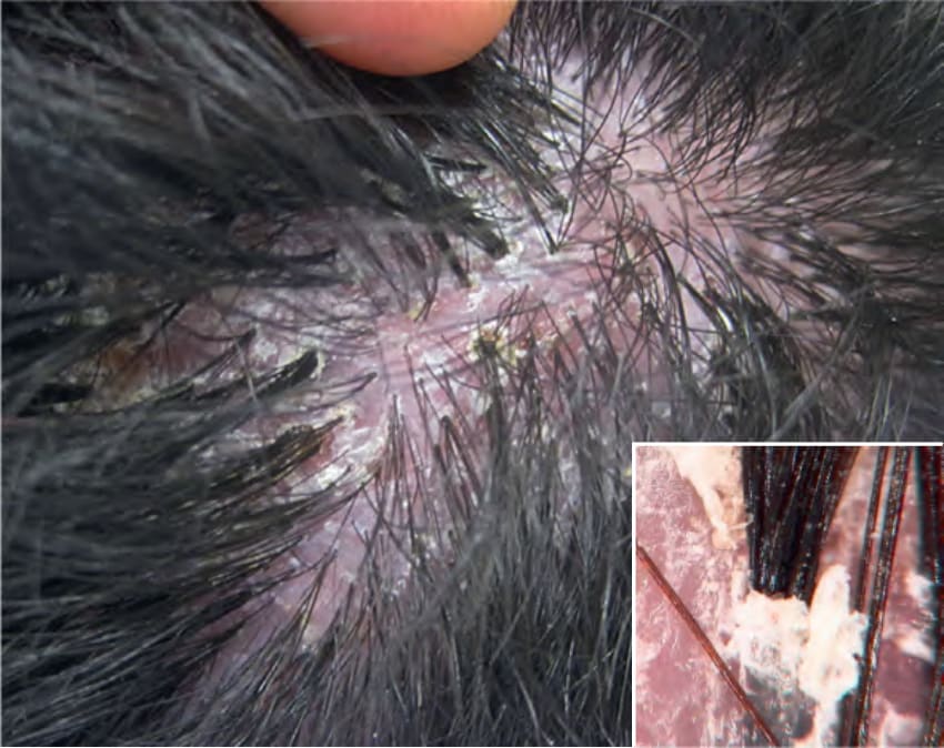
Fig. 69.22 Folliculitis decalvans. Note the multiple hairs arising from a single inflamed follicular orifice (“tufted folliculitis”) within the area of scarring alopecia. Trichoscopy shows hair tufts composed of multiple hairs and tubular perifollicular scaling (inset). Courtesy Damon McClain, MD.
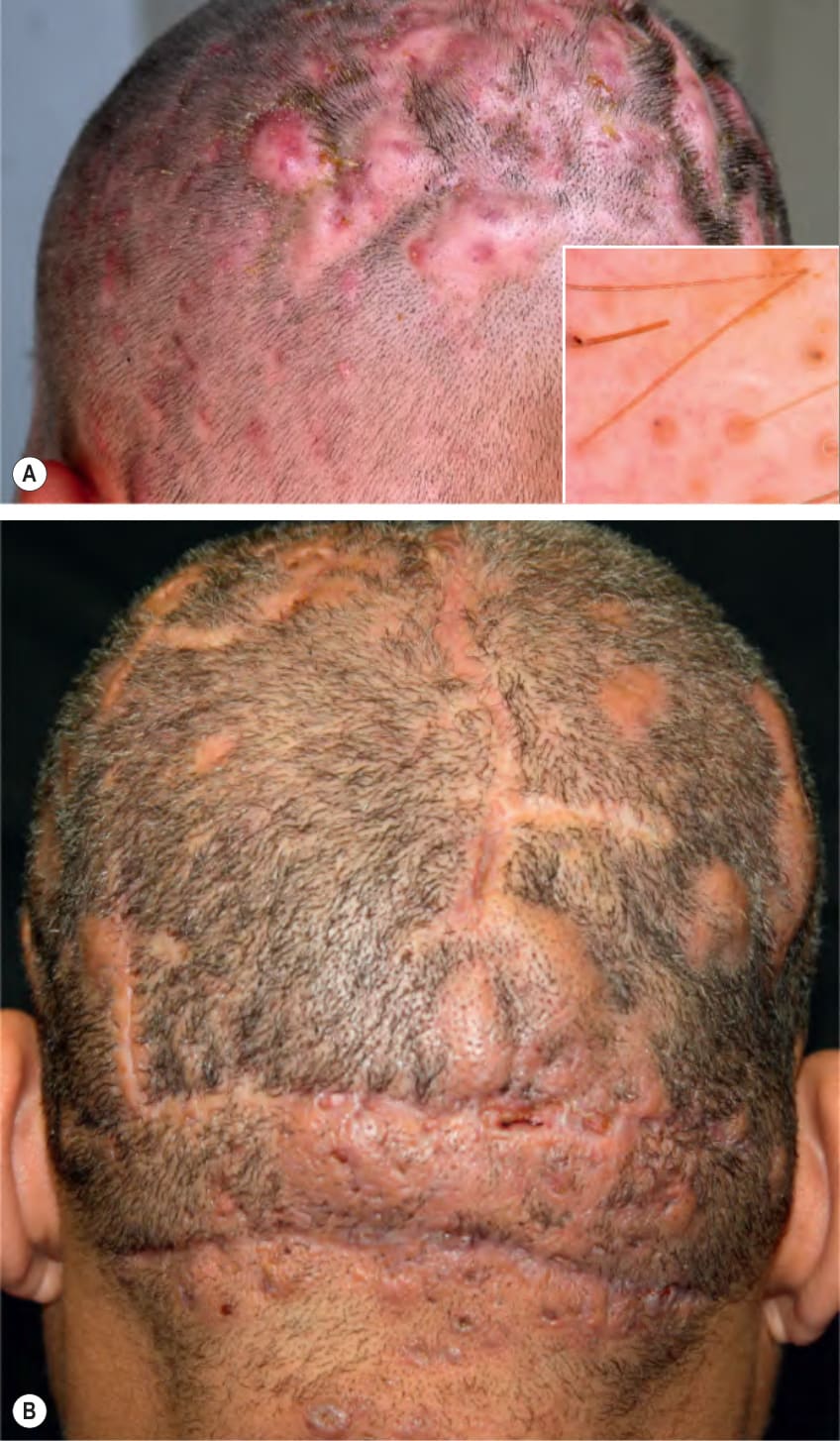
Fig. 69.23 Dissecting cellulitis.A Earlier stage with multiple interconnected, inflammatory, alopecic nodules admixed with pustules and crusts. By trichoscopy, there are 3D (“bubble”) black dots (inset). B Tender nodules with alopecia, draining sinuses, broad scarring, and linear scars. B, Courtesy Jeffrey P. Callen, MD.
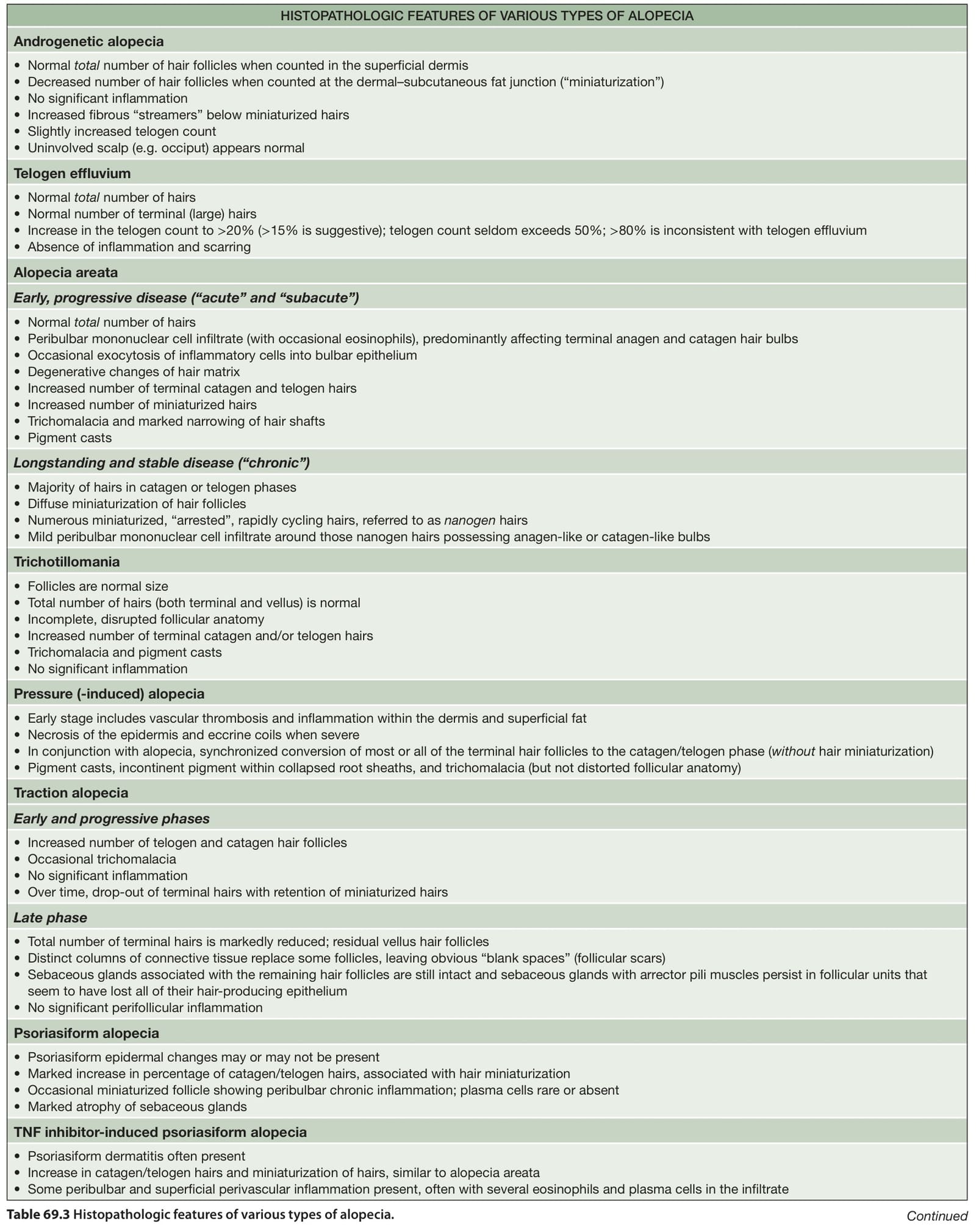
Table 69.3 Histopathologic features of various types of alopecia. Continued
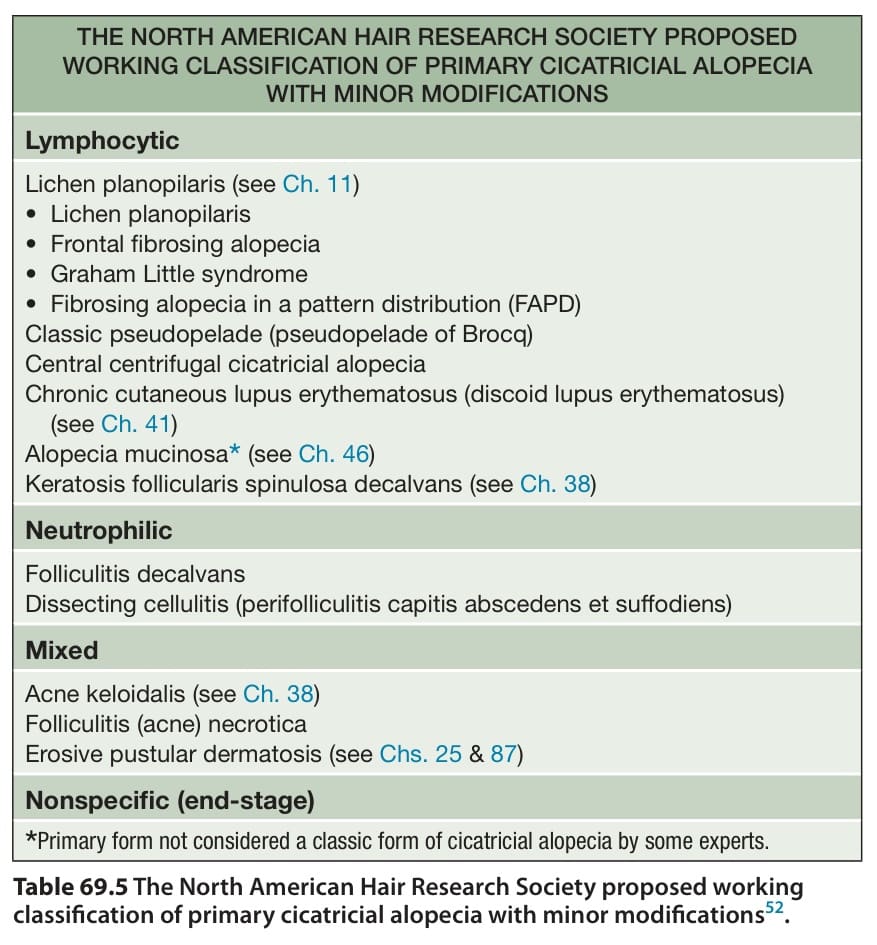
Table 69.5 The North American Hair Research Society proposed working classification of primary cicatricial alopecia with minor modifications.
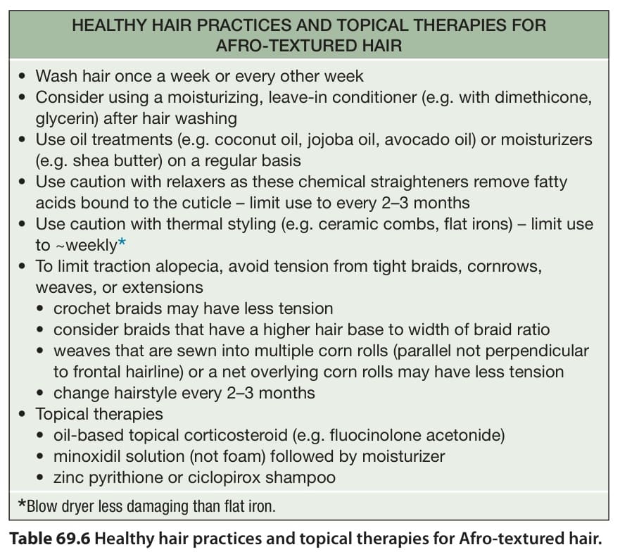
Table 69.6 Healthy hair practices and topical therapies for Afro-textured hair.