HAIR SHAFT ABNORMALITIES
Hair shaft abnormalities, both acquired and inherited (Table 69.7), are divided into two broad categories – those associated with increased hair fragility and those not associated with increased hair fragility (Fig. 69.24). The former group can lead to various degrees of alopecia while alteration of the clinical appearance of the hair (usually in the absence of alopecia) characterizes the latter group. The analysis of hair shaft abnormalities is best performed via trichoscopy (high magnification, if available) or light microscopy of cut hair shafts. Reflectance confocal microscopy and transmission or scanning electron microscopy are additional diagnostic modalities. When trichothiodystrophy is suspected, amino acid analysis can be performed to confirm low sulfur levels. Other than preventive hair care measures (e.g. conditioning, low tension hair styling), there are currently no specific treatments for inherited hair shaft abnormalities. Topical or low-dose oral minoxidil may stimulate hair regrowth but has no documented effect on hair shaft structure.
Hair Shaft Abnormalities Associated With Increased Hair Fragility
“Bubble hair”
This distinctive hair shaft abnormality is usually seen in young women with a localized area of uneven, fragile hairs. By trichoscopy or light microcopy, the hair shafts contain large irregularly spaced “bubbles” that expand and lead to thinning of the hair cortex (see Fig. 69.24F). Hair fractures occur at the site of larger bubbles.
Bubble hairs can develop when wet hair is exposed to hair care techniques that involve heat, including high-temperature hair dryers, hair straighteners or flat irons, and electric curlers. When wet, a hair shaft can expand up to 30% in diameter with retention of water within the shaft; exposure to heat can then lead to formation of expanding and destructive bubbles. Once the damaged hair is trimmed, the condition resolves completely with gentle hair styling.
Monilethrix
Synonym: Beaded hair
In monilethrix, hair shafts have regular, periodic elliptical nodosities that alternate with shaft thinning and a tendency to fracture at the latter sites (see Fig. 69.24C). This disorder is usually inherited in an autosomal dominant fashion with variable expression and is due to heterozygous mutations in one of three hair cortex-specific keratin genes – KRT86, KRT81, or KRT83. There is also an autosomal recessive form due to mutations in DSG4, which encodes desmoglein 4.
Affected individuals usually have normal-appearing hair at birth, but within the first few months of life these fibers are replaced by short, fragile, brittle hair (Fig. 69.25). Patients may state “I have hair that never grows”. The phenotype is variable, ranging from clinically normal hair to mild occipitotemporal hair loss to nearly total alopecia. Additional manifestations include keratotic follicular papules, primarily on the occiput, as well as syndactyly and nail (e.g. brittle, koilonychia), ocular (e.g. juvenile cataracts, decreased visual fields), and dental abnormalities.
By trichoscopy and light microscopy, there is variation in hair shaft thickness with uniformly spaced elliptical nodosities and intervening constrictions. Hairs are bent with a tendency to fracture at constriction sites. Monilethrix-like hair shafts may be observed in other hair disorders, such as alopecia areata or chemotherapy-induced alopecia, but their clinical courses differ. In pseudomonilethrix, there is irregular, square-shaped, flattening of hair shafts. Whether pseudomonilethrix is a true disease or an optical artifact is a matter of debate.
Pili torti
Pili torti is characterized by a flattening and 180° twisting of the hair shaft on its own axis (see Fig. 69.24G). The twists are typically narrow and occur in groups of three to ten twists, giving the hair shaft a spangled appearance. When inherited, pili torti can be an isolated abnormality or it may be a feature of other disorders, including Menkes disease (see Ch. 51), Björnstad syndrome, Bazex–Dupré–Christol syndrome, and several ectodermal dysplasias. Hairs may be sparse or abnormal from birth or early childhood (early-onset, Ronchese type) or they may become brittle and fragile around puberty (late-onset, Beare type).
Acquired pili torti may be drug-induced (e.g. epidermal growth factor receptor inhibitors, retinoids), associated with perifollicular inflammation or fibrosis (e.g. cutaneous T cell lymphoma, cicatricial alopecia), or due to malnutrition. The inherited form of pili torti can improve at puberty while resolution of the acquired form is dependent upon its etiology.
Trichorrhexis invaginata
Trichorrhexis invaginata (“bamboo hair”) is a clinical feature of Netherton syndrome (see Ch. 57). This inherited disorder is due to pathogenic variants in SPINK5, which encodes a serine protease inhibitor, LEKTI. The hair abnormality usually becomes evident during infancy with the development of short, sparse, and very fragile hair. In scalp or eyebrow hairs, the breakage points consist of intussusceptions of the distal shaft (“ball”) into the proximal shaft (“socket”) due to defective cornification of the cortex (see Fig. 69.24B). Fracture of the shaft may leave the proximal shaft with a golf tee-shaped end.
Trichorrhexis nodosa
Trichorrhexis nodosa is the most common of all the structural hair abnormalities. It is characterized by hair shaft fractures in which the individual cortical cells and their fragments splay out, appearing like the ends of two brushes pushed into one another (see Fig. 69.24A). This abnormality is presumably caused by damage to the cuticular cells of the shaft and subsequently to the intercellular cement substance that normally binds cells together. Trichorrhexis nodosa may be congenital or acquired, with the congenital form occurring as an isolated finding (primary genetic) or in association with other inherited disorders, e.g. argininosuccinic aciduria, citrullinemia, trichohepatoenteric syndrome.
The acquired variant is most commonly caused by repeated trauma to the hair shaft. This can result from chemical relaxation, excessive heat (e.g. hair dryers, hair straighteners [flat irons, hot combs]), frequent brushing, or chronic scratching. Three clinical variants of acquired trichorrhexis nodosa have been described: (1) proximal – usually due to damage from chemicals and heat; (2) distal – due to cumulative cuticular damage; and (3) circumscribed – occurs in the scalp, moustache, or beard. Chronic use of masks may explain some cases of beard involvement. Trichorrhexis nodosa can also be seen in patients with other hair shaft abnormalities associated with increased hair fragility.
Trichothiodystrophy
Trichothiodystrophy is primarily an autosomal recessive disorder characterized by sulfur-deficient hair. By light microscopy and trichoscopy, hair shafts have transverse fractures (trichoschisis; see Fig. 69.24D) and alterations in their cuticles. Polarized light is required to visualize the alternating light and dark bands, i.e. the pathognomonic “tiger tail sign” (see Fig. 69.24E). Currently, based upon underlying genetic bases, there are nine types of trichothiodystrophy (TTD; www.omim.org). TTD1–3 have associated photosensitivity and are reviewed in Table 87.4 whereas TTD4–9 do not have photosensitivity, including X-linked recessive TTD5. The acronym PIBIDS has been used to describe possible findings in patients with TTD1–3: P (photosensitivity), I (ichthyosis), B (brittle hair), I (intellectual impairment with low IQ), D (decreased fertility), and S (short stature). Some patients may exhibit spasticity, tremors and ataxia, nail dystrophy, dental caries, cataracts, and bony and immune defects. The hair usually does not improve with age.
Hair Abnormalities Not Associated With Increased Hair Fragility
Acquired progressive kinking of the hair
The term encompasses acquired conditions characterized by curling of the scalp hair. It classically presents with curly, frizzy, and lusterless
hair in the frontotemporal region or vertex of the scalp. There is often progression to AGA, but whether it is a harbinger of very early-onset AGA has been debated. Localized and diffuse forms of acquired hair kinking that are not associated with hair thinning have also been described.
Loose anagen hair syndrome
The classic presentation of loose anagen hair syndrome is a young girl with fairly short blond hair who has diffuse or patchy alopecia. There is no increase in hair fragility, but anagen hairs can be easily and painlessly pulled from the scalp. Because of the limited maximum hair length, patients may state “I have hair that never grows”.
Microscopic examination reveals a ruffled proximal cuticle, absence of the root sheath, and a bent matrix (see Fig. 69.11C). Structural abnormalities that appear to disturb the normal supportive and anchoring functions of the inner root sheath are seen, presumably resulting in a loose attachment of the hair shaft to the anagen follicle. With trichoscopy, rectangular black granular structures are visible. Gradual improvement occurs with aging. In addition to telogen effluvium, alopecia areata, and trichotillomania, the differential diagnosis includes short anagen hair syndrome. The latter is seen primarily in children and is characterized by an increased number of telogen hairs, a shortened anagen phase with limited maximum hair length, and oftentimes a history of not requiring haircuts. If there is no improvement over time, some clinicians prescribe low-dose oral minoxidil (see Alopecia Areata, Treatment).
Pili annulati
Pili annulati (ringed hair) is characterized by hair shafts that have bright and dark bands when viewed by reflected light (see Fig. 69.24H). The bright bands are related to light scattered from periodically occurring clusters of abnormal, air-filled cavities within the hair. The condition may be sporadic or seen as an autosomal dominant trait. On microscopic examination of a hair mount, the air-filled cavities are seen as dark regions. By trichoscopy, there is alternating white and dark bands, with the former subtle, cloudy, and shorter than the remaining dark portions of the hair shaft. Approximately 20%–80% of the hairs are affected and the bands tend to disappear distally.
Pili bifurcati
This hair fiber anomaly is characterized by bifurcation of the hair fiber at multiple irregular intervals along the shaft, forming separate rami, which then again fuse. Each ramus has its own cuticle. As with other hair shaft disorders, the bifurcations may affect only a proportion of hairs and abnormal hairs may not be noticeable clinically.
Pili multigemini
The term pili multigemini is associated with multiple hair shafts arising from one papilla. Each hair fiber has its own inner root sheath, but all the fibers are surrounded by a common outer root sheath. Multi-geminate hairs are usually found in the beard region, often in association with some perifollicular erythema. The differential diagnosis includes trichostasis spinulosa.
Uncombable hair (spun-glass hair)
This entity is also known as pili trianguli et canaliculi and refers to a rare hair abnormality in which hair has a “spun glass” appearance due to the reflection of light from variably oriented, flattened hair surfaces. Hair is characteristically stiff and difficult to comb. Abnormal keratinization of the internal root sheath is postulated to cause the irregularly shaped hair shafts, which have a triangular shape on cross-section and a longitudinal groove that is best seen by scanning electron microscopy (see Fig. 69.24J). Homozygous or compound heterozygous mutations have been identified in three genes that encode proteins involved in
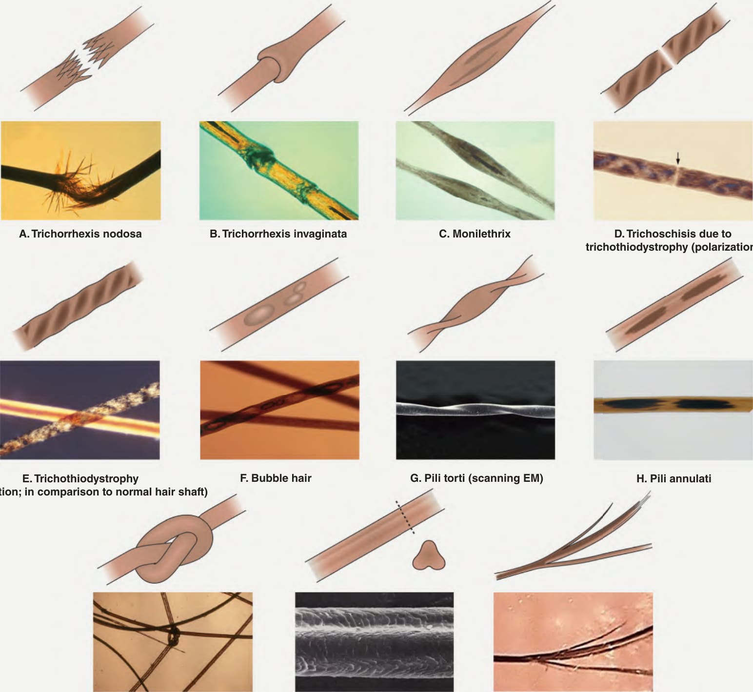
Fig. 69.24 Hair shaft abnormalities. EM, electron microscopy. A, B, Courtesy Maria K. Hordinsky, MD; C–E, G, J, Courtesy Leonard C. Sperling, MD; F, Courtesy Jean L. Bolognia, MD.
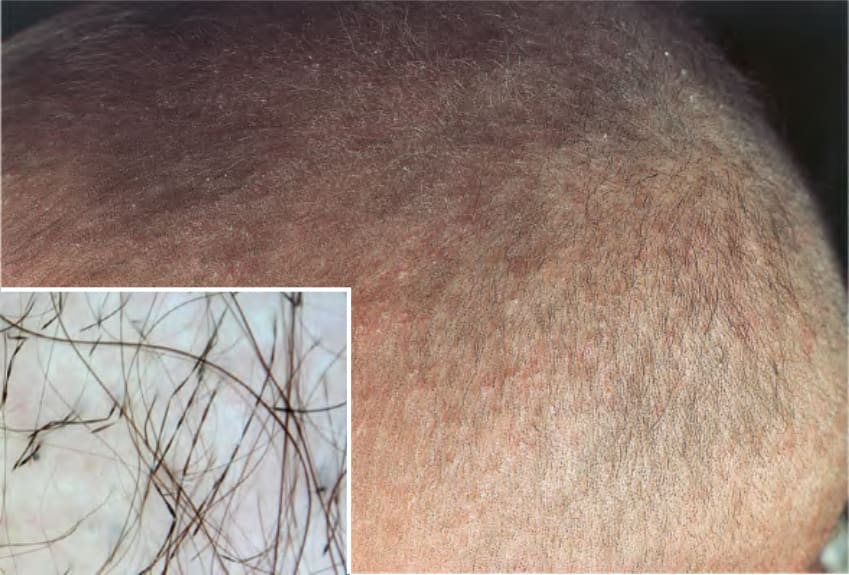
Fig. 69.25 Monilethrix. Fragility of the hair shafts leads to short broken hairs. Small perifollicular papules with scale are also present. By trichoscopy, the hair shafts have a beaded appearance (see Fig. 69.24C).
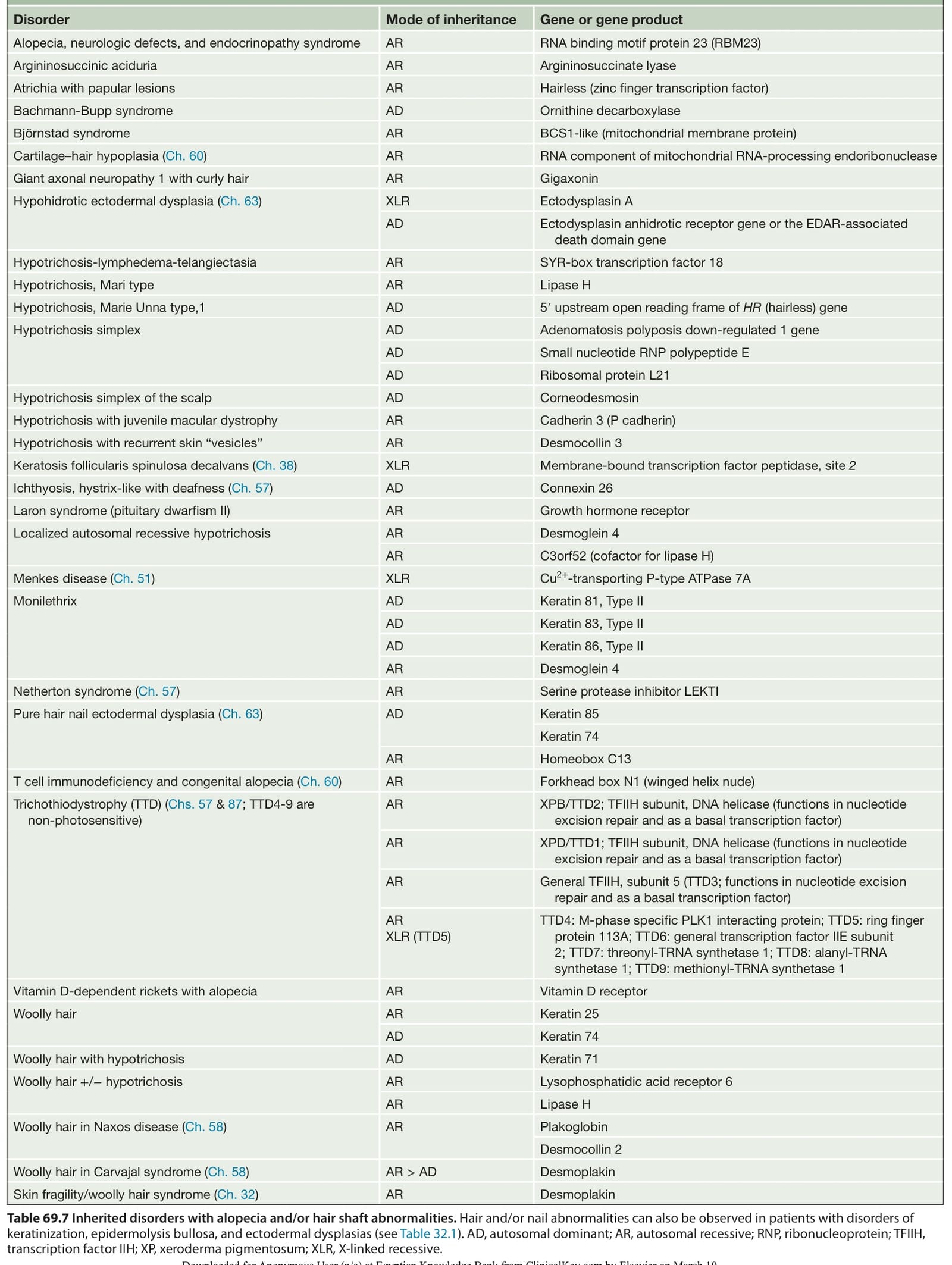
Table 69.7 Inherited disorders with alopecia and/or hair shaft abnormalities. Hair and/or nail abnormalities can also be observed in patients with disorders of keratinization, epidermolysis bullosa, and ectodermal dysplasias (see Table 32.1). AD, autosomal dominant; AR, autosomal recessive; RNP, ribonucleoprotein; TFIIH, transcription factor IIH; XP, xeroderma pigmentosum; XLR, X-linked recessive.