HYPERTRICHOSIS
Introduction
Hypertrichosis describes the growth of excessive hair anywhere on the body. The term is frequently confused with hirsutism, which should only be applied to women with excessive growth of terminal hairs in androgen-dependent sites (i.e. a “male pattern”) due to hyperandrogenemia or increased end-organ sensitivity to androgens. Hypertrichosis can be classified based on its distribution (generalized versus localized), the age of onset (congenital or programmed from birth versus acquired), and the type of hair (lanugo or vellus versus terminal).
Clinical Features
Generalized hypertrichosis
In generalized hypertrichosis, there is lanugo hair, excess vellus hair, or terminal hair over much of the cutaneous surface, including an acquired transformation of terminal hair into lanugo hair (Fig. 70.1). Lanugo is the non-pigmented, non-medullated, fine hair that covers the fetus and can grow up to several centimeters in length; it is normally shed in utero at ~7–8 months’ gestation or during the first few weeks of life and replaced by vellus hair on the body and terminal hair on the scalp. Some neonates, especially if born prematurely, transiently retain lanugo hairs diffusely and shed them during the first few weeks of life.
A number of distinctive but rare genetic syndromes are associated with congenital generalized hypertrichosis (Table 70.1). While the majority of these disorders have extracutaneous manifestations such as gingival hyperplasia or facial dysmorphism, some have primarily hypertrichosis, including universal hypertrichosis. However, this latter entity is sometimes viewed as constitutional, i.e. simply exaggerated normal hairiness that may be familial. Genetic abnormalities associated with congenital generalized hypertrichosis lead to dysfunction of several proteins, ranging from those known to be involved in hair follicle development to membrane transporters (see Table 70.1).
The possibility of intrauterine exposure to medications (e.g. minoxidil) also needs to be considered in infants with congenital generalized hypertrichosis. The differential diagnosis also includes inherited disorders in which hypertrichosis can involve multiple sites and may appear early in life (Table 70.2).
Prepubertal hypertrichosis is a relatively common finding in otherwise healthy infants and children, most often occurring in individuals of Mediterranean or South Asian descent. Pigmented hair is present in a widespread, diffuse distribution and becomes more obvious during childhood. There is involvement of the face (especially the forehead, temples, and preauricular area), proximal extremities, and back; hairs in the latter location assume an “inverted fir tree” pattern. Bushy eyebrows and a low anterior hairline represent additional features.
The facial distribution pattern of prepubertal hypertrichosis can overlap with that of familial hirsutism (see below), and there may be a family history of excessive hairiness. Mildly elevated levels of total and free testosterone have been observed in a subset of girls with prepubertal hypertrichosis, while others have a normal androgen profile. These findings suggest multiple etiologies for this clinical pattern of hypertrichosis, including androgen excess as well as an increased constitutional propensity for hair growth.
Acquired generalized hypertrichosis is most often related to drug ingestion (Table 70.3). Drug-induced hypertrichosis is characterized by slow growth of terminal hair of medium thickness. The findings are most evident on the forehead, temples, flexor aspects of the extremities, and trunk. Drug-related hypertrichosis is usually reversible, and differs in distribution from drug-induced hirsutism.
Acquired generalized hypertrichosis can also represent a sign or consequence of a variety of systemic conditions, including disorders of the CNS (e.g. traumatic brain injuries), juvenile hypothyroidism, juvenile dermatomyositis, acromegaly (especially of the lower face), malnutrition (including anorexia nervosa), POEMS syndrome, and advanced HIV disease.
This is considered to be a paraneoplastic phenomenon as it is associated with internal malignancies, most often of the lung, colon, or breast. Occasionally, acquired hypertrichosis lanuginosa may precede the diagnosis of the neoplasm. In addition, it may be associated with other paraneoplastic dermatoses, such as acanthosis nigricans, palmoplantar keratoderma, the sign of Leser–Trélat, and acquired ichthyosis (see Ch. 53). The lanugo hair appears over the entire body within a short period of time, although in mild forms it may be localized to the face, leading to a “simian” appearance. Lanugo hair may even develop in areas of androgenetic alopecia.
Localized hypertrichosis
Most cases of localized hypertrichosis involve a switch from vellus to terminal hair in sites that do not usually bear terminal hair. Localized hypertrichosis can develop as a component of a hamartoma, as an isolated congenital lesion, as a manifestation of a systemic disease (inherited or acquired), or as a consequence of cutaneous trauma or inflammation.
Hamartomas, including those with delayed clinical presentation, and congenital abnormalities characterized by hypertrichosis of a specific anatomic site (Figs. 70.2 and 70.3; Tables 70.4 & 70.5) are included in this category.
Congenital melanocytic nevi and plexiform neurofibromas Congenital melanocytic nevi often have associated hypertrichosis. The increased hair growth may be noted at birth, but often becomes
more prominent during infancy or early childhood. Hypertrichosis can be seen in all sizes of congenital melanocytic nevi (see Ch. 112). A particularly dense growth of terminal hairs may accompany scalp nevi. Plexiform neurofibromas can also have associated hyperpigmentation and hypertrichosis.
Becker melanosis (nevus) This is a hamartoma characterized by macular hyperpigmentation with irregular borders, most commonly located on the upper lateral trunk (anterior or posterior) (see Ch. 112). Some patients have associated hypertrichosis while others do not. The pigmentation usually arises during the first decade of life, whereas the hypertrichosis typically appears in the second decade (Fig. 70.5). Becker melanosis is most often diagnosed in boys and men. Postzygotic mutations in the ACTB gene encoding β-actin have been found to underlie Becker melanosis and associated anomalies (referred to as Becker nevus syndrome; see below) as well as smooth muscle hamartomas (see Ch. 117 and below).
Occasionally, asymmetry of the extremities and hyperplasia or hypoplasia of the affected areas is evident, especially ipsilateral mammary hypoplasia in women or, less often, the Poland anomaly (unilateral chest wall hypoplasia and ipsilateral hand syndactyly). Rarely, there may be an association with genitourinary tract abnormalities (SNUB syndrome: supernumerary nipples, uropathies, Becker melanosis). Because of its benign nature, surgical removal of Becker melanosis is not recommended. Epilation can be performed by various techniques, but attempts at decreasing pigmentation via laser therapy are often not successful.
Smooth muscle hamartomas, which exist on a spectrum with Becker melanosis, may present as circumscribed hypertrichotic plaques with variable hyperpigmentation. A rare generalized form is characterized by extensive hypertrichosis and folding of the skin which has been reported under the term “Michelin tire baby” (see Table 97.7). Lastly, the hypertrichosis observed in the setting of hemimaxillofacial dysplasia (with facial asymmetry due to unilateral maxillary enlargement, gingival hyperplasia, and hypoplastic teeth) is often associated with Becker melanosis or smooth muscle hamartoma.
Other hamartomas and infantile tumors Hypertrichosis can also overlie the following skin lesions: plaque-type blue nevus, neurocristic hamartoma, fibrous hamartoma of infancy, dermal dendrocyte hamartoma, eccrine angiomatous hamartoma, and tufted angioma.
Nevoid hypertrichosis This is an uncommon congenital alteration characterized by the growth of terminal hairs in a circumscribed area. In general, there are no extracutaneous associations in primary nevoid hypertrichosis; the skin within the affected area is normally pigmented and there is no underlying hamartoma (Fig. 70.6). Table 70.4 outlines specific sites of localized hypertrichosis that can be familial. To date there is a report of one family with primary multifocal localized hypertrichosis. Nevoid hypertrichosis has also been observed to follow the lines of Blaschko, as reported in female carriers of X-linked hypertrichosis (see Table 70.1).
Secondary nevoid hypertrichosis can be associated with lipodystrophy, hemihypertrophy, scoliosis, and abnormalities of the underlying vasculature. Nevoid hypertrichosis also occasionally occurs together with epidermal nevi or nevoid hypopigmentation, and it can represent a manifestation of Proteus and Happle–Tinschert syndromes.
Spinal dysraphism and the hair collar sign Dysraphism is defined as an alteration of the formation of a fold or elevation which is constituted in the midline of the human body during
the union of two lateral portions. In spinal dysraphism, abnormal closure of the neural tube leads to defects in the vertebral column and/ or spinal cord (see Ch. 64). Skin lesions marking a hidden vertebral defect are usually located in the dorsal midline. The faun tail is most often a sign of spina bifida occulta or diastematomyelia (split spinal cord), and is typically located in the lumbosacral region. In addition, a ring of hypertrichosis on the scalp, referred to as the hair collar sign, can surround membranous aplasia cutis or ectopic neural tissue, reflecting the origin of these midline lesions as incomplete neural tube defects.
There are multiple genetic diseases characterized by the presence of localized hypertrichosis as a major or secondary diagnostic feature (see Table 70.2). For example, hypertrichosis within sun-exposed areas is one of the signs of the porphyrias – in particular, porphyria cutanea tarda (Fig. 70.7) and hepatoerythropoietic porphyria. Likewise, the low frontal hairline and synophrys (“unibrow”) in the Cornelia de Lange syndrome are important diagnostic features of that entity. Localized hypertrichosis
may also be observed in acquired systemic diseases, such as infrapatellar hypertrichosis in juvenile dermatomyositis or hypertrichosis overlying pretibial myxedema, indurated plaques of Rosai–Dorfman disease, and areas affected by the complex regional pain syndrome.
After repeated trauma, friction, irritation, or inflammation, the hair within affected areas of skin may become longer and thicker (e.g.
The eyelashes increased in both length and disorder after treatment with erlotinib. Courtesy Edward Cowen, MD.
hypertrichosis of the back in sack carriers, hypertrichosis of a fractured limb after the application of a plaster cast, hypertrichosis of the posterior neck in people who bear heavy weights; Fig. 70.8). Chronic rubbing or scratching can also lead to localized hypertrichosis. Transient localized hypertrichosis has been observed within vaccination sites and varicella scars as well as sites of wart removal and laser epilation. In addition, hypertrichosis has been described overlying lipoatrophy following lupus panniculitis and within resolving lesions of psoriasis and morphea. Extractable hairs mimicking hypertrichosis are often evident within a pilonidal sinus (cyst), which is usually located within the gluteal cleft.
There was no underlying melanocytic nevus or hyperpigmentation. Courtesy Jean L. Bolognia, MD.
Courtesy Luis Requena, MD.
Localized hypertrichosis has been reported after PUVA therapy. Sites of application of potent topical corticosteroids, tacrolimus, and creams containing mercury or iodine can also develop localized hypertrichosis, as can sites of repeated irritation from anthralin. Prostaglandin F-2α analogues (e.g. latanoprost, bimatoprost), which are topical agents used for the treatment of glaucoma, can induce trichomegaly of the eyelashes as well as brown iris pigmentation. The FDA has approved bimatoprost 0.03% solution for the stimulation of eyelash growth, and latanoprost has also been used off-label for this purpose (Fig. 70.9).
Treatment
Methods available to treat hypertrichosis include chemical depilatory agents, electrolysis (see Ch. 140), and laser hair removal (see Ch. 137). Systems utilized for photoepilation include Nd:YAG, diode, and alexandrite lasers as well as intense pulsed light (IPL) sources (see Ch. 137). Barium sulfate depilatory creams have been shown to be especially effective, but they are odiferous and more irritating than calcium thioglycolate depilatory creams. Shaving and other physical methods (e.g. tweezing, waxing, threading, sugaring) may also be employed.
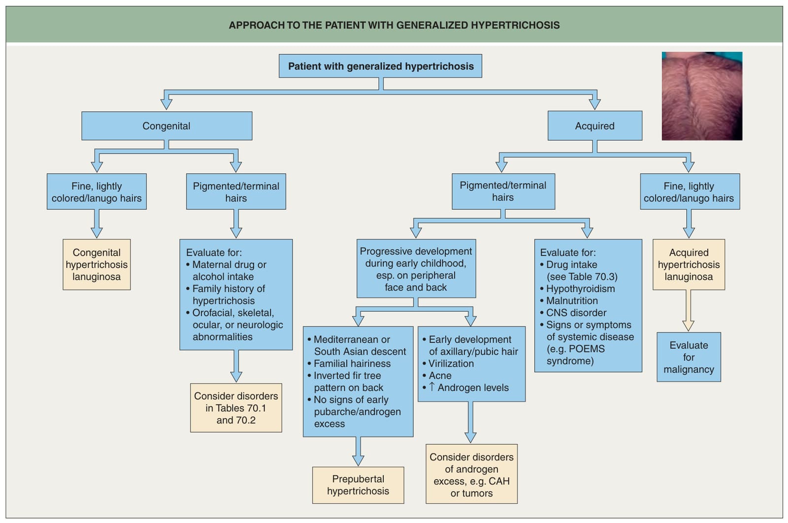
Fig. 70.1 Approach to the patient with generalized hypertrichosis. CAH, congenital adrenal hyperplasia; POEMS, polyneuropathy, organomegaly, endocrinopathy, monoclonal gammopathy, skin lesions. The photograph is an example of universal hypertrichosis, which some authors view as simply exaggerated normal hairiness that may be familial.
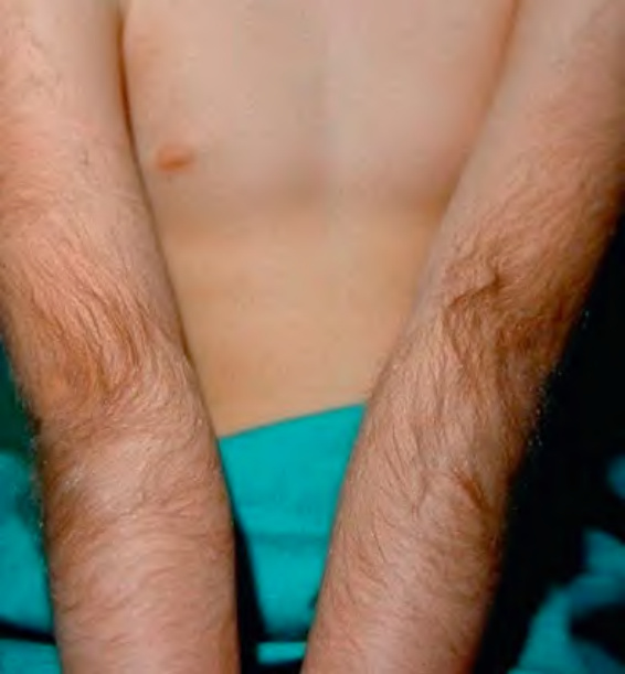
Fig. 70.2 Hypertrichosis cubiti. Multiple terminal hairs on both elbows in a child.
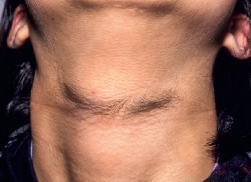
Fig. 70.3 Anterior cervical hypertrichosis.Courtesy Harvey Lui, MD.
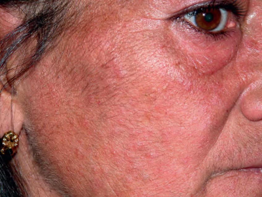
Fig 70.4 Epidermal growth factor receptor inhibitor-induced trichomegaly.
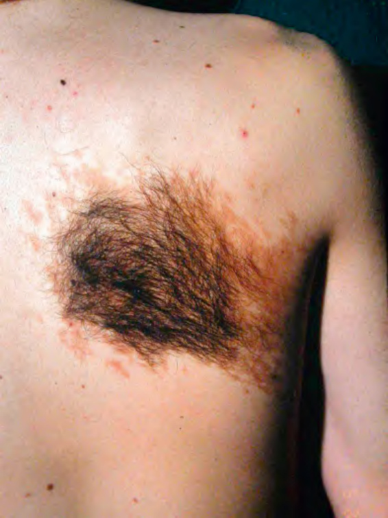
Fig. 70.5 Becker melanosis (nevus) on the mid back in a young man. In addition to hyperpigmentation, there is marked hypertrichosis. The degree of hypertrichosis can vary (see Ch. 112). When it is marked as in this patient, the clinical differential diagnosis includes congenital melanocytic nevus, especially when there is an under-lying smooth muscle hamartoma.
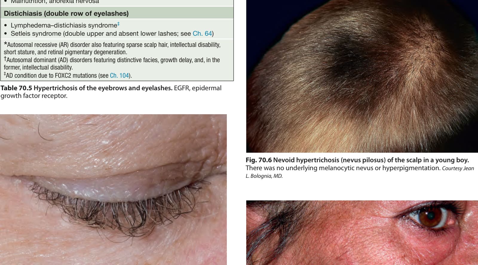
Fig. 70.6 Nevoid hypertrichosis (nevus pilosus) of the scalp in a young boy.
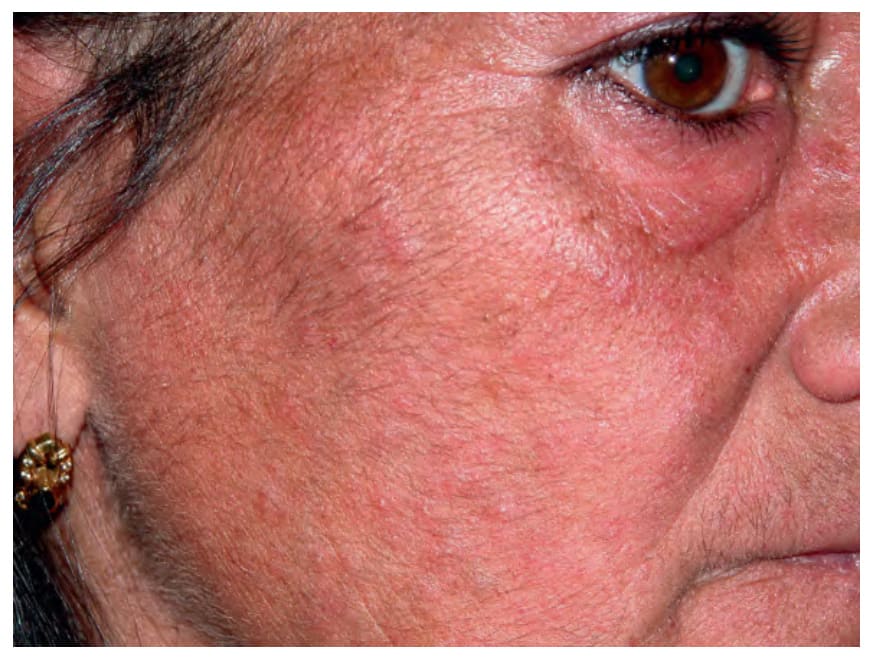
Fig. 70.7 Facial hypertrichosis in a woman due to porphyria cutanea tarda.
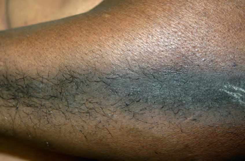
Fig. 70.8 Acquired localized hypertrichosis. Hypertrichosis, hyperpigmentation, and epidermal hyperplasia at the site of friction.
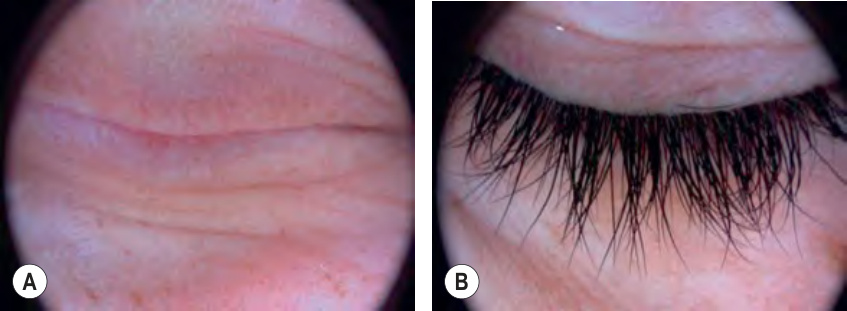
Fig. 70.9 Treatment of eyelash alopecia in a patient with alopecia univer- salis.A Prior to treatment. B Following daily application of topical latanoprost ophthalmic 0.005% solution for 6 months, there was regrowth of eyelashes.
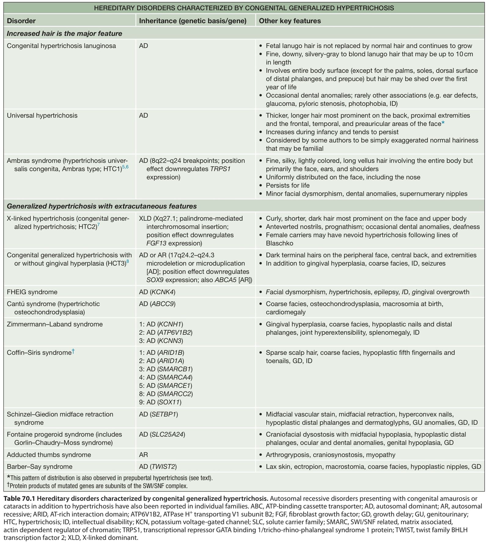
Table 70.1 Hereditary disorders characterized by congenital generalized hypertrichosis. Autosomal recessive disorders presenting with congenital amaurosis or cataracts in addition to hypertrichosis have also been reported in individual families. ABC, ATP-binding cassette transporter; AD, autosomal dominant; AR, autosomal recessive; ARID, AT-rich interaction domain; ATP6V1B2, ATPase H+ transporting V1 subunit B2; FGF, fibroblast growth factor; GD, growth delay; GU, genitourinary; HTC, hypertrichosis; ID, intellectual disability; KCN, potassium voltage-gated channel; SLC, solute carrier family; SMARC, SWI/SNF related, matrix associated, actin dependent regulator of chromatin; TRPS1, transcriptional repressor GATA binding 1/tricho-rhino-phalangeal syndrome 1 protein; TWIST, twist family BHLH transcription factor 2; XLD, X-linked dominant.
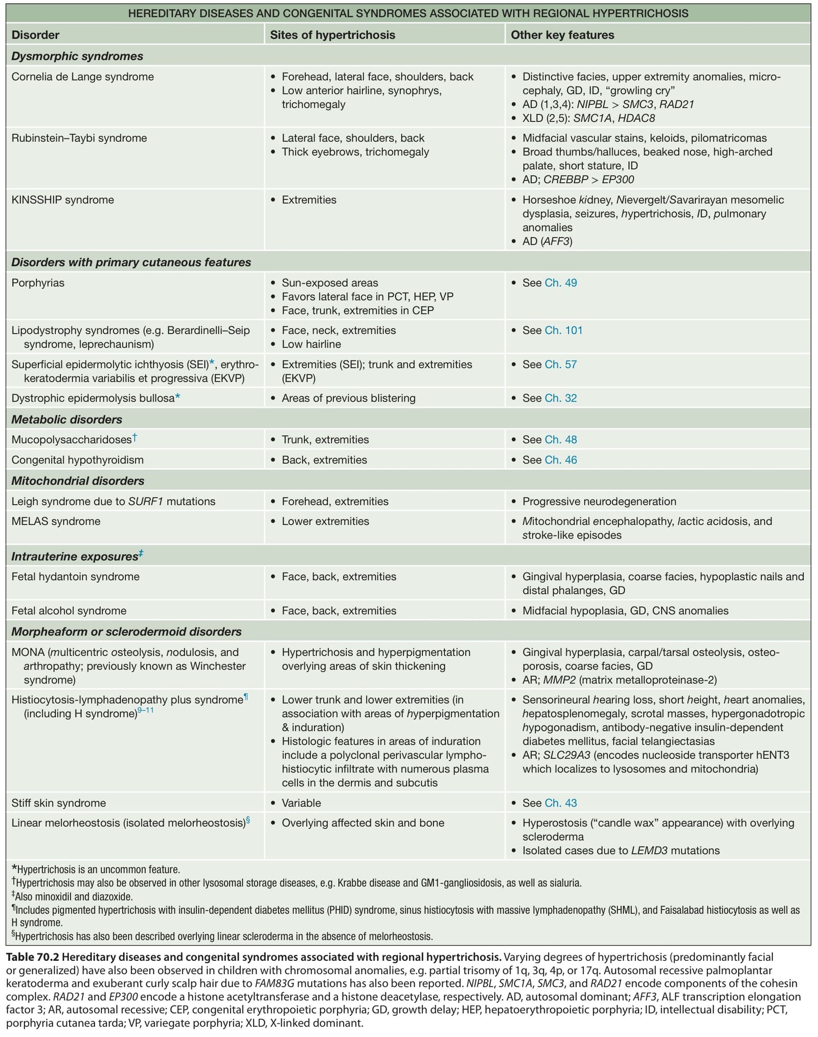
Table 70.2 Hereditary diseases and congenital syndromes associated with regional hypertrichosis. Varying degrees of hypertrichosis (predominantly facial or generalized) have also been observed in children with chromosomal anomalies, e.g. partial trisomy of 1q, 3q, 4p, or 17q. Autosomal recessive palmoplantar keratoderma and exuberant curly scalp hair due to FAM83G mutations has also been reported. NIPBL, SMC1A, SMC3, and RAD21 encode components of the cohesin complex. RAD21 and EP300 encode a histone acetyltransferase and a histone deacetylase, respectively. AD, autosomal dominant; AFF3, ALF transcription elongation factor 3; AR, autosomal recessive; CEP, congenital erythropoietic porphyria; GD, growth delay; HEP, hepatoerythropoietic porphyria; ID, intellectual disability; PCT, porphyria cutanea tarda; VP, variegate porphyria; XLD, X-linked dominant.
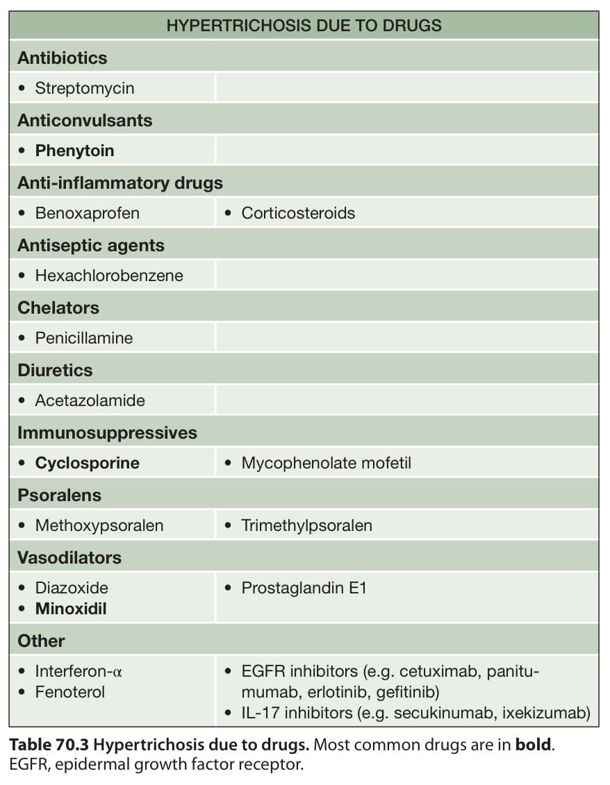
Table 70.3 Hypertrichosis due to drugs. Most common drugs are in bold. EGFR, epidermal growth factor receptor.
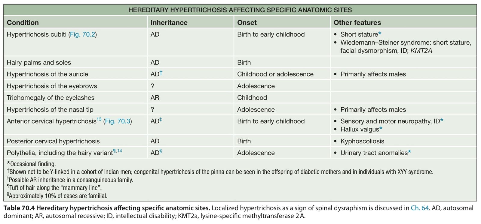
Table 70.4 Hereditary hypertrichosis affecting specific anatomic sites. Localized hypertrichosis as a sign of spinal dysraphism is discussed in Ch. 64. AD, autosomal dominant; AR, autosomal recessive; ID, intellectual disability; KMT2a, lysine-specific methyltransferase 2 A.
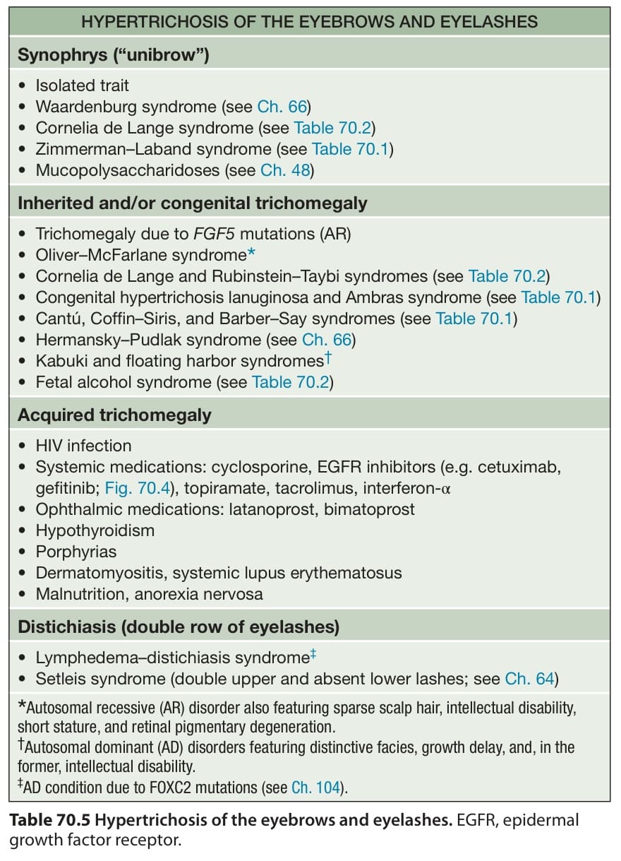
Table 70.5 Hypertrichosis of the eyebrows and eyelashes. EGFR, epidermal growth factor receptor.