HIRSUTISM
Introduction
The term “hirsutism” is defined as excessive terminal hair growth in women that is within androgen-dependent sites (e.g. upper lip, chin, mid sternum, abdomen, back, buttocks; Fig. 70.10), representing a male distribution pattern. It reflects either hyperandrogenemia (an increase in circulating androgens) or an enhanced end-organ response to androgens. Additional signs of hyperandrogenemia may include acne, a female pattern of androgenetic alopecia (see Ch. 69), irregular menstruation, and virilization. Virilization, which is uncommon and occurs with more severe hyperandrogenemia, also manifests with deepening of the voice, clitoromegaly, increased muscle mass, and male pattern hair loss (see Table 70.7).
hirsutism. This patient had seborrhea, acne, hirsutism, and alopecia, termed SAHA syndrome.
Epidemiology
Hirsutism, as defined by an abnormal score on the modified Ferriman and Gallwey scoring system (mFG; Fig. 70.11), affects ~5%–10% of women of reproductive age in the general population and rarely postmenopausal women. In a study of North American women, 35% had terminal hairs on the linea alba, 17% in the periareolar area, 16% in the lumbosacral area, and 10% in the upper pubic triangle. Androgen excess is present in >80% of premenopausal women with hirsutism, and ~70%–80% of these individuals have polycystic ovary syndrome (PCOS). Other women without hyperandrogenemia have idiopathic (constitutional) hirsutism, which tends to be relatively mild. Additional causes of hirsutism include drug intake, non-classic congenital adrenal hyperplasia, other endocrinopathies, and rarely androgen-secreting tumors (adrenal or ovarian; <0.3%) (Table 70.6). Women with rapid onset of hirsutism or signs of virilization are more likely to have an androgen-secreting tumor.
Classification
For clinical and therapeutic purposes, hirsutism can be classified according to the underlying causes (see Table 70.6).
Clinical Features
Idiopathic (constitutional) hirsutism
Idiopathic (constitutional) hirsutism is a diagnosis of exclusion that is characterized by normal menses, normal ovaries on ultrasound, and no evidence of other causes. Androgen levels are usually normal but occasionally minimally elevated. Possible contributing factors include altered peripheral androgen activity and obesity, and this condition may be familial (Fig. 70.12). Approximately 5%–20% of all women with hirsutism and 50% of those with mild hirsutism have the idiopathic form. Some women have acne and androgenetic alopecia as well as hirsutism, in association with slight increases in serum androgen levels (e.g. testosterone and dehydroepiandrosterone sulfate [DHEAS]). This constellation of findings has been referred to as SAHA – seborrhea, acne, hirsutism, and androgenetic alopecia – syndrome (see Fig. 70.10).
Ovarian hirsutism
Ovarian hirsutism can be of non-tumoral or tumoral origin. Non-tumoral etiologies include PCOS and ovarian hyperthecosis, with the former most common in premenopausal women and the latter typically occurring after menopause. A variety of benign or malignant ovarian tumors can also cause hirsutism, with malignant etiologies more likely to present with an abrupt onset of hirsutism, virilization, and markedly elevated testosterone levels.
Polycystic ovary syndrome (PCOS) This common syndrome, first described by Stein and Leventhal in 1935, affects ~5%–15% of women who are of reproductive age. It is characterized by three major features (Rotterdam criteria): (1) oligoor anovulatory cycles; (2) clinical or biochemical signs of hyperandrogenism (Fig. 70.13); and (3) ultrasonographic evidence of polycystic ovaries. Diagnosis requires two of these three criteria as well as exclusion of other causes of hirsutism (Fig. 70.14). Biochemically, there is typically a decrease in follicle-stimulating hormone (FSH) and an increase in luteinizing hormone (LH), as well as elevated serum levels of estrone and testosterone. Occasionally, serum prolactin levels are also increased.
After a normal menarche, patients develop persistent oligo- or amenorrhea that is frequently accompanied by infertility. Acne is present in 70% of patients and hirsutism in 90%. The associated hirsutism is usually lateral (i.e. on the breasts and lateral aspects of the face and neck) as well as on the central abdomen (see Fig. 70.13). Androgenetic alopecia in a female or male pattern is also commonly evident, but there are no signs of genital virilization. At least 50% of patients are obese, and some develop acanthosis nigricans and acrochordons in association with insulin resistance (see below). Patients with PCOS have large ovaries with a pearly gray surface, a thickened capsule and cysts with hyperplasia of the internal theca, but no signs of activity in the granulosa.
Women with PCOS have a higher prevalence of insulin resistance, dyslipidemia, hypertension, and obesity, which represent components of the “metabolic syndrome” that confers increased risk of atherosclerotic cardiovascular disease. Patients should be screened and followed for these conditions as well as non-alcoholic fatty liver disease (recently renamed metabolic dysfunction-associated steatotic liver disease), sleep apnea, endometrial cancer, and mood disorders, depending upon risk assessment (see Fig. 70.14). Although obesity, insulin resistance, and hyperandrogenemia are interconnected components of PCOS, it is important to note that insulin resistance also frequently occurs in non-obese patients with PCOS. HAIR-AN syndrome characterized by hyperandrogenemia (HA), insulin resistance (IR), and acanthosis nigricans (AN) is now considered to represent a more severe subtype of PCOS that is more likely to result in type 2 diabetes, hypertension, and cardiovascular disease.
PCOS is oftentimes familial, but it is clearly a heterogeneous disorder. Attempts to identify its genetic basis have led to several possible candidate genes that could play a role in insulin resistance and androgen production, including those which encode cytochrome (CYP) 17, CYP 11A1, and insulin receptor substrate 1.
Ovarian hyperthecosis This rare entity, which occurs primarily in postmenopausal women, is characterized by severe hyperandrogenism and insulin resistance. It
results from differentiation of ovarian interstitial cells into steroidogenically active luteinized theca cells that overproduce testosterone. Women typically present with acne and hirsutism and are often virilized (Fig. 70.15). Serum testosterone levels are greatly elevated and result in increased peripheral estrogen production, which can increase the risk of endometrial carcinoma.
Androgen-secreting ovarian tumors (e.g. Sertoli–Leydig cell, Leydig cell, steroid cell, and granulosa cell tumors) account for <5% of all ovarian neoplasms and can result in markedly elevated serum testosterone levels. Patients typically present with sudden onset and rapid progression of virilization in addition to hirsutism.
Adrenal hirsutism
Adrenal hirsutism can also be of non-tumoral or tumoral origin. The most frequent non-tumoral etiology is non-classic (mild) congenital adrenal hyperplasia. Androgen-secreting adrenal tumors are much less common and typically present in older women with abrupt onset and rapid progression of hirsutism and virilization.
Congenital adrenal hyperplasia (CAH) refers to a group of autosomal recessive disorders caused by genetic defects in enzymes involved in adrenal hormone production (Fig. 70.16). The most common type (≥95%) is 21-hydroxylase deficiency (21-OHD) due to CYP21A2 mutations, which has a broad range of clinical manifestations depending on the underlying CYP21A2 defect. The classic (severe) form of 21-OHD CAH occurs in ~1 : 15 000 live births worldwide, whereas the non-classic (milder) form has a prevalence of ~1 : 200 to 1 : 2000 and is more common in certain ethnic groups, including women of Ashkenazi Jewish, Middle Eastern, and Indian heritage. Other rare forms of CAH include those caused by defects in 11β-hydroxylase and 3β-hydroxysteroid dehydrogenase. Classic CAH presents during the first 2 weeks of life with dehydration and electrolyte abnormalities (salt wasting) due to aldosterone as well as cortisol deficiency in ~75% of affected neonates. Female neonates with both salt-wasting and simple virilizing forms of classic CAH typically have ambiguous genitalia, while male neonates may manifest with subtle hyperpigmentation of the genitals, flexural areas, and palmoplantar creases. The US and many other countries have universal newborn screening programs for classic CAH based on detection of elevated levels of 17-hydroxyprogesterone, the preferred substrate for 21-hydroxylase. As a result, affected neonates rarely present to a dermatologist.
Non-classic CAH (NCAH) presents postnatally, typically with onset during childhood or early adulthood; 17-hydroxyprogesterone levels are elevated but not high enough to be detected via newborn screening. Hirsutism represents the only manifestation in ~40%–60% of women with NCAH, which accounts for ~4% of all women with hirsutism. Premature pubarche is the most common presentation in children with NCAH, whereas female adolescents and women often develop acne, androgenic alopecia, irregular menses, and infertility as well as hirsutism.
Polycystic ovarian morphology may also be observed in NCAH; although virilization (Table 70.7) is more frequently evident in women with NCAH than in those with PCOS, measuring the serum 17-hydroxyprogesterone level is typically required to distinguish these two entities. Boys and men with NCAH less frequently seek medical attention for signs of androgen excess (e.g. early beard growth, large phallus).
Virilizing adrenal adenomas and carcinomas most often develop in postmenopausal women and may result in rapid onset and progression
of hirsutism and virilization. Androgen-secreting carcinomas are more common than adenomas and typically produce both androgens and cortisol. Clinical findings may also include acne, androgenetic alopecia, amenorrhea, and features of Cushing syndrome (see below). Serum
levels of testosterone and DHEAS levels are markedly elevated, and serum cortisol may also be increased.
Other endocrinopathies associated with hirsutism
Other endocrinopathies in which women may develop hirsutism together with disease-specific manifestations include Cushing syndrome, hyperprolactinemia, acromegaly, thyroid disorders, and insulin resistance disorders. Pregnant women may also present with hirsutism as a result of ovarian and placental androgen production.
Cushing syndrome may be associated with either high plasma ACTH levels (pituitary hyperproduction or “ectopic ACTH syndrome”) or a nearly complete lack thereof (adrenal primary nodular hyperplasia, adrenal adenoma, or adrenal carcinoma). All patients have an increase
in plasma cortisol, which is the cause of the major clinical features: central obesity with “moon facies” and “buffalo hump”, hypertension, glucose intolerance, purple striae, and ecchymoses. Patients with adrenal hyperplasia (usually due to ACTH hyperstimulation by a pituitary adenoma) or adrenal adenomas typically have an insidious onset of symptoms, but with an adrenal carcinoma or ectopic ACTH production by a malignant tumor, manifestations appear more rapidly. If there is significant production of androgens, which is not the norm for adrenal adenomas, virilization with hirsutism will be present in addition to the typical manifestations of Cushing syndrome.
Hirsutism represents a rare manifestation of hyperprolactinemia, which is most often caused by pituitary adenomas and drugs (e.g. dopamine antagonists). This etiology of hirsutism should be considered in women <50 years of age who present with galactorrhea together with oligomenorrhea, amenorrhea, or infertility (“amenorrhea–galactorrhea syndrome”).
Acromegaly results from excess secretion of growth hormone (GH), most commonly by a pituitary adenoma. The clinical features reflect elevated serum concentrations of both GH and insulin-like growth factor 1 (IGF-1), which is GH dependent. Long-term excess of GH
The most common cause of congenital adrenal hyperplasia is 21-hydroxylase deficiency. This leads to an accumulation of 17-OH-progesterone.
and IGF-1 results in overgrowth of many tissues and organs, including skin thickening as well as the classic findings of enlarged hands and feet, macrognathia, and macroglossia. Additional cutaneous manifestations can include hirsutism, hypertrichosis, hyperhidrosis, acanthosis nigricans, acrochordons, and cutis verticis gyrata.
Thyroid dysfunction interferes with sex hormone metabolism and can result in irregular menstruation. Hypothyroidism can also cause cystic ovarian changes and decreased levels of sex hormone-binding globulin (SHBG), which increases the amount of free circulating testosterone.
Disorders of severe insulin resistance (e.g. obesity-related, insulin receptor mutations [type A syndrome], anti-insulin receptor antibodies [type B syndrome]) are often associated with hyperandrogenism, hirsutism, and even virilization. Marked hyperinsulinemia results in ovarian hyperandrogenism, presumably via receptors for insulin and IGF-1 on ovarian theca cells. Insulin also decreases SHBG levels, thereby increasing the amount of free circulating testosterone. Ovaries often show a polycystic pattern on ultrasound.
Drug-induced hirsutism
Several drugs have been associated with the development of hirsutism (see Table 70.6). Identifying such culprit drugs and stopping them will typically result in reversal of the hirsutism.
Diagnosis
Hirsutism is a clinical diagnosis and a potential indicator of a hyperandrogenic disorder that may require treatment and have associated comorbidities such as metabolic syndrome or fertility issues. In a premenopausal woman with hirsutism, the mFG score (see Fig. 70.11) should be determined and assessment for associated findings initiated (Fig. 70.17). Additional evaluation of women with hyperandrogenemia for signs of PCOS are outlined in Figs. 70.18 and 70.14.
It is important to inquire about the age of onset (premenopausal vs postmenopausal), progression (rapid vs gradual), and emotional and psychosocial impact of the hirsutism. Patients should also be asked about their menstrual history (e.g. oligo- or amenorrhea), medications (see Table 70.6), comorbidities (e.g. insulin resistance disorders), ethnicity, and family history. Physical examination should determine if true hirsutism (versus hypertrichosis) is present as well as its severity (see Fig. 70.11). In addition, patients should be evaluated for additional findings of hyperandrogenism (e.g. acne, androgenic alopecia), virilization (see Table 70.7), acanthosis nigricans, central obesity, and manifestations of other endocrinopathies associated with hirsutism (see Table 70.6). An abrupt onset and rapid progression of hirsutism or the presence of virilization should raise suspicion for a more serious cause of androgen excess (e.g. an androgen-secreting tumor). Premenopausal women with hirsutism are most likely to have PCOS, idiopathic hirsutism or NCAH, whereas postmenopausal women with new onset hirsutism are more likely to have an androgen-secreting tumor or ovarian hyperthecosis.
In a premenopausal woman with hirsutism, serum total testosterone is recommended as the initial biochemical study. Additional considerations include measuring the serum 17-hydroxyprogesterone level (e.g. in women at higher risk of NCAH based on ethnicity or family history) and transvaginal pelvic ultrasonography to assess for PCOS. Additional evaluation based upon these results and the clinical presentation are outlined in Figs. 70.17 and 70.18. Of note, in women taking an oral contraceptive (OCP) or spironolactone, the results of laboratory testing
will not be reliable. If an evaluation is desired, then discontinuation of the medications for at least 8–12 weeks is recommended.
The most useful biochemical tests to evaluate a postmenopausal woman with hirsutism are serum total testosterone and DHEAS levels. Serum total testosterone levels >150 ng/dl are suggestive of an androgen-secreting tumor, with higher testosterone levels typically associated with an ovarian source and DHEAS >700 mcg/dl suggestive of an adrenal source. Hyperandrogenism due to PCOS or NCAH can persist into menopause, and a serum 17-hydroxyprogesterone level should be considered if not evaluated previously. Other laboratory testing depends upon the presence of signs or symptoms suggestive of another endocrinopathy (see Table 70.6).
Treatment
The impact of hirsutism on a woman’s quality of life should be assessed and regardless of the mFG score, treatment should be initiated if it is causing distress to the patient (“patient-important” hirsutism). Management of premenopausal and postmenopausal hirsutism involves a combination of systemic pharmacologic therapies (Table 70.8) and direct hair removal methods, both of which should be offered to women with “patient-important” hirsutism. Bilateral oophorectomy can be considered in the management of ovarian hyperthecosis. In the case of ovarian and adrenal tumors, surgical intervention is usually required.
Systemic therapy
The 2018 Endocrine Society clinical guidelines recommend initial treatment with systemic pharmacologic therapy in premenopausal “patient-important” hirsutism that persists despite cosmetic measures. They suggest a combined estrogen–progesterone OCP as first-line treatment, as long as fertility is not immediately desired and there are no contraindications (e.g. history of venous thromboembolism [VTE], inherited thrombophilia, smoking if age >35 years, migraine with aura, estrogen-dependent neoplasm). Combined OCPs (cOCPs) suppress LH secretion and thus ovarian androgen production. They also stimulate the production of SHBG, which results in a decrease of circulating free androgen levels. The Endocrine Society guidelines do not recommend one cOCP over another for treating hirsutism based on a meta-analysis of effectiveness. Although cOCPs containing antiandrogenic progestins (e.g. drospirenone, cyproterone acetate [not available in the US]) have been shown in some studies to lower mFG scores slightly more than other combined OCPs, the difference is not thought to be clinically important. A cOCP containing the lowest effective dose of ethinyl estradiol (usually 20 mcg) and a low VTE-risk progestin (e.g. norgestimate, norethindrone) is recommended in women at higher risk for VTE (e.g. obesity, age ≥40 years). Although levonorgestrel is associated with a low VTE risk, this progestin is the most androgenic and may have adverse metabolic effects, making it less suitable for patients with PCOS.
If the clinical response is not achieved after 6 months of a cOCP, then an antiandrogen (e.g. spironolactone) can be added (see Ch. 36). A trial of at least 6 months is suggested before making dose adjustments or medication changes (Fig. 70.19). Antiandrogen monotherapy is not advised unless the patient is using adequate contraception, given the risk of impact on a developing fetus. If the hirsutism is severe or previous use of a cOCP has been unsuccessful, then initial therapy with both a cOCP and antiandrogen can be considered.
Additional systemic treatment options for hirsutism are summarized in Table 70.8. Flutamide and bicalutamide are nonsteroidal androgen receptor blockers that are approved for the treatment of prostate cancer. Although both of these agents have been utilized for the treatment of hirsutism, flutamide is not recommended for this indication because of its higher potential for hepatotoxicity. Insulin-lowering medications (e.g. metformin) have not shown efficacy as monotherapy for hirsutism. Management of PCOS comorbidities are outlined in Fig. 70.14. Lifestyle modifications (e.g. diet, exercise) are recommended for all hirsute women with obesity. Lastly, for women with NCAH, glucocorticoids may be utilized to treat hirsutism that does not respond adequately to a cOCP ± antiandrogen or to induce ovulation.
In postmenopausal women with “patient-important” hirsutism and no evidence of an androgen-secreting tumor, treatment typically includes an antiandrogen plus direct hair removal methods (see Table 70.8).
Topical therapy
Topical antiandrogen therapy (e.g. canrenone [active metabolite of spironolactone], finasteride) is generally not recommended for treatment of hirsutism because of limited evidence of efficacy. A newer topical antiandrogen, clascoterone, has been approved for the treatment of acne but has yet to be studied for hirsutism.
Eflornithine HCL 13.9% cream is FDA-approved for the reduction of unwanted facial hair in women; it was discontinued by the original manufacturer in 2023. It irreversibly inhibits ornithine decarboxylase, an enzyme that catalyzes the rate-limiting step in follicular polyamine synthesis, which is necessary for hair growth. In a randomized vehiclecontrolled study in 594 women, it was shown to reduce hair length and mass by ~25%. Because eflornithine HCL slows hair growth rather than removes hair, it is best used and more effective in conjunction with other direct hair removal methods (e.g. laser or intense pulsed light [IPL]). Results can take up to 8 weeks to become evident, and hair gradually returns to pretreatment levels upon discontinuation.
Direct hair removal methods
Direct hair removal methods can either remove a portion of the hair from above the skin’s surface (depilation) or the entire hair shaft (epilation). Epilation methods may result in temporary or potentially
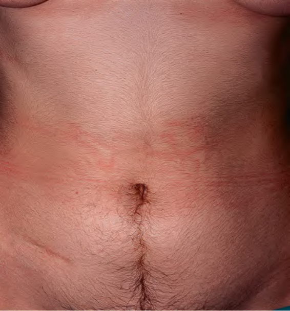
Fig. 70.10 Idiopathic (constitutional)
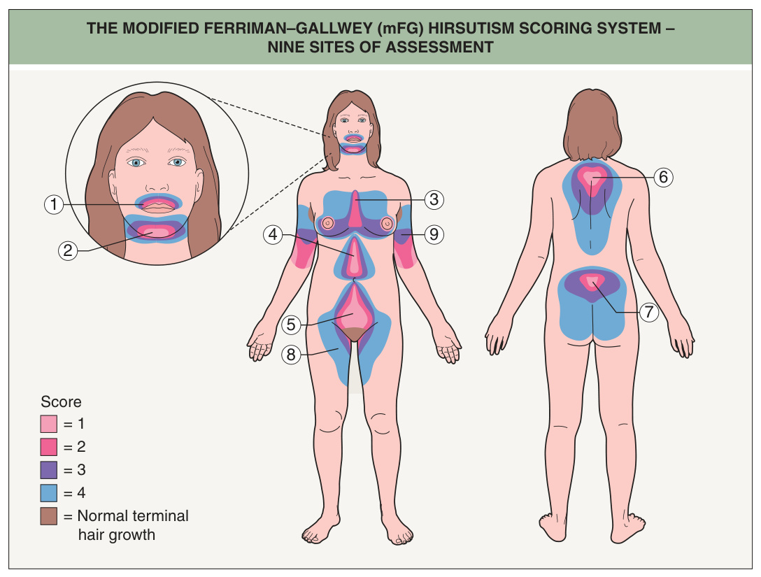
Fig. 70.11 The modified Ferriman–Gallwey (mFG) hirsutism scoring system – nine sites of assessment. The modified Ferriman and Gallwey score is based upon the sum of the values (graded from 0 to 4 points) for each of the nine body areas illustrated as follows: 0 points (no terminal hairs); 1 point (minimal terminal hairs); 2 points (hair growth is more than minimal but not yet that of a man); 3 points (hair growth is that of a man who is not very hairy); and 4 points (equivalent to a very hairy man). Total mFG scores that define hirsutism in reproductive-aged women vary depending upon race/ ethnicity and are as follows: US and United Kingdom White or Black women, ≥8 (8–15 generally considered mild, 16–25 moderate, and >25 severe hirsutism); Mediterranean, Middle Eastern and Hispanic women, ≥9–10; South American women, ≥6; and Asian women, ranging from ≥2 for Han Chinese women to ≥7 for Southern Chinese women.
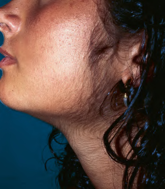
Fig. 70.12 Familial hirsutism. Prolongation of the preauricular hair implantation line with terminal hairs of the lateral lower face and neck.
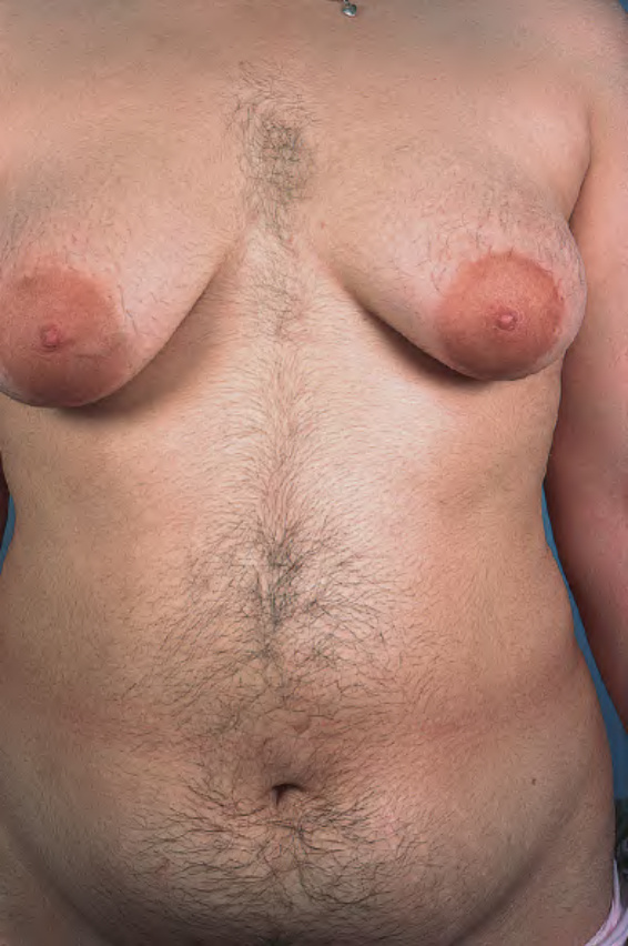
Fig. 70.13 Hirsutism in a young woman with polycystic ovary syndrome. Both lateral and central patterns are present.

Fig. 70.14 Evaluation of a patient with polycystic ovary syndrome. Hb, hemoglobin; HDL, high-density lipoprotein; LDL, low-density lipoprotein. Adapted from Ehrman DA. Polycystic ovary syndrome. N Engl J Med 2005;352:1223–36.
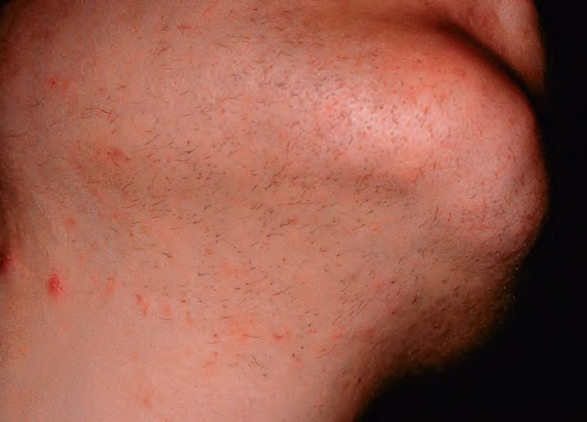
Fig. 70.15 Facial hirsutism due to ovarian hyperthecosis.Courtesy Robert Hartman, MD.
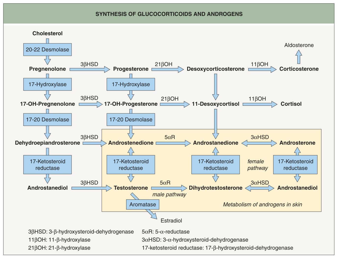
Fig. 70.16 Synthesis of glucocorticoids and androgens.
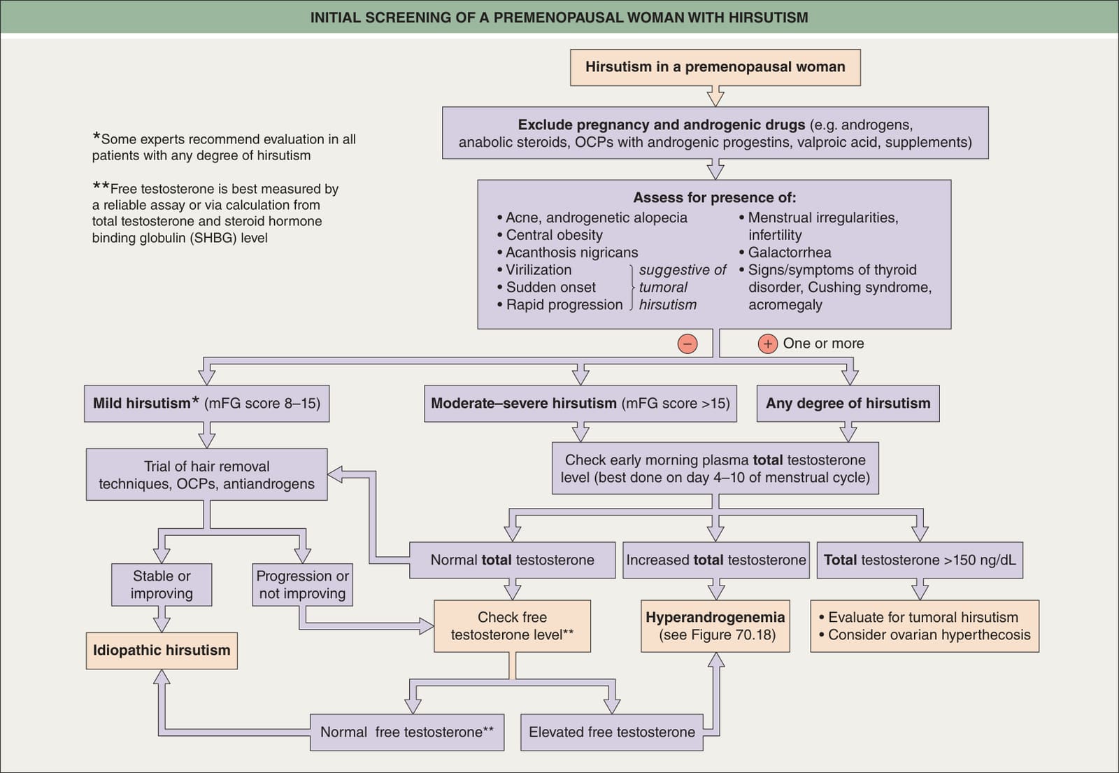
Fig. 70.17 Initial screening of a premenopausal woman with hirsutism. mFG, modified Ferriman and Gallwey; OCPs, oral contraceptives.
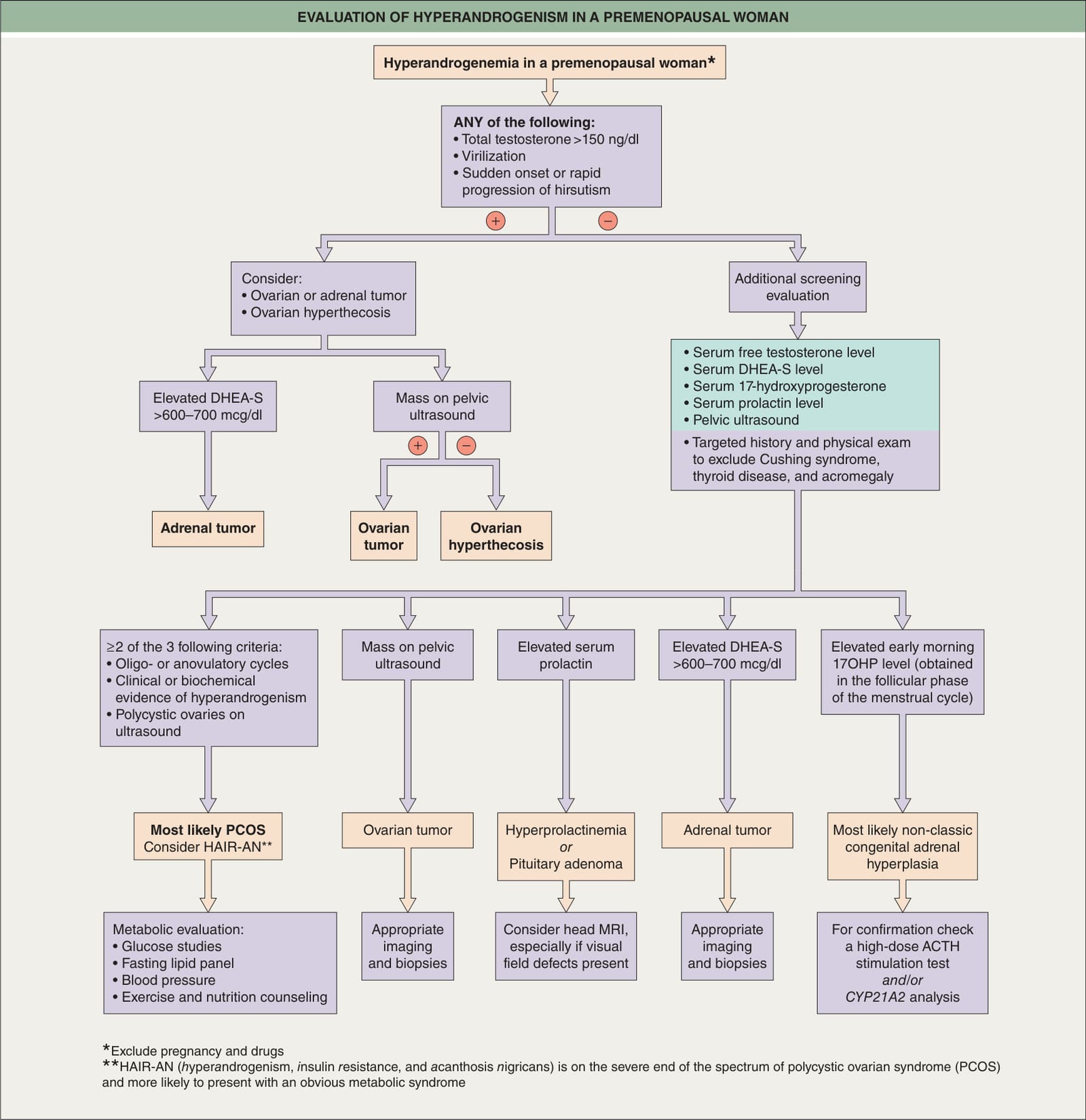
Fig. 70.18 Evaluation of hyperandrogenemia in a premenopausal woman.
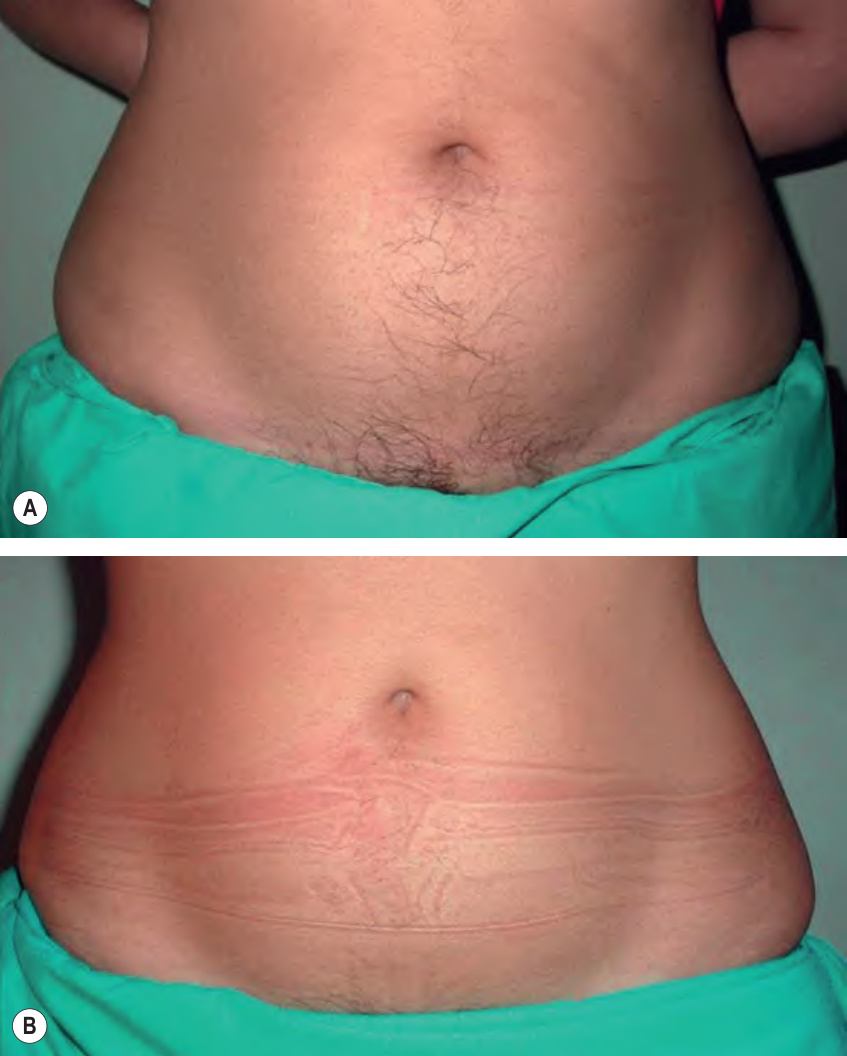
Fig. 70.19 Hirsutism before (A) and after treatment (B) with antiandrogen therapy. This patient had the SAHA (seborrhea, acne, hirsutism, and alopecia) syndrome and achieved clinical improvement in hirsutism after one year of antiandrogen therapy.
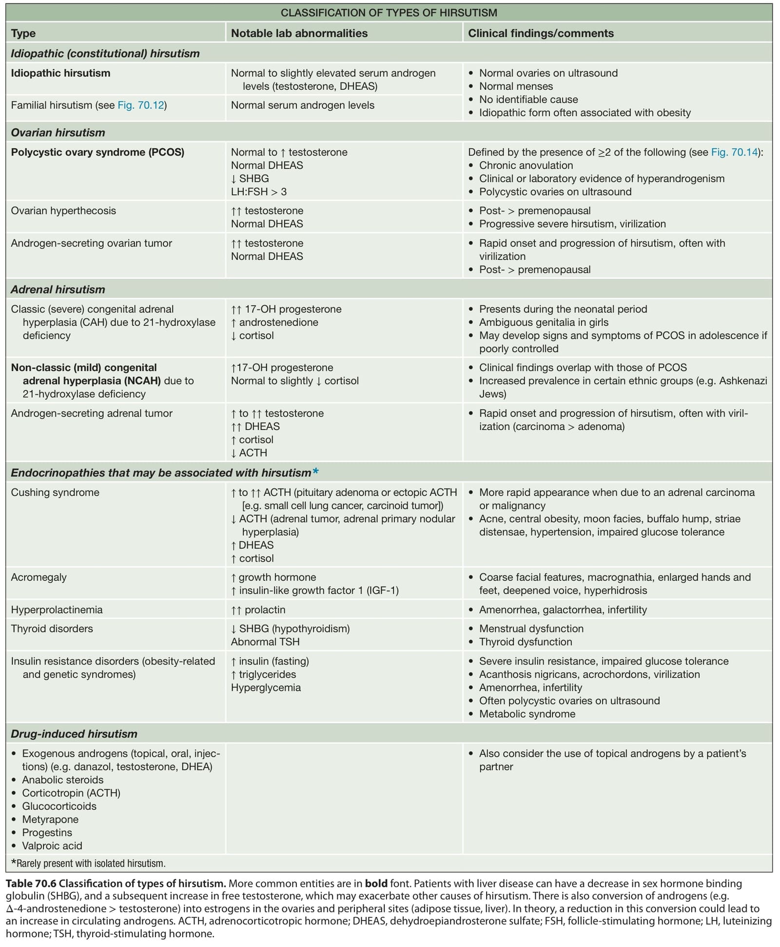
Table 70.6 Classification of types of hirsutism. More common entities are in bold font. Patients with liver disease can have a decrease in sex hormone binding globulin (SHBG), and a subsequent increase in free testosterone, which may exacerbate other causes of hirsutism. There is also conversion of androgens (e.g. Δ-4-androstenedione > testosterone) into estrogens in the ovaries and peripheral sites (adipose tissue, liver). In theory, a reduction in this conversion could lead to an increase in circulating androgens. ACTH, adrenocorticotropic hormone; DHEAS, dehydroepiandrosterone sulfate; FSH, follicle-stimulating hormone; LH, luteinizing hormone; TSH, thyroid-stimulating hormone.
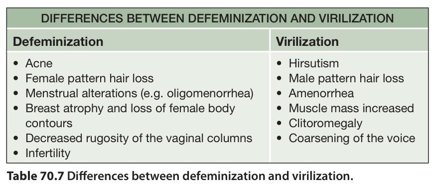
Table 70.7 Differences between defeminization and virilization.
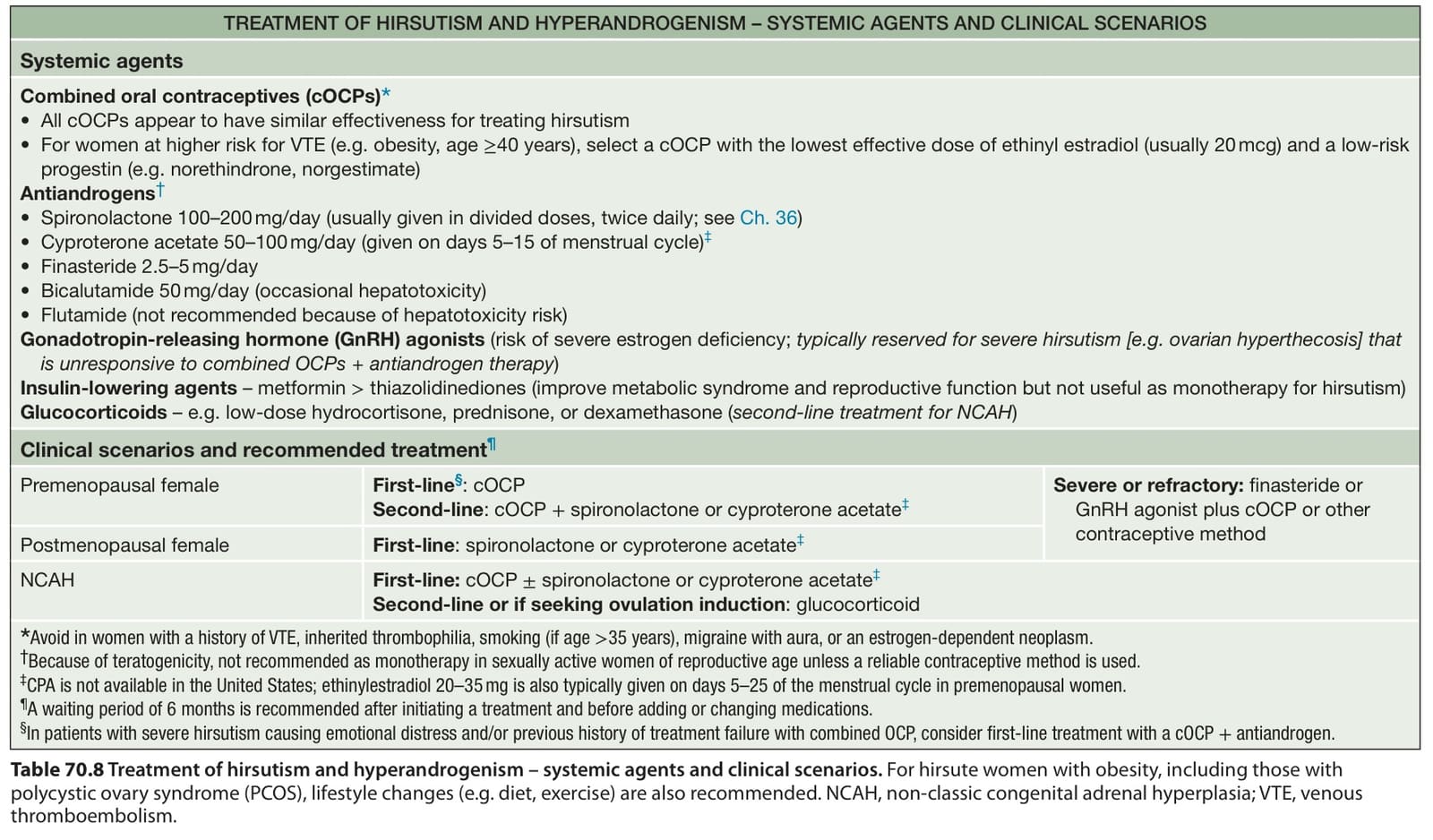
Table 70.8 Treatment of hirsutism and hyperandrogenism – systemic agents and clinical scenarios. For hirsute women with obesity, including those with polycystic ovary syndrome (PCOS), lifestyle changes (e.g. diet, exercise) are also recommended. NCAH, non-classic congenital adrenal hyperplasia; VTE, venous thromboembolism.
permanent hair reduction. Each hair removal option has advantages and disadvantages.
Depilation methods include trimming, shaving, dermaplaning, and chemical depilatories. Shaving is easy and inexpensive but must be done frequently and can cause skin irritation. Contrary to popular belief, shaving does not change the texture, color, or growth rate of hair. It does, however, create a blunt tip upon regrowth that can give the illusion of thicker hair. Chemical depilatory agents, which dissolve the hair shafts via breakage of disulfide bonds, can be used for large areas but often cause skin irritation.
The results of epilation typically last longer than those of depilation. Tweezing, threading, waxing, and sugaring are effective temporary epilation techniques, but they are painful and often impractical for larger areas. Potentially permanent options include electrolysis and photoepilation utilizing a laser or IPL. Although effective, electrolysis is painful, time-consuming, and not practical for treating large areas (see Ch. 140). Photoepilation, which can treat larger areas in a shorter amount of time, represents the preferred option for long-term hair removal. It utilizes pulses of light absorbed by melanin to destroy pigmented terminal hair follicles through thermal damage. Photoepilation systems include alexandrite, diode, and Nd:YAG lasers and various IPL sources, which are selected based upon the patient’s skin and hair color (see Ch. 137). Clinical trials have shown ~40%–80% hair reduction, depending upon the device used and the number of treatments provided. Photoepilation is ineffective for blond, white, or gray hairs and can be costly and painful, with potential side effects including pigmentary changes and rarely scarring or paradoxical hypertrichosis.
Additional figures and tables on Abraham’s classification of hirsutism, and Methods to reduce unwanted hair, available in our eBook (see inside front cover for access code).
W, eds. Trichology. Diseases of the Pilosebaceous Follicle. Madrid: Aula Médica; 1997:243–263.2. Olsen E. Hypertrichosis. In: Olsen E, ed. Disorders of Hair
Growth. Diagnosis and Treatment. New York: McGraw-Hill; 1994:315–336.3. Camacho FM. Hypertrichosis. In: Blume-Peytavi U, Tosti A, syndrome. In: Camacho FM, Randall VA, Price VH, eds. Hair and its Disorders. Biology, Pathology and Management. London: Martin Dunitz; 2000:359–367.30. Camacho FM. SAHA syndrome. In: Camacho FM,
Whiting DA, Trüeb R, eds. Hair Growth and Disorders. Berlin: Springer-Verlag; 2008:333–356.4. Camacho FM. Hypertrichosis. In: Camacho FM, Tosti A,
Montagna W, eds. Trichology. Diseases of the Pilosebaceous Follicle. Madrid: Aula Médica; 1997:673–690.31. Escobar-Morreale HF, Santacruz E, Luque-Ramirez M,
Randall VA, Price VH, eds. Montagna Trichology. Diseases of the Pilosebaceous Follicle. Madrid: Aula Médica; 2014:431–473.5. Ramot Y, Zlotogorski A. Recent discoveries in genetic hair
disorders. In: Camacho FM, Tosti A, Randall VA, Price VH, eds. Montagna Trichology. Diseases of The Pilosebaceous Follicle. Madrid: Aula Médica; 2014:420–427.6. Fantauzzo KA, Tadin-Strapps M, You Y, et al. A position
Whiting DA, Trüeb R, eds. Hair Growth and Disorders. Berlin: Springer-Verlag; 2008:357–377.24. Camacho FM. Hirsutism. In: Camacho FM, Tosti A,