ANATOMY
Key features
Nail matrix → nail plate production proximal matrix → dorsal nail plate distal matrix (lunula) → ventral nail plate
Proximal nail fold → nail matrix protection
Nail bed and hyponychium → nail plate adhesion and distal detachment
Nail growth rate: fingernails: 3 mm/month; toenails: 1 mm/month
The nail plate is a fully keratinized structure produced by the germinative epithelium of the nail matrix. As it grows, the nail plate emerges from the proximal nail fold and progresses distally, lying across and strictly adhering to the nail bed (Fig. 71.1). As the nail plate approaches the tip of the digit, it detaches from the underlying tissues, forming the hyponychium. Proximally and laterally, the nail plate is surrounded by the nail folds. The proximal nail fold consists of two layers of epithelium – a dorsal layer, which is the distal continuation of the skin of the dorsum of the digit, and a ventral layer, which is continuous with the nail matrix.
The nail matrix is responsible for the production of the nail plate and consists of an epithelium that keratinizes without the formation of a
Nail plate production occurs continuously, proceeding from the 15th week of embryonic life until death. Under normal conditions, the mean growth rate of a fingernail is 3 mm/month and that of a toenail is 1 mm/ month. Nail growth rate can be influenced by several factors, including age, systemic diseases, and medications.
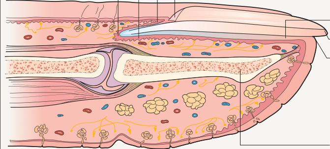
Fig. 71.1 Schematic drawing of the nail apparatus in longitudinal section. For additional details, see Figs. 149.1 & 149.2 granular layer. Nail matrix keratinization occurs along an oblique axis, reflecting the upward and distal movement of cells during the process of maturation and differentiation. For this reason, the proximal portion of the nail matrix produces the dorsal portion of the nail plate, while the distal matrix is responsible for the production of the ventral nail plate (see Fig. 149.2). The distal portion of the nail matrix is visible through the transparent nail plate as a white, distally convex half-moon known as the lunula. The matrix contains melanocytes that are normally quiescent; however, they may become activated and synthesize melanin, which is transferred to the surrounding keratinocytes. Distal migration of melanin-containing keratinocytes then gives rise to a hyperpigmented nail plate (melanonychia).