NAIL SIGNS
The clinical signs of various nail disorders depend upon the component of the nail apparatus that has been affected: nail plate alterations are due to matrix damage whereas nail plate detachment and/or uplifting arise from dysfunction of the hyponychium or nail bed (Table 71.1). Paronychia represents inflammation of the nail fold, which can lead to secondary matrix damage. The nail plate can develop a wide range of structural changes, from thickening to fissuring to atrophy, in addition to discoloration (Fig. 71.2).
Nail signs can be schematically divided into three major categories:
●signs due to abnormal nail matrix function
●signs due to nail bed disorders
●signs due to deposition of pigment within the nail plate.
Nail Signs Due to Abnormal Nail Matrix Function
Beau’s lines
Key features
Transverse depressions that are more evident in the central portion of the nail plate
Most often due to trauma
Involvement of multiple digits suggests a systemic cause
First described by Beau in 1846, these transverse depressions of the nail plate surface result from a temporary interruption of the mitotic activity of the proximal nail matrix. The depth of the depression indicates the extent of the damage within the matrix; the width of the depression indicates the duration of the insult. Beau’s lines migrate distally along with nail plate growth, with multiple lines indicating repeated damage (Fig. 71.3). Most commonly, Beau’s lines are caused by mechanical trauma (e.g. manicures, onychotillomania) or dermatologic disease of the proximal nail fold (e.g. eczema, chronic paronychia). The presence of Beau’s lines at the same level in all nails suggests a systemic cause (e.g. severe or febrile illness, cytotoxic drugs, erythroderma).
Multiple transverse depressions due to repeated traumatic insults to the proximal nail matrix from aggressive manicures. The depressions are more evident in the central portion of the nail plate.
Onychomadesis (nail shedding)
Key features
Proximal detachment of the nail that is most often due to trauma
Similar involvement of multiple digits suggests a systemic cause
The term onychomadesis describes the detachment of the nail plate from the proximal nail fold and is due to a severe insult that produces a complete arrest of nail matrix activity. Onychomadesis appears as a sulcus that replaces the proximal nail plate. The causes of onychomadesis include those that produce Beau’s lines (see above). Retrospectively, it can indicate a previous infection, most typically hand-foot-andmouth disease (Fig. 71.4), but also scarlet fever or Kawasaki disease. In addition, onychomadesis may appear in the setting of pemphigus vulgaris or Stevens–Johnson syndrome.
Pitting
Pitting describes the presence of punctate depressions of the nail plate surface that can have an irregular outline (see Fig. 71.2). Pits are due to foci of abnormal keratinization of the proximal nail matrix which result in clusters of parakeratotic cells within the dorsal nail plate. In the proximal plate, these clusters appear as nontransparent collections
of scales. Over time, shedding of the parakeratotic cells leads to the appearance of pits. These pits migrate distally with nail growth.
Diseases that produce nail pitting include psoriasis, alopecia areata, and eczema. Fig. 71.2 demonstrates how patterns of the pits can differ. Psoriatic pits vary in size and depth and have an irregular distribution, whereas in patients with alopecia areata, the pits are small, superficial, and geometrically distributed.
Onychorrhexis
Onychorrhexis is characterized by longitudinal ridging and fissuring of the nail plate (see Fig. 71.2). The depth of the ridges can vary as can the extent of involvement, i.e. a portion versus the entire nail plate, and one or several nails may be involved. It is often associated with nail thinning, indicating damage to the nail matrix. Diseases commonly responsible for onychorrhexis include lichen planus (several nails), conditions that lead to impaired vascular supply, trauma, and tumors that compress the nail matrix (typically a single nail). The differential diagnosis includes the common phenomenon of increased longitudinal ridging of the nail plate that is age-related.
Trachyonychia (twenty-nail dystrophy, sandpapered nails)
Key features
Nail roughness
Associated with thinning, koilonychia, and cuticular hyperkeratosis
Conditions that may cause trachyonychia include alopecia areata
(common), lichen planus (uncommon), psoriasis (rare), and eczema (very rare)
Trachyonychia is a specific nail plate surface abnormality characterized by diffuse homogeneous roughness (Fig. 71.5). In most patients, the affected nails are opaque, lusterless, and rough; the nail plate surface has longitudinal ridging due to fine superficial striations distributed in a regular, parallel pattern (sandpapered nails). A less common variant, referred to as shiny trachyonychia, is characterized by multiple small punctate depressions distributed in a geometric pattern within parallel, longitudinal lines. Trachyonychia is a reflection of mild, diffuse damage to the proximal nail matrix by inflammatory disorders (see above). It can also be a presenting sign of systemic AL amyloidosis.
True leukonychia
Key features
White opaque discoloration that is punctate, striate, or diffuse
Punctate and striate leukonychia most often due to trauma to the distal nail matrix
Needs to be distinguished from apparent leukonychia (nail bed discoloration that fades with pressure) and friable pseudoleukonychia due to superficial white onychomycosis or nail fragility
The nail plate has a normal surface but loses its transparency and looks white because of the presence of parakeratotic cells within its ventral portion. True leukonychia is caused by diseases that disturb distal nail matrix keratinization and it presents with three morphologic variants:
●Punctate leukonychia. The nail plate has small opaque white spots that move distally with nail growth and sometimes disappear before reaching the distal nail. It is caused by trauma and is most commonly observed in the fingernails of children.
●Striate leukonychia. The nail plate has one or more transverse, opaque white, parallel lines that migrate distally with nail growth. It is frequently observed in the fingernails of women, due to matrix trauma secondary to manicures. Striate leukonychia may also occur in great toenails as a consequence of trauma from shoes. It is also typical of Mees’ lines, the white transverse bands seen in arsenic and thallium poisoning.
●Diffuse leukonychia (porcelain nails, leukonychia totalis). The nail plate is completely or almost completely opaque and white (Fig. 71.6). Diffuse true leukonychia is rare and may result from mutations in PLCD1 (encodes phosphoinositide-specific phospholipase C delta 1 subunit, a key enzyme in phosphoinositide metabolism expressed in the nail matrix) or GJA1 (encodes connexin 43). Patients with GJA1 mutations also have keratoderma and hypotrichosis. Bart–Pumphrey syndrome, in which patients have deafness, knuckle pads, and palmoplantar keratoderma in addition to leukonychia, is due to mutations in GJB2 (encodes connexin 26; see Ch. 58).
Koilonychia (spoon nails)
The nail plate is thinned and depressed centrally with upward eversion of its lateral and distal edges, leading to a concave spoon-like shape (see Fig. 71.2). Koilonychia, especially of the 2nd–4th toes in young children (1–4 years of age), is physiologic and eventually resolves spontaneously. Isolated koilonychia is unusual and occurs in association with severe iron deficiency or from contact with chemicals that damage the nail plate. More commonly, koilonychia accompanies other nail abnormalities (e.g. trachyonychia), including in the setting of inflammatory diseases such as lichen planus.
Nail Signs Due to Nail Bed Disorders
Onycholysis
The distal nail plate is detached from the nail bed and usually appears white because of the presence of air in the subungual space. If an exogenous pigment is present, the nail may appear yellow (fungi and exudate) to green–black (pyocyanin). In addition to environmental trauma, the nail bed disorders that most commonly produce onycholysis are psoriasis and onychomycosis (Fig. 71.7). Drug-induced onycholysis/ photo-onycholysis involves several nails and may have associated subungual hemorrhage (e.g. taxanes).
Subungual hyperkeratosis
The nail plate appears thickened due to an accumulation of subungual scales. Subungual hyperkeratosis results from excessive proliferation of the keratinocytes in the nail bed and hyponychium. It is commonly seen in inflammatory disorders such as psoriasis and contact dermatitis as well as in distal subungual onychomycosis.
Apparent leukonychia
Key features
White discoloration that fades with pressure
Nail plate transparency maintained
Often due to drugs (e. g. chemotherapeutic agents) or systemic diseases (e. g. hypoalbuminemia)
The nails appear white because of abnormalities in the color of the nail bed and this is usually due to nail bed edema. Apparent leukonychia does not move distally with nail growth and the white discoloration fades with pressure (see Fig. 71.2). See section on Systemic Diseases for discussion of the three major forms.
Splinter hemorrhages
Splinter hemorrhages appear as one or more red–brown to purple– black, thin longitudinal lines, and they are most commonly seen in the distal portion of the nail (see Fig. 71.2). The shape of the subungual hemorrhages is due to the longitudinal orientation of nail bed capillaries. Onychoscopy allows better visualization of the hemorrhages which typically have a deep red to black color with peripheral fading.
The most common cause of splinter hemorrhages is trauma, followed by psoriasis, onychomycosis, and occasionally medications. Proximal splinters are rare and possible indicators of systemic diseases, including endocarditis (infectious and marantic), vasculitis (including septic vasculitis), trichinosis, and the antiphospholipid antibody syndrome.
Approximately 30% of patients with tuberous sclerosis complex have “red comets”, i.e. short red longitudinal streaks of the nail with an enlarged distal end (see Fig. 61.17). They are partially blanchable and are thought to represent telangiectasias plus extravasated blood.
Nail Signs Due to Deposition of Pigment
Key features
Exogenous → convex proximal border
Endogenous → concave proximal border (distally convex)
Subungual deposition → nail bed discoloration with a transparent nail plate
Causes of nail pigmentation include: (1) staining from external pigment; (2) pigment under the nail plate or in the nail bed dermis; and (3) deposition of pigment within the nail plate. Nail pigmentation from external staining typically follows the shape of the proximal nail fold. Common examples are yellow–brown nail discoloration due to nicotine in smokers or darkening of the nail plate due to hair dyes in hairdressers. Subungual deposition of pigment is often seen with Pseudomonas colonization, where the green discoloration of the nail results from the production of pyocyanin by the bacteria. Dermatophytes usually cause a yellow–white subungual color. Occasionally, there can be deposition of heavy metals or medications (e.g. minocycline) within the nail bed dermis. In melanonychia, the nail appears brown–black due to the incorporation of nail matrix-produced melanin into the nail plate itself (see below).
Longitudinal melanonychia
Key features
Longitudinal brown to black band extending from the proximal nail fold to the distal margin
Common in darkly pigmented individuals
Single band may be a sign of melanocyte activation or proliferation
(nevus, melanoma)
Multiple bands point to physiologic, trauma/onychotillomania, drugs, or systemic disease
Longitudinal melanonychia results from melanin production by nail matrix melanocytes and is most commonly due to simple activation of these melanocytes. It can also result from melanocyte hyperplasia as is found in a lentigo, nevus, or melanoma. Clinically, melanonychia presents as one or more longitudinal pigmented bands extending from the proximal nail fold to the distal margin (Fig. 71.8). The band of melanonychia can vary in color from light brown to black and the pigmentation may be homogeneous or variable. The width ranges from a few millimeters to the width of the whole nail. Melanonychia of several digits is usually due to melanocyte activation and is common in patients with darkly pigmented skin (Table 71.2). When melanonychia presents during adulthood in a single digit and there are no systemic or local causative factors, it is important to consider the possibility of nail melanoma. Fig. 71.9 outlines an approach to the patient with longitudinal melanonychia.
Dermoscopic criteria for benign and malignant nail melanocytic lesions have been proposed, but nail dermoscopy is not always feasible nor is it completely reliable in the evaluation of nail pigmentation. For example, it cannot be performed when the nail is totally black or very thick such that the lines and borders appear blurred. Additionally, dermoscopic evaluation may give false results, as irregular lines may sometimes be seen with benign pigmentation, especially in children. In addition, hyponychial brush pigmentation (distal fibrillar pattern) is seen in 25%–30% of pediatric patients with longitudinal melanonychia, especially if congenital or early-onset. Histopathologic examination, when clinically warranted, remains the gold standard for the evaluation of nail pigmentation. A detailed description of biopsy procedures for longitudinal melanonychia is found in Chapter 149.
Green nail syndrome
The yellow–green to green–black nail plate discoloration is due to the production of the pigments pyocyanin and pyoverdin by Pseudomonas aeruginosa (see Ch. 74). This discoloration is most often present in the subungual space created by onycholysis and less often in the lateral nail plate in the setting of chronic paronychia. Predisposing factors include prolonged exposure to water, use of detergents and soaps, nail trauma, and other causes of onycholysis or paronychia. Not surprisingly, this disorder is seen in barbers, dishwashers, bakers, and medical personnel.
Pseudomonas colonization can be eliminated from the nail surface by applying a few drops of 2% sodium hypochlorite solution (or household chlorine bleach diluted 1 : 4 in water) and from the subungual space by soaking the affected digits in the same solution for 5 minutes per day for 20–30 days. The underlying nail disorder must also be addressed, e.g. clipping back the detached nail plate if there is onycholysis.
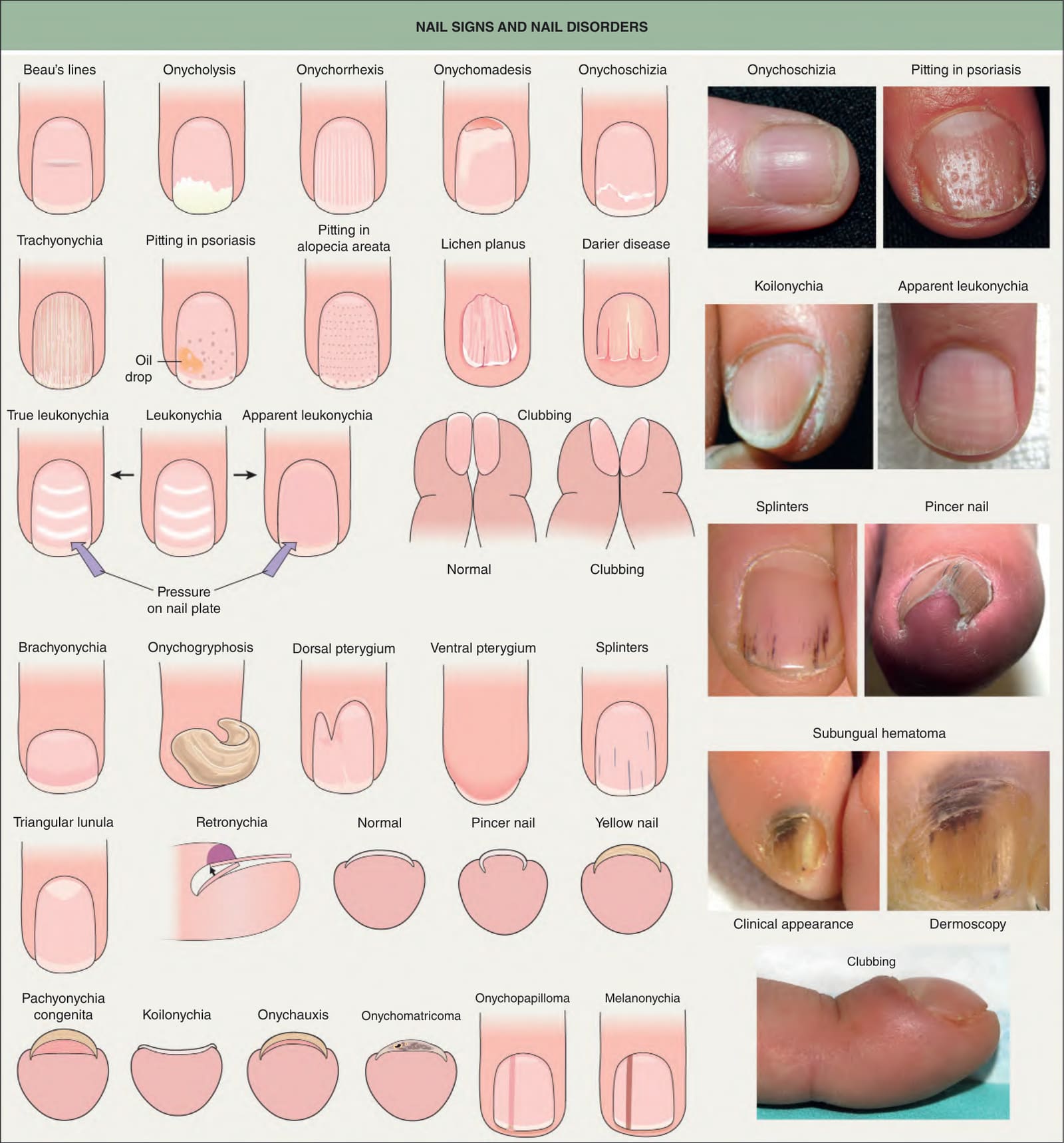
Fig. 71.2 Nail signs and nail disorders. In apparent leukonychia (e. g. Muehrcke’s lines due to hypoalbuminemia and presumed secondary edema), the transverse white lines disappear with pressure, but not in true leukonychia (e. g. Mees’ lines due to parakeratosis). Terry’s nails and half-and-half nails are additional examples of apparent leukonychia. Dermoscopy of a subungual hematoma shows small red longitudinal splinter hemorrhages and dark purple pigmentation under the proximal nail fold and nail plate due to accumulated blood, with a typical fringed distal edge. Photographs courtesy Jean L. Bolognia, MD; Leonard J. Swinyer, MD; Kalman Watsky, MD; and authors.
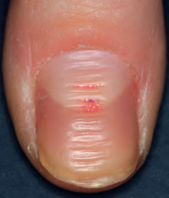
Fig. 71.3 Beau’s lines.
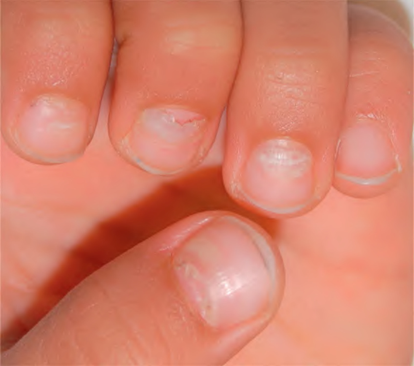
Fig. 71.4 Onychomadesis of the fingernails following hand-foot-and-mouth disease due to coxsackievirus A6 infection.
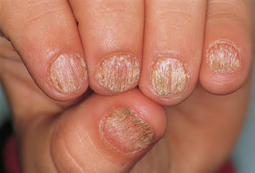
Fig. 71.5 Trachyonychia (twenty-nail dystrophy). The nails appear to be sandpapered in a longitudinal direction.
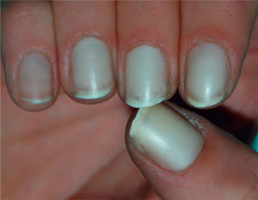
Fig. 71.6 True leukonychia – diffuse variant. The nail plate is completely or almost completely opaque and white. It may be inherited due to mutations in PLCD1, GJA1 (also keratoderma and hypotrichosis), or GJB2 (also deafness, keratoderma and knuckle pads); the latter two genes encode connexins 43 and 26, respectively.
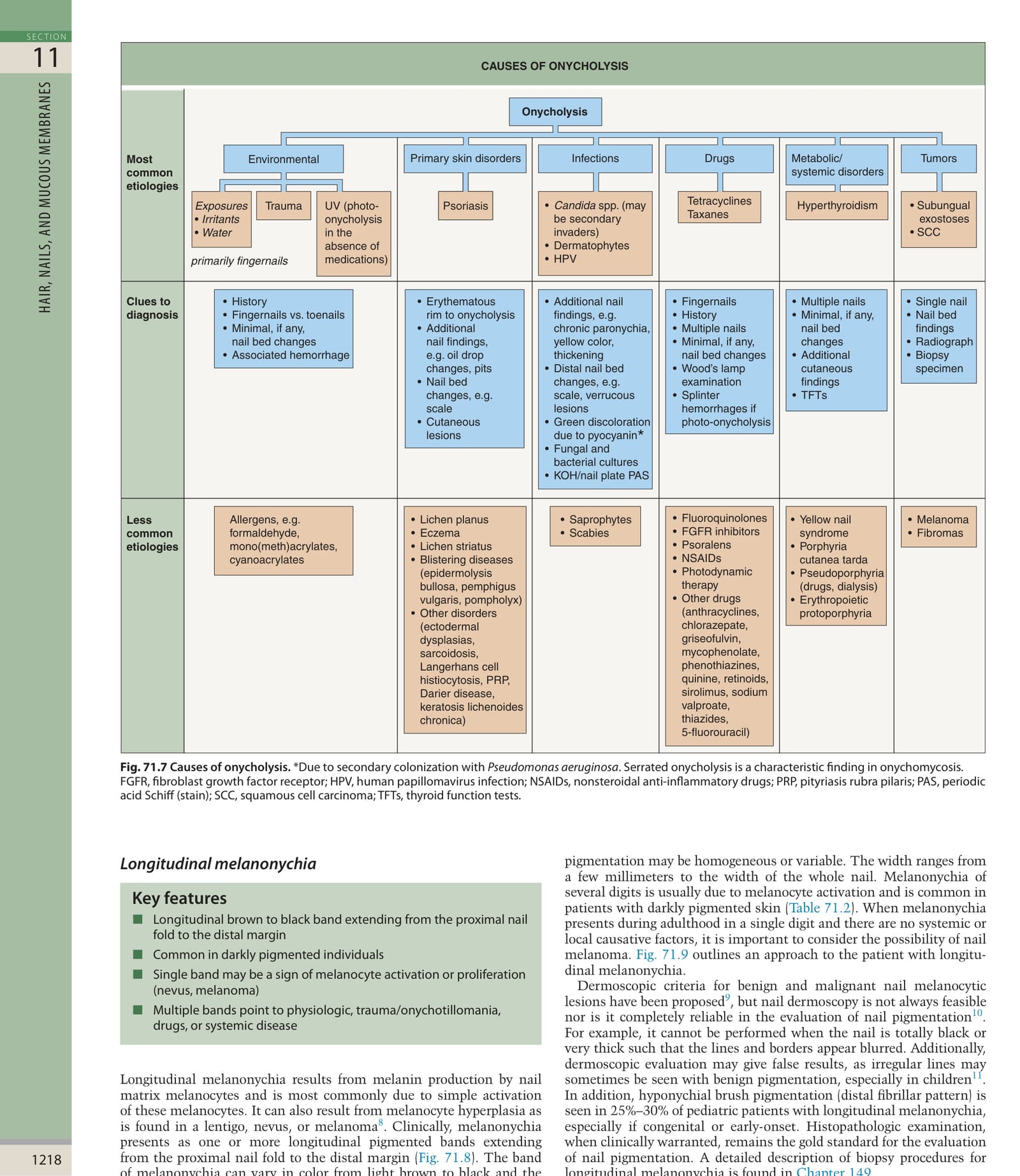
*Fig. 71.7 Causes of onycholysis. Due to secondary colonization with Pseudomonas aeruginosa. Serrated onycholysis is a characteristic finding in onychomycosis. FGFR, fibroblast growth factor receptor; HPV, human papillomavirus infection; NSAIDs, nonsteroidal anti-inflammatory drugs; PRP, pityriasis rubra pilaris; PAS, periodic acid Schiff (stain); SCC, squamous cell carcinoma; TFTs, thyroid function tests.
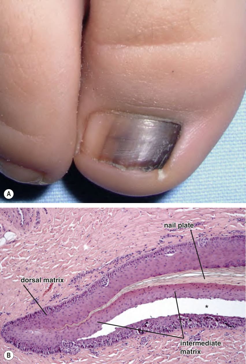
Fig. 71.8 Longitudinal melanonychia due to a congenital melanocytic nevus of the nail matrix.A The pigmentation involves approximately 75% of the nail plate and the color varies from light brown to black. B The biopsy specimen shows nests of melanocytes within the basal layer and lower portion of the nail matrix epithelium. The cleft (*) represents an artifact.
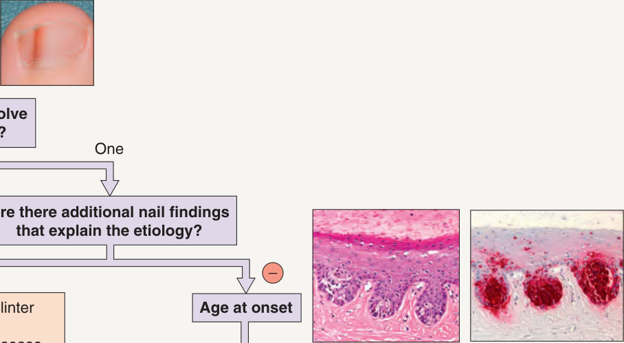
Fig. 71.9 Approach to the patient with longitudinal melanonychia. See Table 71.2 for a more extensive list of etiologies.
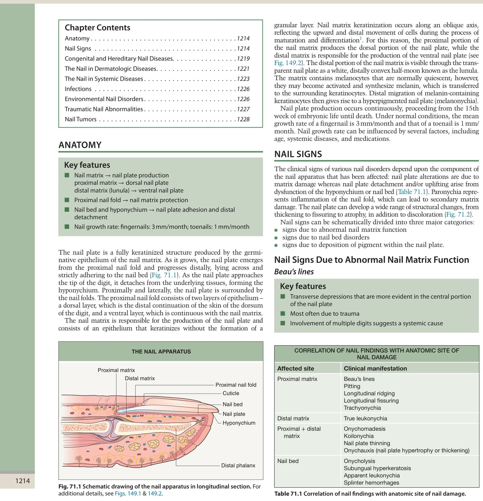
Table 71.1 Correlation of nail findings with anatomic site of nail damage.
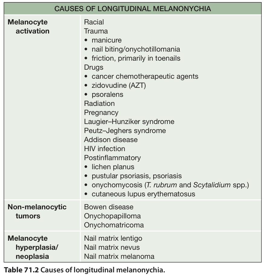
Table 71.2 Causes of longitudinal melanonychia.