ENVIRONMENTAL NAIL DISORDERS
Brittle Nails (Fragility, Onychoschizia)
Brittle fingernails are very common and usually affect women. Nail brittleness results from dehydration of the nail plate as a result of environmental factors such as frequent handwashing. In onychoschizia,
there is lamellar exfoliation in which the distal nail plate splits horizontally into multiple layers (see Fig. 71.2). Other signs of nail brittleness include splitting, softening, onychorrhexis, and keratin degranulation. The latter occurs in persons who wear nail polish continuously; it is characterized by whitish discoloration and fragmentation of the super-ficial layers of the nail (Fig. 71.19).
Treatment
●Avoidance of water and chemical exposure.
●Biotin (2.5–5 mg/day). Inform the patient that biotin should be held for several days prior to having bloodwork done as it can interfere with tests that use biotin–streptavidin technology (e.g. TSH).
●Topical moisturizers and humectants, e.g. 12% lactic acid, 10% urea.
Chronic Paronychia
Chronic paronychia commonly involves the fingernails of adult women. Although the pathogenesis is still debated, there is accumulating evidence that the condition represents a contact reaction to irritants or allergens. Occupational chronic paronychia is common in food handlers. Chronic paronychia is characterized clinically by longstanding inflammation of the proximal nail fold with erythema, edema, and absence of the cuticle. One or several fingernails are affected, in particular the thumb and second or third fingers of the dominant hand. Damage to the nail matrix results in nail plate surface abnormalities such as Beau’s lines. Pseudomonas colonization of the brittle nail surface may induce a green discoloration. Recurrent, self-limited episodes of acute exacerbation are common.
Treatment
●Avoidance of water and chemical exposure.
●Topical corticosteroids and topical imidazoles, but not systemic antifungals.
●Topical antiseptics (e.g. 4% thymol in 95% ethanol).
Idiopathic Onycholysis
Idiopathic onycholysis frequently involves the fingernails as a consequence of repetitive water immersion and exposure to irritants (see Fig. 71.7). The affected nail is detached from the nail bed and usually appears white because of air in the subungual space. Secondary microbial colonization of the subungual space can lead to discoloration, e.g. green due to pyocyanin. An absence of subungual hyperkeratosis is characteristic.
Treatment
●Avoidance of water and chemical exposures.
●Clipping of the detached nail plate and topical antiseptics.
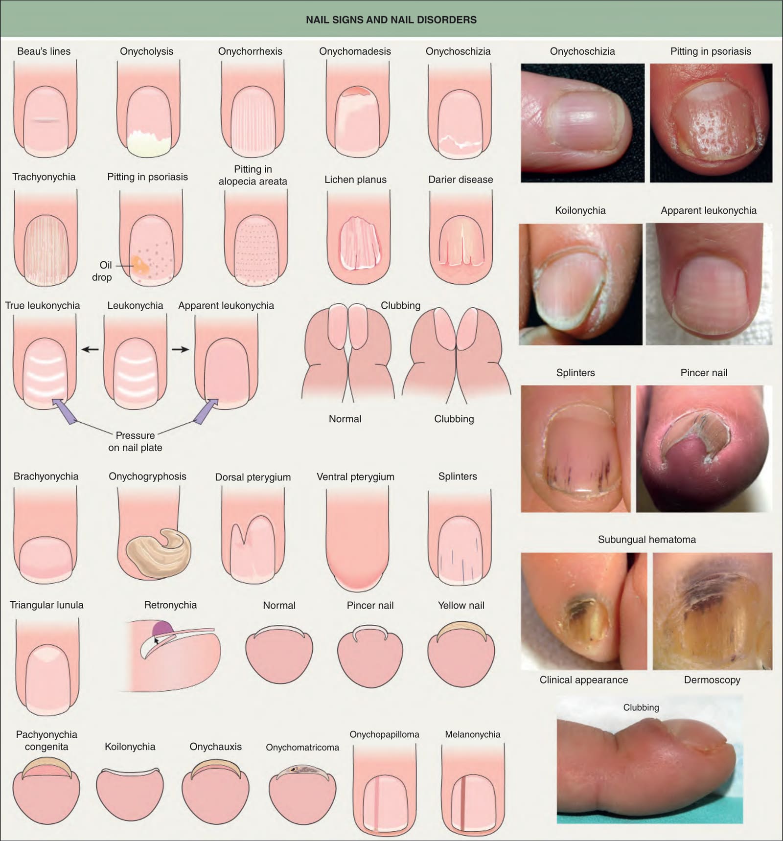
Fig. 71.2 Nail signs and nail disorders. In apparent leukonychia (e. g. Muehrcke’s lines due to hypoalbuminemia and presumed secondary edema), the transverse white lines disappear with pressure, but not in true leukonychia (e. g. Mees’ lines due to parakeratosis). Terry’s nails and half-and-half nails are additional examples of apparent leukonychia. Dermoscopy of a subungual hematoma shows small red longitudinal splinter hemorrhages and dark purple pigmentation under the proximal nail fold and nail plate due to accumulated blood, with a typical fringed distal edge. Photographs courtesy Jean L. Bolognia, MD; Leonard J. Swinyer, MD; Kalman Watsky, MD; and authors.
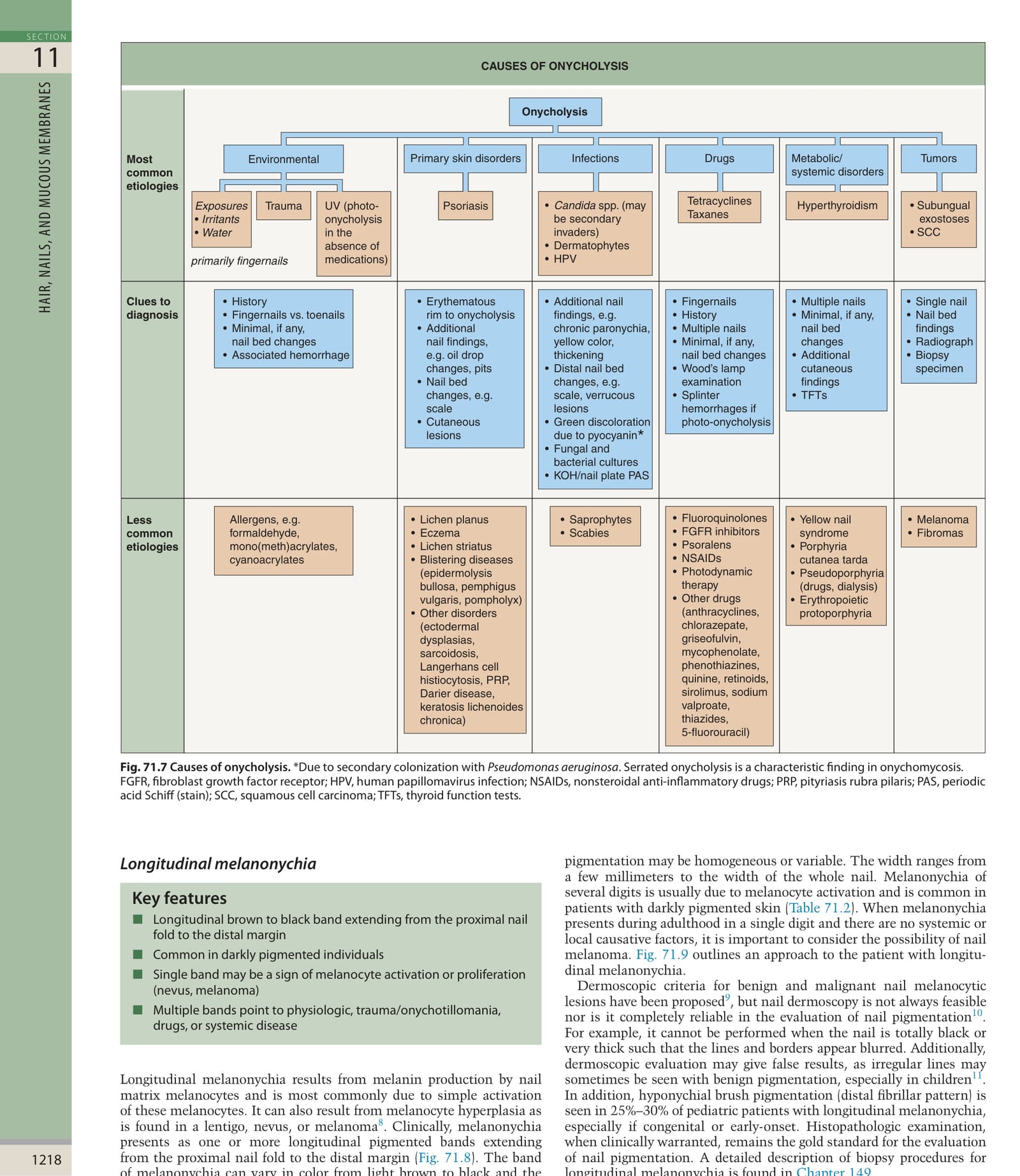
*Fig. 71.7 Causes of onycholysis. Due to secondary colonization with Pseudomonas aeruginosa. Serrated onycholysis is a characteristic finding in onychomycosis. FGFR, fibroblast growth factor receptor; HPV, human papillomavirus infection; NSAIDs, nonsteroidal anti-inflammatory drugs; PRP, pityriasis rubra pilaris; PAS, periodic acid Schiff (stain); SCC, squamous cell carcinoma; TFTs, thyroid function tests.
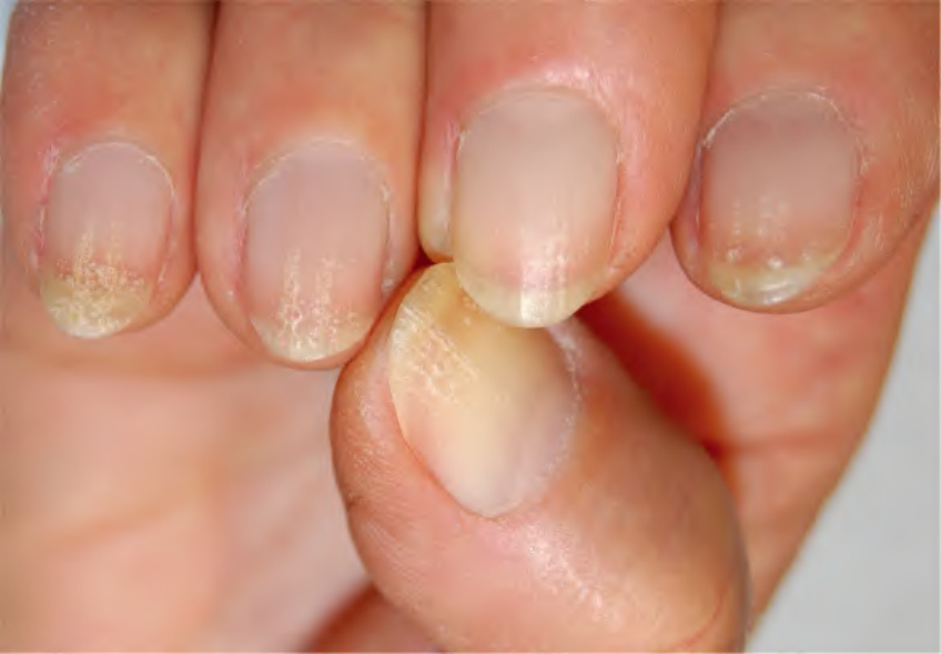
Fig. 71.19 Keratin degranulation of the fingernails due to prolonged wearing of nail lacquer. The distal nail plate is opaque and fragile due to multiple small areas of breakage on its surface.
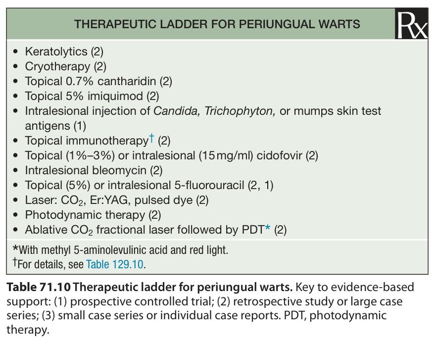
Table 71.10 Therapeutic ladder for periungual warts. Key to evidence-based support: (1) prospective controlled trial; (2) retrospective study or large case series; (3) small case series or individual case reports. PDT, photodynamic therapy.
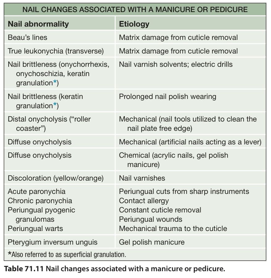
Table 71.11 Nail changes associated with a manicure or pedicure.