TRAUMATIC NAIL ABNORMALITIES
Mechanical insults or exposure to chemicals can lead to nail injury. These may be accidental, self-induced, or due to attempts at adornment (e.g. manicures, pedicures; Table 71.11).
Onychotillomania
Key features
Cuticle absent and proximal nail fold inflamed
Nail plate surface abnormalities, e. g. midline transverse ridging
Melanonychia
Hemorrhages and crusts
Self-induced nail abnormalities are common, but often not recognized due to the wide range of nail signs and the difficulty in obtaining a correct clinical history.
●Nail biting may affect the nail plate and/or the proximal nail fold; the trauma often produces nail matrix damage with secondary nail plate abnormalities, including surface irregularities and longitudinal melanonychia.
●Habit-tic deformity affects the thumb and is due to the nervous habit of rubbing and pushing back the midportion of the cuticle of the thumb with the index finger. The nail plate of the thumb shows multiple midline Beau’s lines that resemble a washboard (Fig. 71.20A). Median canaliform dystrophy of Heller (solenonychia) is considered a separate entity in which there is central longitudinal split with an inverted fir tree pattern (Fig. 71.20B); it is also probably caused by trauma.
●Nail destruction associated with psychiatric disorders. The clinical picture is variable, with nail plate destruction, hemorrhages, and periungual crusts and erosions (Fig. 71.21). Instruments (scissors, clippers, etc.) are often utilized to destroy the nails.
Treatment
●Topical preparations that taste unpleasant and bandages.
●Serotonin reuptake inhibitors (fluoxetine, paroxetine, sertraline), N-acetylcysteine.
(B).A The multiple midline Beau’s lines resemble a washboard. B A central longitudinal split with an inverted fir tree pattern. In both, deposits of exogenous pigments can be seen.
Subungual Hematoma
Acute hematomas occur after trauma severe enough to damage subungual blood vessels, leading to accumulation of blood under the nail plate. Acute hematomas are typically associated with pain, and compression of the matrix may cause secondary nail plate dystrophy. An X-ray of the digit may be advisable to exclude a bone fracture. Repeated microtrauma or a bleeding diathesis can lead to asymptomatic small subungual hematomas of the nails that are incorporated within the ventral nail plate.
Subungual hematomas migrate distally with nail growth and this may be demonstrated by gently pushing back the cuticle. The color of subungual hematomas ranges from purple–red to black. Onychoscopy is useful for differentiating hematomas from melanin deposition (see Fig. 71.2).
Treatment
Acute hematomas require prompt drainage of the blood by creating a hole through the nail plate.
Traumatic Toenail Abnormalities
Traumatic toenail abnormalities are most commonly seen in women who wear high-heeled pointed shoes, but can also be seen in athletes. They are often bilateral.
●Traumatic onycholysis of the hallux is the most common clinical presentation (Fig. 71.22). The onycholysis may be distal, where the detachment is caused by compression of the distal pulp by poorly fitting shoes, or lateral, when there is overlapping of the second toe onto the first. Clipping of the detached nail plate reveals a normal
nail bed. The differential diagnosis includes onychomycosis, but here the onycholysis is associated with subungual hyperkeratosis and the proximal margin of the detachment has a jagged edge by onychoscopy.
●Transverse leukonychia of the hallux results from repeated microtrauma by shoes to untrimmed, long, great toenails. Multiple bands of true leukonychia move distally with nail growth.
●Frictional melanonychia affects the toenails of the fourth and/or fifth digits of women. It is due to activation of nail matrix melanocytes by friction from shoes or from the adjacent digit. The bands may be multiple and black in color.
●Retronychia is caused by embedding of the nail into the proximal nail fold following trauma and may present with inflammation of the proximal nail fold.
Onychogryphosis
Onychogryphosis is common in elderly persons and almost exclusively affects the toenails, usually the hallux. The nail acquires a typical ram’s horn shape due to asymmetric growth (see Fig. 71.2). The nail plate is thick, hard, and yellow–brown. Multiple transverse striations are often present. Nail hardness and self-neglect may lead to monstrous deformities.
Pincer Nails (Trumpet Nails)
Pincer nails are usually seen on the toes and may be hereditary or acquired; the latter is most commonly due to ill-fitting shoes. The nail plate displays an excessive transverse overcurvature, especially in its distal portion (see Fig. 71.2). This results in compression (pinching) of the distal nail bed and can be associated with severe pain. Subungual exostosis should be excluded by X-ray examination. Consider druginduced pincer nails (e.g. from β-blockers) when several nails, including fingernails, are involved.
Treatment
Lateral chemical or surgical matricectomy is the treatment of choice for pincer nails (see Ch. 149).
Ingrown Toenails (Onychocryptosis)
Lateral ingrowing usually affects the hallux of young adults with congenital malalignment of the great toenails. Precipitating factors in all patients include improper or aggressive nail cutting and trauma. In addition, hyperhidrosis contributes to fragmentation of the distolateral edge. Penetration of nail plate spicules into the lateral nail fold epithelium causes painful inflammation. Chronic inflammation then leads to formation of granulation tissue that eventually undergoes epithelialization.
Distal embedding is a common complication of nail avulsion. The nail plate growth is blocked by the pulp, which forms a distal rim. Distal embedding can also occur in young infants but it resolves spontaneously.
Retronychia describes ingrowth of the proximal nail plate into the proximal nail fold, with one to three nail plates misaligned beneath the uppermost nail plate (see Fig. 71.2). There may be associated proximal nail fold erythema and swelling as well as proximal periungual pyogenic granulomas.
Treatment
●Prevention through patient education.
●Removal of the embedded spicule.
●Uplifting of the lateral nail plate with cotton, dental floss, slit tubes, or cotton casts.
●Granulation tissue may be reduced by topical antibiotics and topical corticosteroids or chemical cauterization (e.g. silver nitrate) or removed via curettage.
●Chemical (phenol, sodium hydroxide or trichloroacetic acid), laser, or excisional surgical removal of the lateral matrix is advisable in severe cases.
●In retronychia, surgical avulsion of the nail plate/s is necessary.
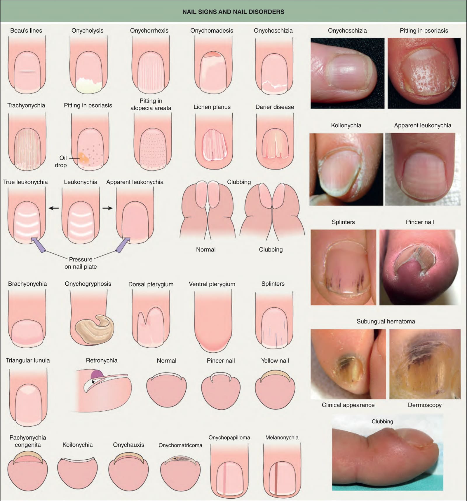
Fig. 71.2 Nail signs and nail disorders. In apparent leukonychia (e. g. Muehrcke’s lines due to hypoalbuminemia and presumed secondary edema), the transverse white lines disappear with pressure, but not in true leukonychia (e. g. Mees’ lines due to parakeratosis). Terry’s nails and half-and-half nails are additional examples of apparent leukonychia. Dermoscopy of a subungual hematoma shows small red longitudinal splinter hemorrhages and dark purple pigmentation under the proximal nail fold and nail plate due to accumulated blood, with a typical fringed distal edge. Photographs courtesy Jean L. Bolognia, MD; Leonard J. Swinyer, MD; Kalman Watsky, MD; and authors.
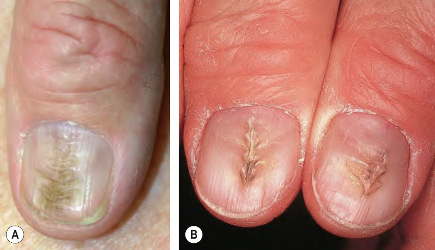
Fig. 71.20 Habit-tic deformity (A) and median canaliform dystrophy of Heller
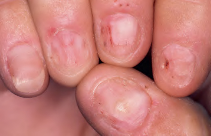
Fig. 71.21 Onychotillomania. In four of the nails, the nail plate is damaged or absent. The hemorrhagic crusts on the nail bed and proximal nail fold serve as a clue to the diagnosis.
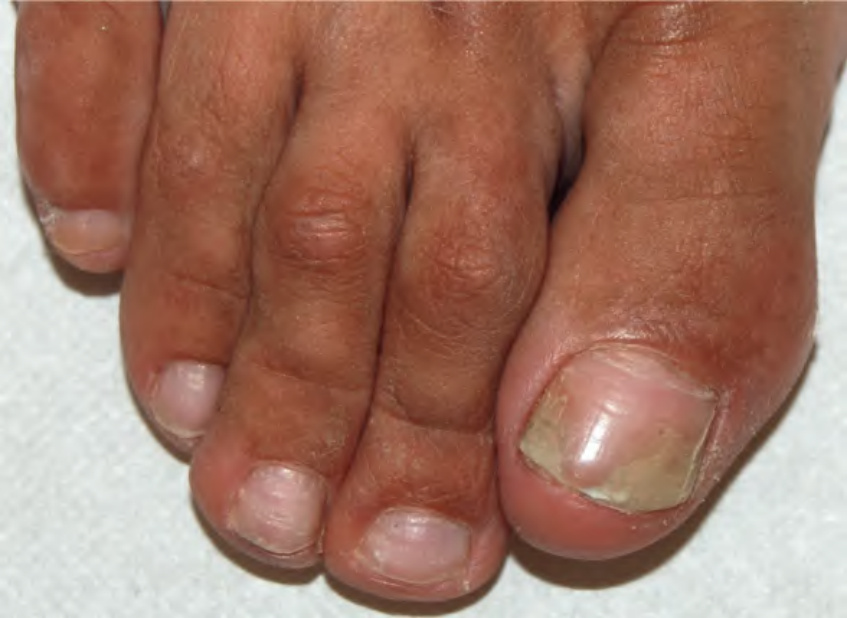
Fig. 71.22 Traumatic onycholysis of the nail of the first toe. Age-related lateral deviation of the hallux (hallux valgus) plays a role. Although the two entities can coexist, it is often misdiagnosed as lateral onychomycosis.
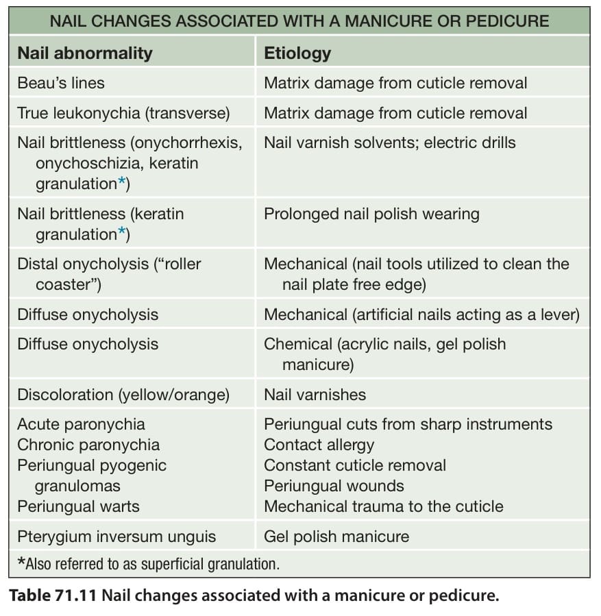
Table 71.11 Nail changes associated with a manicure or pedicure.