NAIL TUMORS
Nail surgery is discussed in Chapter 149.
Benign Tumors and Proliferations
Pyogenic granuloma (botryomycoma)
Pyogenic granulomas commonly appear within the nail apparatus, where they may be periungual or subungual and rarely under the proximal nail fold. While pyogenic granulomas of the nail unit often follow penetrating trauma, other common causes include ingrown toenails, systemic drugs (e.g. retinoids, EGFR inhibitors), peripheral nerve damage, and frictional onycholysis due to prolonged walking. The tumor appears as a bleeding, friable, soft red nodule (see Fig. 71.18). When subungual, it is associated with onycholysis. The differential diagnosis includes amelanotic melanoma and treatment is surgical.
Fibromas/fibrokeratomas
Isolated periungual and subungual fibromas are not rare in the general population. Multiple lesions occur in 50% of patients with tuberous sclerosis complex (Koenen tumors). Periungual fibromas appear as pink or skin-colored, fusiform-shaped papules originating from the proximal nail fold (see Ch. 61). The fibroma may compress the nail matrix and produce a longitudinal groove in the nail plate. Subungual fibromas that grow underneath the nail plate produce longitudinal erythronychia or onycholysis. A fibrokeratoma is characterized by a hyperkeratotic tip and may be surrounded by a collarette of raised skin.
Subungual exostosis
Subungual exostoses, first described by Dupuytren in 1847, are benign bony proliferations associated with nail abnormalities. Subungual exostoses are commonly precipitated by trauma and usually seen on the great toe of young patients. An exostosis produces a hard, tender, subungual nodule that elevates the nail plate (Fig. 71.23A). The nodule may ulcerate or become hyperkeratotic. The diagnosis is confirmed by X-ray examination (Fig. 71.23B) and the treatment is surgical.
Myxoid cyst (mucous cyst, digital mucous cyst)
Myxoid cysts are a common finding and favor middle-aged women (see Ch. 110). Typically located in the proximal nail fold of the fingernails, they appear as small, soft, skin-colored papulonodules that often spontaneously drain a viscous jelly-like fluid. Compression of the matrix produces nail plate depression and longitudinal grooves (Fig. 71.24). Occasionally, the cysts are subungual. These cysts are connected to the distal interphalangeal joint by a tract, and osteoarthritis of the distal joint is a frequent association.
Possible treatments for myxoid cysts include sclerotherapy, cryosurgery, and intralesional corticosteroid injections. All of these procedures are
The longitudinal nail groove is a result of the compression of the nail matrix by the cyst.
associated with a high frequency of relapses. Definitive cure of myxoid cysts requires a surgical procedure that includes ligation of the pedicle that connects the cyst to the joint.
Glomus tumor
A glomus tumor arises from the neuromyoarterial glomus cells of the nail bed dermis. It is typically associated with severe pain that radiates proximally and is often aggravated by physical (e.g. compression) or thermal stimuli (especially cold). Subjective symptoms typically exceed clinical signs. The tumor may not be clinically apparent or may appear as a red–bluish nail bed macule visible through the transparent nail plate (see Fig. 114.33). MRI allows the diagnosis (see Fig. 114.34).
Onychomatricoma
First described by Baran and Kint in 1992, onychomatricoma is an uncommon neoplasm that produces localized or diffuse thickening of the nail plate in which multiple longitudinal hollows contain the digitating tumor (Fig. 71.25). The affected nail is thickened and yellow– white in color, with transverse overcurvature and multiple splinter hemorrhages. In an en face view of the distal nail, multiple holes in the thickened free margin are typically seen (see Fig. 71.2).
Histologically, the tumor is characterized by multiple fibroepithelial projections that extend into the thickened nail plate. The tumor epithelium is identical to that of the normal nail matrix and keratinizes without a granular layer. Histopathologic examination of a simple nail clipping is sufficient for diagnosis.
Onychopapilloma
Onychopapilloma is a benign tumor that grows under the nail plate and has a thin filiform shape. It arises from the distal matrix/proximal
nail bed and extends to the distal nail margin. Small tumors produce thin longitudinal erythronychia while larger onychopapillomas induce thinning of the overlying nail plate and distal fissuring. Splinter hemorrhages are an associated finding. Examination of the free margin reveals a keratotic mass that adheres to the ventral nail plate (Fig. 71.26). Less often the tumor manifests as longitudinal leukonychia or melanonychia.
By dermoscopy, the origin (lunula) and the end (distal margin) of the thin red band can be appreciated, as can splinter hemorrhages and the distal keratotic mass. A nail clipping demonstrates a localized asymmetric keratotic thickening on the underside of the nail plate, corresponding to the location of the filiform tumor.
Melanocytic nevi of the nail matrix
Melanocytic nevi of the nail matrix are an uncommon cause of longitudinal melanonychia (see Table 71.2). They usually develop during childhood and often involve the fingers, especially the thumb. Clinically, a band of melanonychia that is brown to black in color
involves a portion of the nail or its entirety. The color, width, and pigment distribution may vary considerably and it is not unusual to observe fading or darkening of the pigmentation over time in an individual patient. Pigmentation of periungual tissues is also possible because nests of nevus cells can be present in the proximal nail fold or hyponychium. When dark brown or black in color, bands are often visible through the cuticle (pseudo-Hutchinson sign). The age at onset represents the most important clue to the diagnosis. Histologically, a lentigo or junctional nevus is usually seen (see Fig. 71.8B).
Optimal management of nail matrix nevi is still debated. See Fig. 71.9 for an approach to longitudinal melanonychia, including in children.
Malignant Tumors
Malignant tumors of the nail are summarized below. For further details regarding these malignancies and their treatment, see Chapters 108 and 113.
Bowen disease (squamous cell carcinoma in situ)
Bowen disease of the nail is uncommon and is seen most often in middle-aged men. Fingers of the left hand are most commonly involved. Clinically, it may be difficult to differentiate Bowen disease from warts. The affected digit shows periungual or subungual verrucous lesions with onycholysis and/or longitudinal melanonychia (Fig. 71.27). Predisposing factors include HPV infection and chronic X-ray exposure.
Keratoacanthoma
Keratoacanthoma of the nail is extremely rare and usually affects a single digit. It appears as a painful, subungual, keratotic nodule that grows rapidly over a period of weeks. Deep invasion with bone destruction frequently occurs and typically there is evidence of osteolysis on X-ray examination. A history of trauma is often reported. In contrast to cutaneous keratoacanthomas, keratoacanthomas of the nail do not regress spontaneously.
Squamous cell carcinoma
Squamous cell carcinoma (SCC) is the most common malignant tumor of the nail apparatus. It often affects the fingernails of middle-aged men. A number of studies have confirmed a causative role for HPV, usually HPV-16, in the development of this tumor. Clinically, it may present as slowly growing periungual or subungual verrucous mass or nodule that may ulcerate and bleed. Compared to cutaneous SCC elsewhere, nail SCC tends to more quickly become invasive, but metastases are rare. Bone invasion occurs in <20% of patients and the osteolysis detected on X-ray examination is more often due to compression rather than true bone invasion.
Verrucous carcinoma (carcinoma cuniculatum,
Verrucous carcinoma is a rare, low-grade variant of SCC characterized by locally aggressive clinical behavior but low potential for metastasis. It rarely involves the nail apparatus. Clinically, the tumor appears as a rapidly growing verrucous nodule that often destroys the nail. Bone resorption is common.
Melanoma
Nail melanoma is rare, accounting for 0.7%–3.5% of all melanomas. It most frequently involves the thumb of middle-aged individuals, and patients often report a history of trauma. Diagnosis is often delayed, and in 25% of patients the tumor is amelanotic. Historically, the 5-year survival is only 15%. From a clinical standpoint, nail melanoma may have the following presentations:
●Longitudinal melanonychia. Melanoma should be suspected when a single band arises during adulthood without another plausible explanation (see Fig. 71.9). The pigmented band is usually dark brown or black in color, with blurred margins and a width >3–4 mm. An ABCDEF “rule” has been proposed to aid in diagnosis: A) Age – peak incidence fifth to seventh decades with Africans, Asians, and Native Americans (more darkly pigmented individuals) accounting for up to one-third of all cases; B) Black or brown with band width of 3 mm or more; C) Change in the nail or lack of improved change with treatment; D) Digit most commonly involved (thumb, great toe, index); E) Extension of pigment onto the proximal or lateral nail fold (Hutchinson sign); and F) Family or personal history of dysplastic nevus or melanoma. Except for criterion F, these represent validated clinical criteria. When longitudinal melanonychia is due to nail melanoma, dermoscopically the band typically has nonhomogeneous pigmentation, blurred borders, and nonparallel interrupted lines of different colors. However, for diagnosis, onychoscopy alone is not completely reliable, especially if the nails are thick or the bands are black. Moreover, sometimes the lines are regular in nail melanoma. Therefore, integration of the history, clinical appearance, and dermoscopic findings is required.
●Amelanotic nodule that ulcerates and bleeds, resembling a pyogenic granuloma (Fig. 71.28). The presence of: (1) Hutchinson sign – pigmentation of periungual tissues due to radial spread of the tumor; or (2) micro-Hutchinson sign – pigmentation visible only by dermoscopy is a strongly suggestive clinical feature.
Additional figures and table on the ABCDEF rule for subungual melanoma, available in our eBook (see inside front cover for access code).
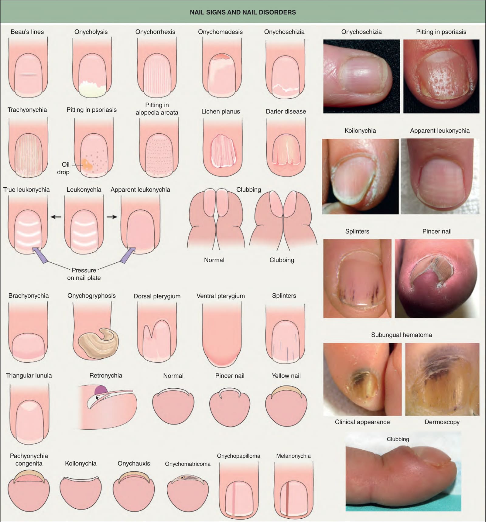
Fig. 71.2 Nail signs and nail disorders. In apparent leukonychia (e. g. Muehrcke’s lines due to hypoalbuminemia and presumed secondary edema), the transverse white lines disappear with pressure, but not in true leukonychia (e. g. Mees’ lines due to parakeratosis). Terry’s nails and half-and-half nails are additional examples of apparent leukonychia. Dermoscopy of a subungual hematoma shows small red longitudinal splinter hemorrhages and dark purple pigmentation under the proximal nail fold and nail plate due to accumulated blood, with a typical fringed distal edge. Photographs courtesy Jean L. Bolognia, MD; Leonard J. Swinyer, MD; Kalman Watsky, MD; and authors.
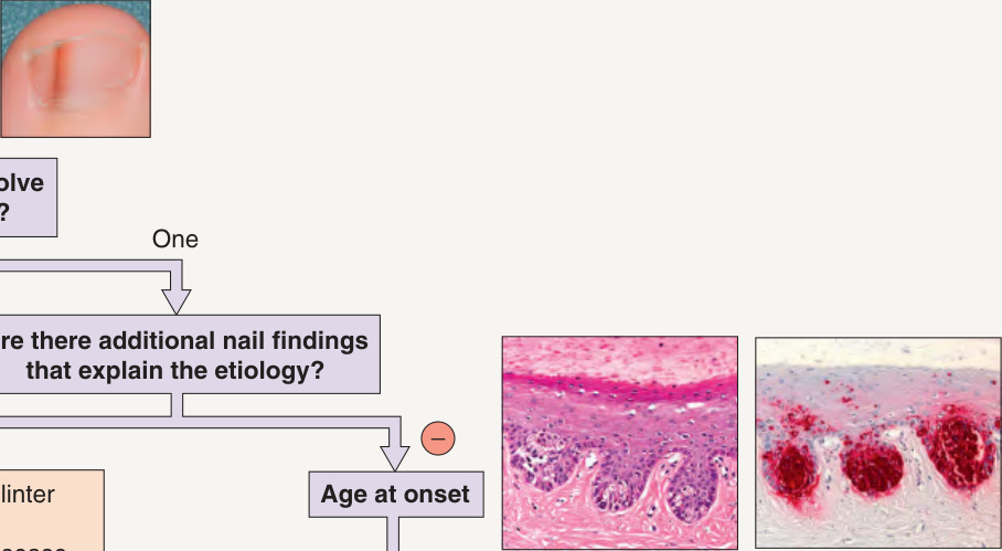
Fig. 71.9 Approach to the patient with longitudinal melanonychia. See Table 71.2 for a more extensive list of etiologies.
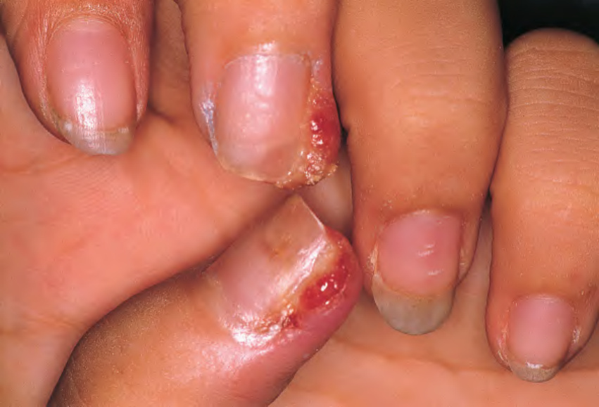
Fig. 71.18 Multiple periungual pyogenic granulomas in a patient taking indinavir.
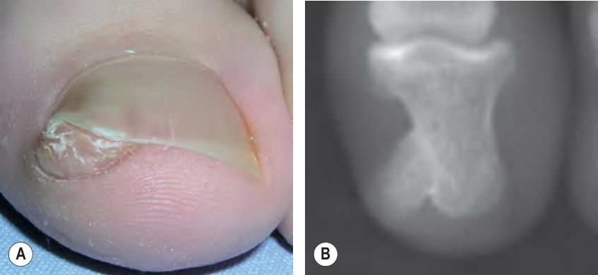
Fig. 71.23 Subungual exostosis.A Skin-colored subungual nodule elevating the nail plate. B Radiograph of the digit demonstrating the subungual bony proliferation.
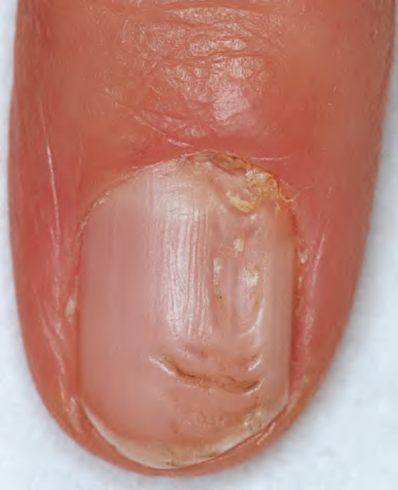
Fig. 71.24 Myxoid cyst.
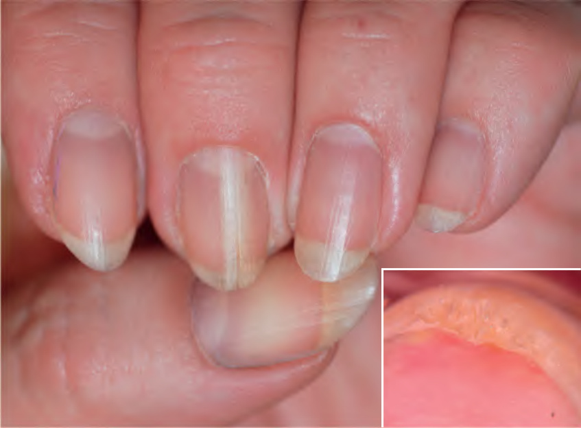
Fig. 71.25 Onychomatricoma of the left third fingernail. A longitudinal, thickened yellowish band is seen extending from the cuticle to the distal nail plate. Proximal splinter hemorrhages are also visible. Onychoscopy of the distal nail plate reveals the diagnostic finding of localized nail thickening with a honeycomb appearance (inset).
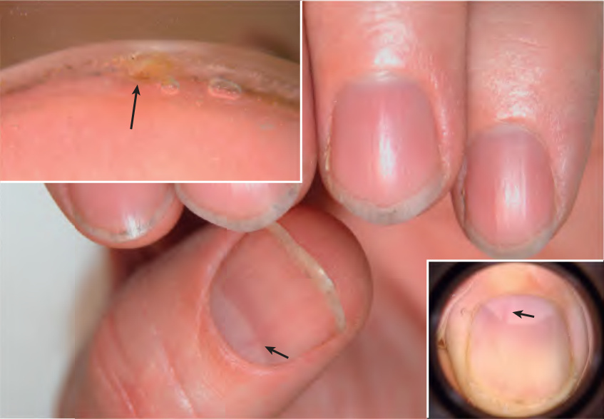
Fig. 71.26 Onychopapilloma of the left first fingernail. A thin longitudinal band of erythronychia is seen extending from the lunula. Onychoscopy reveals the crescent-shaped proximal origin of the erythronychia (lower inset) and a small subungual mass can be appreciated on the free edge of the nail (upper inset).
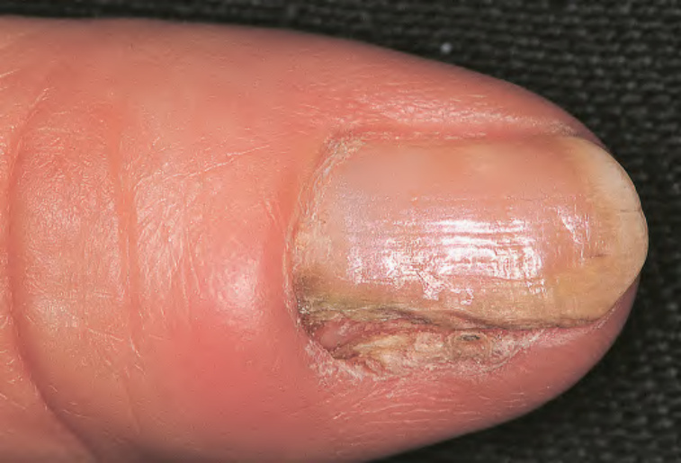
Fig. 71.27 Bowen disease. The lateral portion of the nail plate is absent. The nail bed shows hyperkeratosis with scaling and fissuring of the epithelium.
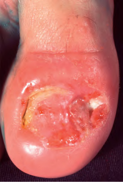
Fig. 71.28 Amelanotic melanoma. Note the diffuse nail destruction and ulceration as well as swelling of the proximal nail fold. The tumor can resemble granulation tissue. Courtesy Lorenzo Cerroni, MD.
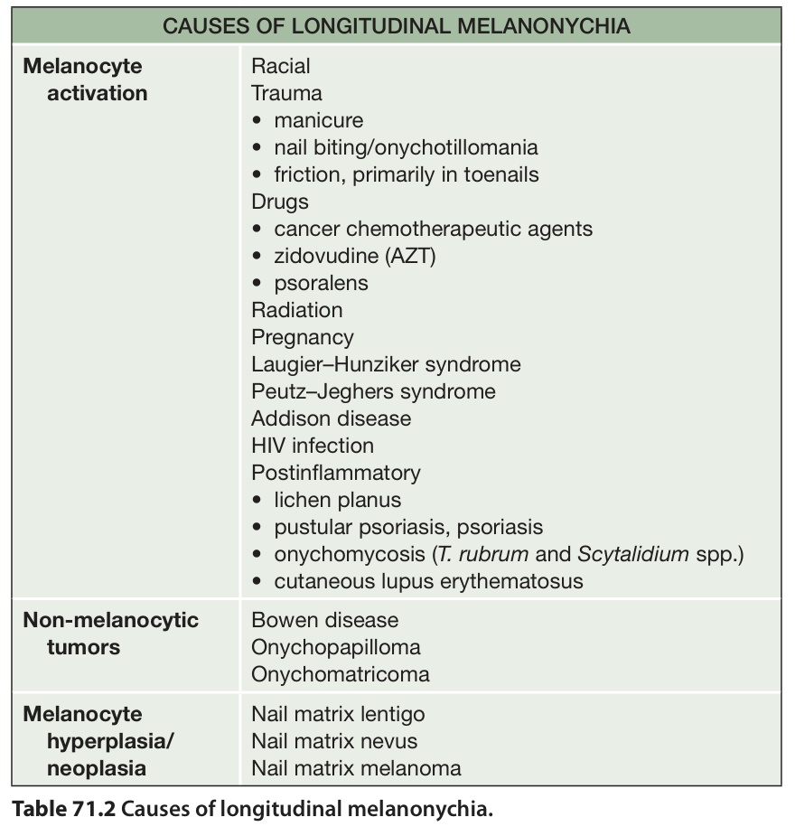
Table 71.2 Causes of longitudinal melanonychia.
Connecticut: Appleton & Lange; 1990.2. Hardin J, Haber RM. Onychomadesis: literature review.