Introduction
Maryam Jessri, Kristin K. McNamara and Nathaniel Treister Oral Diseases
Introduction
From both a clinical and histologic standpoint, the oral cavity is a diverse anatomic region reflecting its various functions and environmental interactions. Sites that are exposed to the friction of mastication, such as the hard palate and attached gingivae, are bound to the underlying periosteum and are referred to as attached mucosa or keratinized mucosa (Fig. 72.1). Histologically, these anatomic sites have prominent rete ridge formation and a thick layer of keratin. The dorsal tongue has the thickest epithelium within the oral cavity and its specialized functions include taste, mastication, and deglutition. The filiform, fungiform, foliate, and circumvallate papillae are distributed on the dorsal tongue, with pigmentation of the fungiform papillae a normal variant (Fig. 72.2A). All other oral mucosal sites, including the soft palate, labial mucosa, and floor of the mouth, are “unattached” and represent non-keratinized mucosa. Numerous salivary glands are present within the connective tissue (submucosa) beneath the non-keratinized mucosa. In the buccal mucosa, there are variable numbers of heterotopic sebaceous glands (Fordyce granules).
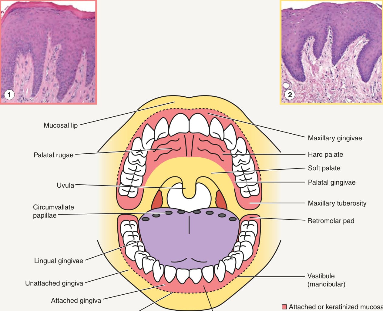
Fig. 72.1 The oral cavity – anatomy and histopathologic findings. Pink represents attached (keratinized) mucosa, yellow represents movable (non-keratinized) mucosa, and purple the dorsal tongue. Inset 1: Histologic features of the palate with prominent rete ridge formation, a granular layer, and orthohyperkeratosis; the attached gingivae have similar findings. Inset 2: Histologic features of the buccal mucosa with less pronounced rete ridges, minimal parakeratosis, and no orthokeratosis; the soft palate, labial mucosa, ventral tongue, and floor of mouth exhibit similar histologic features.