PERIODONTAL AND DENTAL DISEASE
Necrotizing Ulcerative Gingivitis
Synonyms: Trench mouth Acute necrotizing ulcerative gingivitis (becoming historic given lack of chronic form) Necrotizing gingivostomatitis Vincent infection Fusospirochetal gingivitis
Key features
Painful, hemorrhagic, necrotic gingivae with classic “punched-out”
interdental papillae
Mixed bacterial infection in susceptible hosts with predisposing risk factors
Predisposing risk factors include immunosuppression, malnutrition, stress, smoking, and poor oral hygiene
Introduction
Necrotizing ulcerative gingivitis (NUG) is characterized by pain, bleeding, and necrosis of interdental gingival papillae. The infection is due to a mixed population of bacteria, including spirochetes (e.g. Treponema), Prevotella intermedia, Fusobacterium and Selenomonas spp., and Porphyromonas gingivalis.
Epidemiology
NUG affects <0.1% of the population, is primarily seen in young to middle-aged adults, and does not have a sex predilection. Predisposing factors include immunosuppression, malnutrition, poor oral hygiene, physical stress, and smoking.
Clinical features
NUG is characterized by inflamed, edematous, and hemorrhagic inter-dental papillae. However, unlike generalized severe gingivitis, the affected papillae in NUG have a blunted appearance with “punched-out”, crater-like areas of necrosis and ulceration. Generally, patients present with halitosis, severe pain, spontaneous bleeding from the gingivae, and accumulation of necrotic debris on the teeth and gingival surfaces. Systemic signs and symptoms may include fever, malaise, and lymphadenopathy.
Pathology
The histopathologic features are nonspecific and include ulceration of the epithelium that is covered by fibrinous debris and a mixed inflammatory infiltrate in the lamina propria. Bacterial colonization is common but because the primary etiologic agents represent normal oral flora, bacterial culture results are not very helpful.
Treatment
The most appropriate therapy is provided by a periodontist and includes debridement of the necrotic tissue to reduce inflammation and microbial load as well as investigation of the underlying cause(s). Chlorhexidine oral rinses (0.12%) may reduce the bacterial load. Broad-spectrum antibiotics are prescribed, especially when there are systemic signs of infection.
Desquamative Gingivitis
Key features
A clinical descriptor for oral manifestations of an underlying immune-mediated vesiculoerosive disease
Characterized by diffusely smooth, red, and sometimes painful gingivae
Histopathologic and immunofluorescence studies are required for appropriate diagnosis and subsequent treatment of the underlying condition
Introduction
Desquamative gingivitis is a nonspecific clinical term used to describe superficial sloughing of the gingival epithelium. It is nearly always a manifestation of a vesiculoerosive disease such as lichen planus (see Ch. 11), mucous membrane pemphigoid (see Ch. 30), or pemphigus vulgaris (see Ch. 29). Fig. 72.7 outlines the multiple causes of desquamative gingivitis.
Epidemiology
Desquamative gingivitis primarily affects women over the age of 40 years.
Clinical features
Desquamative gingivitis primarily affects the maxillary and mandibular gingivae, either focally or diffusely (see Fig. 72.1). Affected areas are friable, denuded, and erythematous with occasional ulceration (Fig. 72.8). The gingivae may be the sole site of involvement or the gingivitis may be accompanied by erosions elsewhere in the mouth, on the lips, or in other mucosal sites. Patients often report soreness and sensitivity and, depending on the severity, diet and maintaining oral hygiene may be adversely impacted. Intact vesicles of the gingivae are rarely seen.
Pathology
The specific histopathologic and immunopathologic features of desquamative gingivitis depend on the underlying vesiculoerosive disease. Diagnosis is established via an incisional biopsy processed for both routine histology and direct immunofluorescence (DIF). In addition, when indicated, indirect IF (IIF) testing or an enzyme-linked immunosorbent assay (ELISA) can be utilized to detect circulating autoantibodies (see Fig. 72.7).
Treatment
Once a diagnosis is established, appropriate treatment for the responsible condition can follow. Meticulous dental prophylaxis (cleaning), oral hygiene instructions, and longitudinal evaluation are recommended. Reducing bacterial plaque often decreases the severity of lesions and is an important adjunct to medical therapy.
Sequelae of Dental Caries
Key features
Dental infections can spread beyond the periapical region of teeth
Periapical abscess, osteomyelitis, cellulitis, intraoral dental sinus, and cutaneous sinus are potential sequelae
Emergent cases require immediate treatment to avoid airway obstruction or possible sepsis
Introduction
Dental caries is due to a microbial process that causes localized destruction and loss of mineralized tissues. This may result in infection of the pulp with extension to the periapical region of the tooth. In immunocompromised hosts or with particularly virulent bacteria, there may be spread of the infection to bone and soft tissues or further localized destruction. The latter can lead to a cutaneous sinus of dental origin (Fig. 72.9).
Clinical features
Dental caries is defined as localized loss of the mineralized structure of the tooth (enamel, dentin, or cementum) due to an infectious process. It may present as any one or a combination of the following: cavitation of enamel, brown discoloration, whitened chalky appearance, or soft scrapable tooth structure. Sequelae from spread of the bacterial infection beyond the tooth structure vary from contained asymptomatic bone destruction to severe facial cellulitis.
A periapical abscess forms when there is a localized accumulation of inflammatory cells and bacterial debris in the periapical space of a non-vital tooth; the abscess may be acute or chronic and symptoms range from none to severe pain. When the infection erodes through cortical bone to create a path for drainage, a “sinus tract” or “parulis” forms. An intraoral dental sinus tract is characterized by a soft, non-tender, erythematous papule that develops on the alveolar process, typically in the same region as the apex of the offending non-vital tooth. If the infection tunnels through the skin, a cutaneous sinus tract of dental origin forms which can present as an ulceration, a fibrosed depression, or an erythematous, sometimes eroded, papule (see Fig. 72.9).
Erosions and prominent, fairly well-defined areas of erythema of the attached (keratinized) gingivae. Histopathologic examination plus DIF (and occasionally IIF or ELISA) are needed to establish the etiology (see Fig. 72.7).
In addition to sinus tracts, cellulitis and occasionally osteomyelitis may develop and both usually have associated pain. Patients with cellulitis can also have fever, chills, and lymphadenopathy while a foul-smelling drainage may accompany osteomyelitis. There are two potential complications that represent a particular danger to patients and demand immediate attention. Ludwig’s angina presents as swelling of the submandibular, submental, and sublingual spaces. Urgent attention is required for this form of cellulitis as it can rapidly spread to the retropharyngeal space, obstructing breathing, and even spread to the mediastinum. Cavernous sinus thrombosis due to a dental abscess may initially present as swelling of the canine space. Infraorbital, ocular, and eventually CNS involvement may develop.
Treatment
Treatment involves elimination of the focus of infection, which often requires incision and drainage, endodontic “root canal” therapy or extraction of the involved tooth, appropriate antibiotic therapy, and airway management, if necessary. Microbiological testing assists in the choice of antibiotics and allows exclusion of cervicofacial actinomycosis.
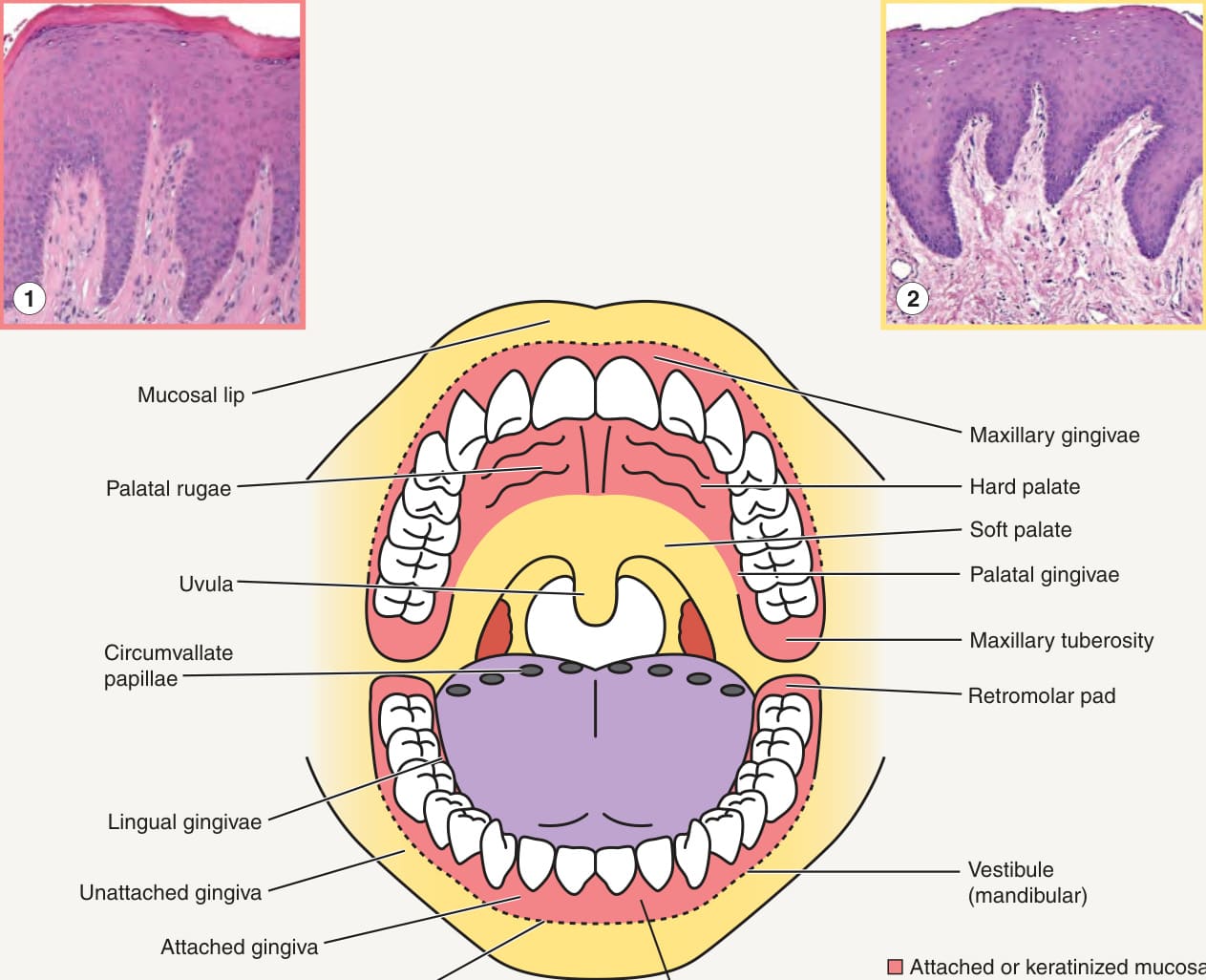
Fig. 72.1 The oral cavity – anatomy and histopathologic findings. Pink represents attached (keratinized) mucosa, yellow represents movable (non-keratinized) mucosa, and purple the dorsal tongue. Inset 1: Histologic features of the palate with prominent rete ridge formation, a granular layer, and orthohyperkeratosis; the attached gingivae have similar findings. Inset 2: Histologic features of the buccal mucosa with less pronounced rete ridges, minimal parakeratosis, and no orthokeratosis; the soft palate, labial mucosa, ventral tongue, and floor of mouth exhibit similar histologic features.
Fig. 72.7 Differential diagnosis of desquamative gingivitis. If the gingivae are painful, hemorrhagic, and necrotic with punched-out interdental papillae, then necrotizing ulcerative gingivitis is the most likely clinical diagnosis. For presumed chronic ulcerative stomatitis, the detection of circulating anti-p63 antibodies via ELISA may assist in diagnosis. BP, bullous pemphigoid; BMZ, basement membrane zone; DIF, direct immunofluorescence (IF); EBA, epidermolysis bullosa acquisita; GVHD, graft-versus-host disease; IIF, indirect IF; LABD, linear IgA bullous dermatosis.
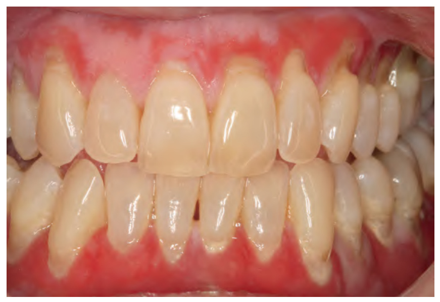
Fig. 72.8 Desquamative gingivitis due to oral erosive lichen planus.
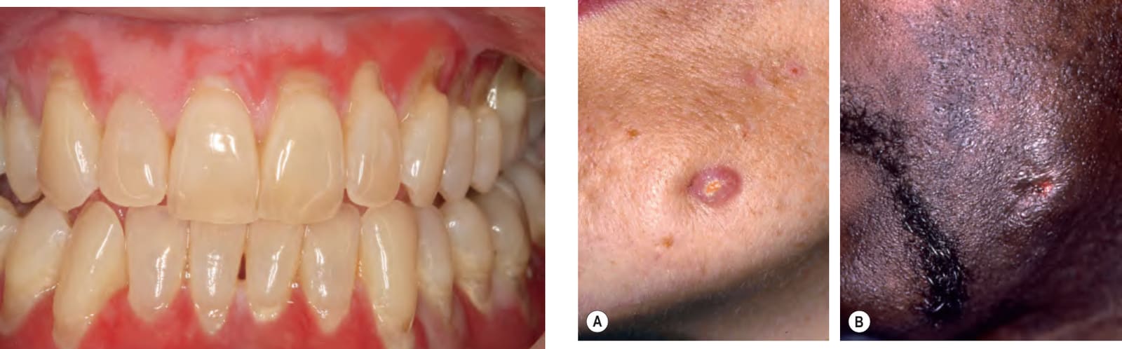
Fig. 72.9 Cutaneous sinus of dental origin.A Dull pink papule and adjacent indentation on the jawline. B An ulceration in a similar location. These sinuses originated from periapical dental abscesses in the mandible that then channeled through the overlying skin to drain. They may be mistaken for a pyogenic granuloma, neoplasm, or cutaneous infection. A, Courtesy Carl M. Allen, DDS, MSD and Charles Camisa, MD.