PHYSICAL AND CHEMICAL INJURIES
Fibroma
Synonyms: Bite fibroma Irritation or traumatic fibroma Fibrous hyperplasia or nodule
Key features
Smooth, pink, soft to rubbery nodule, typically on the buccal mucosa
Asymptomatic unless persistently irritated or traumatized
Introduction
A fibroma is the most common “tumor” of the oral cavity. It most likely represents reactive hyperplasia of fibrous connective tissue in response to local trauma, including repeated biting.
Epidemiology
Fibromas most commonly occur during the fourth to sixth decades, with no sex predilection.
Clinical features
The typical lesion is a sessile, soft, smooth-surfaced papule or nodule similar in color to the surrounding normal mucosa. Depending on the location and extent of acute and chronic trauma, fibromas may be ulcerated or have a white keratotic surface. These papulonodules are usually <1.5 cm in diameter and are most commonly observed along the bite line of the buccal mucosa. However, they can also occur in other easily traumatized sites such as the lower labial mucosa and the anterolateral aspects of the tongue.
Pathology
Histologically, there is an unencapsulated mass of hyperplastic fibrous connective tissue with varying vascularity and minimal inflammation. The overlying epithelium may be ulcerated, atrophic, or hyperkeratotic secondary to friction or biting.
Treatment
Surgical excision is curative and histopathologic examination allows confirmation of the clinical diagnosis.
Contact Stomatitis
Key features
Can be allergic or irritant
Irritant contact stomatitis is the result of direct contact with a caustic agent
Allergic contact stomatitis can be acute or chronic and has a range of signs and symptoms
Introduction
Contact stomatitis is a broad term that encompasses clinical findings secondary to exposure of the oral mucosa to a known contactant. It is subdivided into irritant contact stomatitis (ICS) and allergic contact stomatitis (ACS). As in the skin, the former results from direct mucosal injury by an irritant and when pronounced is referred to as a chemical burn; the latter is characterized by delayed type hypersensitivity to an allergen.
Epidemiology
Contact stomatitis may present at any age, but is more common in adults. While ICS does not have a sex predilection, ACS has a female predominance.
Clinical features
In mild ICS, erythema and textural changes of the mucosa may be seen. Direct placement of a caustic agent such as an aspirin or alendronate onto the mucosa can lead to more severe disease, with erythema appearing shortly after exposure. Depending on the duration of exposure and irritancy potential of the contactant, ICS may present as edema, sloughing of the epithelium, and even painful necrotic ulcers (Fig. 72.10).
A wide range of allergens can induce ACS including flavorings such as cinnamon and peppermint in dentifrices, mouthwashes, and chewing gum and dental restorative materials (e.g. amalgam) (Fig. 72.11; Table 72.1). The clinical findings of acute ACS range from varying degrees of erythema with or without edema to superficial ulcerations and in rare cases, vesicle formation. In chronic ACS, sites of involvement may be erythematous, keratotic, or erosive and in some patients resemble lichen planus. The most common symptom of both ACS and ICS is burning within the mouth which may be generalized or limited to the affected area.
Pathology
The range of clinical presentations results in varying histopathologic findings. In ACS, epithelial spongiosis and submucosal inflammatory infiltrates that contain eosinophils and plasma cells can be seen. Ulcerations from ICS or ACS have coagulative necrosis of the epithelium with variable extension into the underlying connective tissue. In lichenoid contact stomatitis, e.g. due to contact with metals contained in dental amalgam, a lichenoid inflammatory reaction is seen that is indistinguishable from oral lichen planus. Metal particles within the lamina propria (particularly amalgam), if present, are a clue to lichenoid contact stomatitis.
Treatment
A thorough history may identify the responsible contactant and its elimination is often curative. In the case of ACS, patch testing can
identify the responsible allergen (see Ch. 14). Application of emollient pastes or topical anesthetics as a palliative measure may provide temporary relief.
Morsicatio Mucosae Oris
Synonyms: Chronic bite keratosis Variants: Morsicatio buccarum (buccal mucosa) Morsicatio labiorum (labial mucosa) Morsicatio linguarum (tongue)
Key features
Factitial habit
Shaggy white lesions of the anterior buccal mucosa, lateral tongue, or lower labial mucosa
No treatment indicated
Introduction
Morsicatio mucosae oris (MMO) is a benign condition that is due to repetitive chewing, biting, or nibbling of the oral mucosa.
Epidemiology
MMO usually affects individuals during or after their fourth decade of life.
Clinical features
The most common site for MMO is the buccal mucosa followed in frequency by the ventrolateral tongue and lower labial mucosa. MMO classically presents as bilateral shaggy white lesions of the anterior buccal mucosa that approximate the area of chronic trauma (Fig. 72.12).
Pathology
Histopathologic findings are nonspecific and include acanthosis, marked parakeratosis, an irregular, shaggy surface of the epithelium with fissures and clefts, and varying degrees of vacuolization and spongiosis in the superficial portion of the epithelium (Fig. 72.12 inset). Surface colonization by bacteria is also a frequent feature.
Treatment
Because MMO is a benign condition, it requires no treatment.
Traumatic Ulcer
Key features
Acute or chronic injury to the oral mucosa may cause a traumatic ulcer
Commonly occurs on the tongue, lips, or buccal mucosa
Simple traumatic ulcers are self-limiting and heal within a week following removal of the etiologic agent
Traumatic ulcerative granuloma with stromal eosinophilia may mimic oral squamous cell carcinoma
Introduction
Acute or chronic mechanical injury, e.g. biting or trauma from sharp foods or objects such as the sharp edge of a fractured tooth, may result in laceration of the oral mucosa. Simple traumatic ulcers (STUs) usually heal within days. However, sometimes there is progression to a chronic variant which is referred to as traumatic ulcerative granuloma with stromal eosinophilia (TUGSE) as well as eosinophilic ulceration, traumatic granuloma, and eosinophilic granuloma of the tongue. These ulcers may mimic oral squamous cell carcinoma (OSCC).
Epidemiology
TUGSE has a bimodal peak prevalence: (1) infants between 1 week and 1 year of age (Riga–Fede disease); and (2) adults. TUGSE is uncommon in older children and adolescents.
Clinical features
Traumatic ulcers most commonly affect the tongue, followed by the lips and buccal mucosa. An STU presents as a central ulcer with a variable degree of surrounding erythema and it heals within 7–10 days. TUGSE often presents as a central ulcer with a yellow fibrinous base whose border is rolled, white, and keratotic with mild erythema (Fig. 72.13). Because these ulcers may persist for months, they can mimic OSCC.
Riga–Fede disease is associated with neonatal teeth and most commonly involves the anterior ventral or dorsal surface of the tongue due to mandibular or maxillary incisors, respectively.
Pathology
In an STU, there is a sharply demarcated ulcer with overlying fibrin and neutrophils and a variable nonspecific inflammatory infiltrate at its base. The adjacent epithelium is normal or hyperplastic. TUGSEs are characterized by a dense, diffuse inflammatory response composed of eosinophils, lymphocytes, plasma cells, and pleomorphic mononuclear cells that extend deeply into the submucosal tissue and underlying striated muscle. Although CD30+ cells are a common finding, TUGSE is not associated with EBV infection and should be distinguished from EBV+ mucocutaneous ulcer.
Treatment
For STU, avoidance of injury or elimination of the obvious causative agent(s) is usually curative. Given the clinically suspicious presentation, TUGSEs often undergo incisional or excisional biopsy which may trigger healing. Application of topical corticosteroids can accelerate healing and provide symptomatic relief; however, some cases may require more aggressive therapy including intralesional corticosteroids or surgical excision.
Drug-Related Gingival Hyperplasia
Synonyms: Medication-related gingival hyperplasia Drug-related gingival overgrowth Drug-related gingival enlargement
Key features
Enlargement of the gums occurs during the first year of drug administration
Most commonly associated with phenytoin, nifedipine, and cyclosporine
Severity correlates with patient susceptibility and level of oral hygiene
Introduction
Drug-related gingival hyperplasia is characterized by abnormal growth of the gingivae in response to a systemic medication. Multiple medications have been associated with gingival enlargement, in particular phenytoin, nifedipine, and cyclosporine (Table 72.2). Although a dosedependent association is still controversial, the overgrowth definitely correlates with poor oral hygiene. Other causes of gingival hyperplasia are outlined in Table 72.2.
Epidemiology
The most common age for presentation depends upon the epidemiology of the condition being treated by the culprit medication. While drug-related gingival hyperplasia due to the use of phenytoin is more common in those under the age of 25, cases secondary to calcium channel blockers mainly occur in middle-aged or older adults.
Clinical features
Evidence of gingival hyperplasia can be noted as early as one month after drug initiation, with the interdental papillae the most common initial site of involvement. The hyperplasia may extend over part or all of the involved teeth. Although edentulous areas are usually spared, significant hyperplasia under poorly maintained dentures has been reported. The enlarged gingiva is usually normal in color, firm, and nodular or lobulated. However, associated inflammation can lead to a red, edematous, or friable appearance.
Pathology
Microscopically, the redundant tissue is of normal composition or exhibits fibrovascular hyperplasia. When the specimen is clinically inflamed, there is increased vascularity and a chronic inflammatory cell infiltrate.
Treatment
A rigorous regimen of oral hygiene consisting of frequent professional cleaning and home plaque control utilizing chlorhexidine rinses is recommended. Discontinuation of the suspected offending drug or even substitution with another drug of the same class may result in cessation or some regression of the gingival hyperplasia. If discontinuation of the suspected medication does not lead to improvement, gingivectomy may be considered.
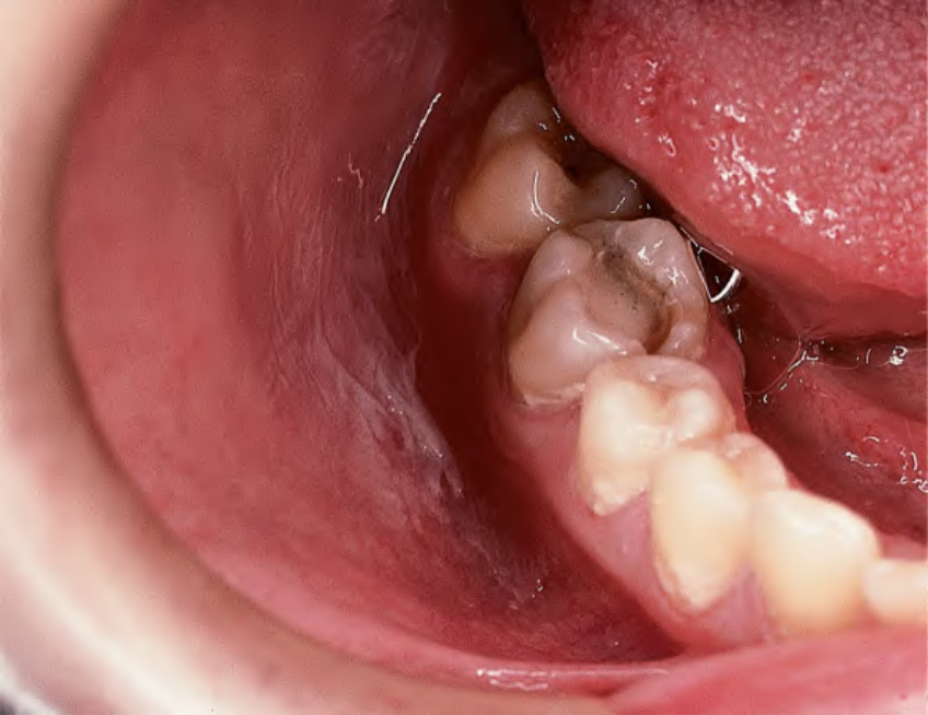
Fig. 72.10 Irritant contact stomatitis (aspirin burn). To relieve a toothache, this patient placed an aspirin in the mouth adjacent to the symptomatic tooth. Note the wrinkled white membrane that represents superficial epithelial necrosis. Courtesy Carl M. Allen, DDS, MSD and Charles Camisa, MD.
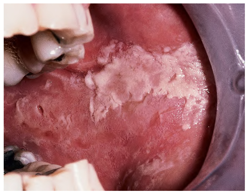
Fig. 72.11 Allergic contact stomatitis from artificial cinnamon flavoring. Shaggy, white keratotic plaque of the left buccal mucosa caused by the use of artificial cinnamon-flavored gum. Courtesy Carl M. Allen, DDS, MSD and Charles Camisa, MD.
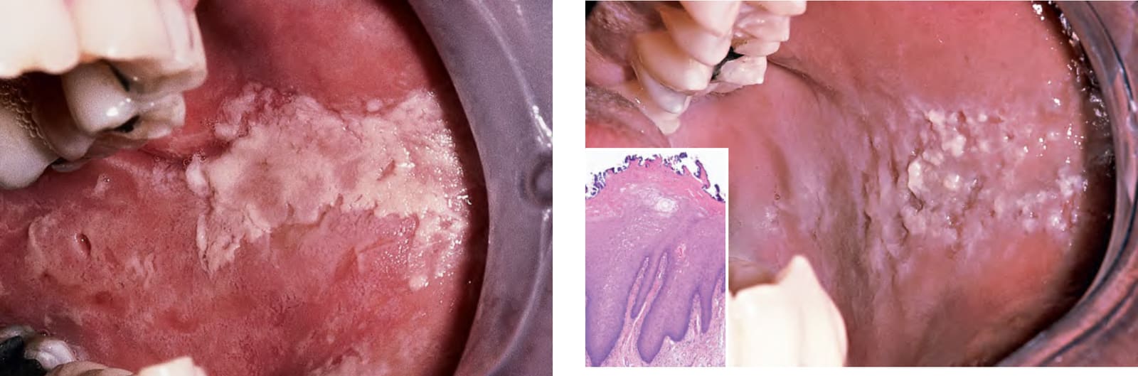
Fig. 72.12 Cheek chewing (morsicatio buccarum) – clinical and histopathologic findings. Repetitive nibbling of the superficial layers of the epithelium resulted in these changes. Note that the characteristic shaggy, white lesion approximates the area where the upper and lower teeth meet. Inset: Oral epithelium exhibiting acanthosis and prominent parakeratosis, with a ragged surface architecture and superficial colonization by bacteria. Courtesy Carl M. Allen, DDS, MSD and Charles Camisa, MD.
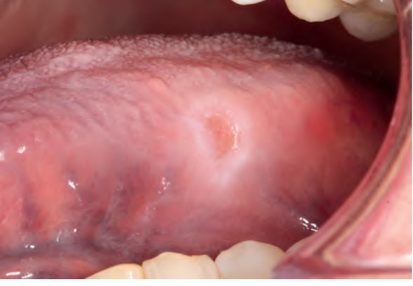
Fig. 72.13 Traumatic ulcer of the lateral tongue. The ulceration has a yellow fibrinous base with a blended white hyperkeratotic border. Compare this to the ulcerated leukoplakia with well-defined keratotic borders depicted in Fig. 72.21A and the exophytic, ulcerated mass depicted in Fig. 72.21B, which represent oral squamous cell carcinomas. Courtesy Carl M. Allen, DDS, MSD, Charles Camisa, MD, and Kristin K. McNamara, DDS, MS.
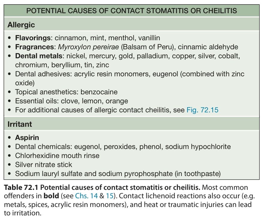
Table 72.1 Potential causes of contact stomatitis or cheilitis. Most common offenders in bold (see Chs. 14 & 15). Contact lichenoid reactions also occur (e.g. metals, spices, acrylic resin monomers), and heat or traumatic injuries can lead to irritation.
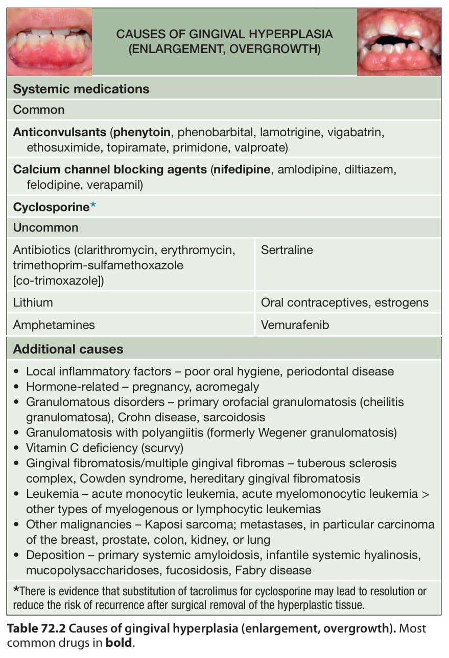
Table 72.2 Causes of gingival hyperplasia (enlargement, overgrowth). Most common drugs in bold.