CHEILITIS
Key features
A general term used to describe inflammation of the lips
Depending on the underlying cause, the clinical presentation and treatment may vary
Introduction
Cheilitis is an umbrella term used to describe inflammation of the lips, which may involve both the vermilion zone and the perioral region. It is caused by a variety of internal and external factors, most commonly contact dermatitis (irritant > allergic) and chronic UV exposure (Fig. 72.15 & 72.16).
Eczematous cheilitis
Eczematous cheilitis can be due to atopic dermatitis as well as irritant and allergic contact dermatitis. A combination of these dermatitides is also commonly seen. It typically presents with xerosis, scaling, erythema, and fissuring of the lips. Burning and pruritus are common symptoms.
Irritant contact cheilitis often results from chronic lip licking (liplicker’s dermatitis) as well as from environmental factors such as cold, wind, and low humidity (see Fig. 72.16A). Additional causative factors include irritants in cosmetics, foods, and oral hygiene products (see Ch. 15). Typically, the upper and lower lips are involved, with frequent
extension onto the cutaneous lip. Atopic cheilitis occurs in individuals with atopic dermatitis or an atopic diathesis and characteristically presents with xerosis and angular fissures in addition to erythema and scaling (see Fig. 12.14A). A number of allergens (see Fig. 72.15) can lead to allergic contact cheilitis (see Fig. 72.16B). Women are more commonly affected than men, presumably due to greater exposure to lipsticks and cosmetics. Patients typically present with erythema, scaling, edema, fissuring, and superficial erosions on both the upper and lower lips, oftentimes with extension to the perioral cutaneous regions. In addition to careful history-taking, patch testing can assist in identifying culprit allergens (see Ch. 14).
Treatment involves removal and future avoidance of any known or suspected irritants or allergens. Simple emollients, such as white petrolatum, should be used frequently. Low- to mid-potency topical corticosteroids or topical calcineurin inhibitors can be applied twice daily for 2–3 weeks. Prolonged use of topical corticosteroids should be avoided, as it can exacerbate perioral dermatitis.
Actinic Cheilitis (see below) Angular Cheilitis
In angular cheilitis (perlèche), there is inflammation of the skin and adjacent labial mucosa at the lateral commissures (angles or corners) of the mouth. Moist, macerated erythematous patches may be accompanied by erosions, angular fissures, and soreness (see Fig. 77.22C); oral thrush and candidal cheilitis may also be present. It is caused by excessive moisture that accumulates in the oral commissure grooves along with secondary infection with Candida albicans, and less commonly Staphylococcus aureus. It can occur at any age but is most often seen in older individuals with deep oral commissure grooves due to recession of the alveolar ridges, including those who are edentulous and wear dentures. In young children, excessive drooling and thumb sucking are common causes.
Diagnosis is usually made clinically, but in more difficult cases a lesional swab for bacterial and fungal culture can be obtained. Treatment involves addressing predisposing factors (e.g. poorly fitting dentures) as well as treating secondary fungal and bacterial infections. A topical antifungal cream (e.g. ketoconazole, clotrimazole) +/− topical mupirocin ointment applied twice a day for 1–3 weeks is often used, and oral thrush, if present, is treated. Maintenance therapy includes nighttime use of a barrier emollient such as white petrolatum or zinc oxide paste.
Drug-Induced Cheilitis
Several medications have been associated with cheilitis, most notably systemic retinoids, indinavir, sorafenib, and anticholinergic medications. Systemic retinoids routinely cause xerosis, peeling, and fissuring of the lips, and the effect is dose-dependent (see Fig. 126.8). Some implicated medications cause xerostomia with associated dryness of the lips. Management while on the medication includes liberal use of emollients (e.g. white petrolatum).
Exfoliative Cheilitis
In exfoliative cheilitis, there is persistent exfoliation of the vermilion lips, with scaling, desquamation, crusting, and hyperkeratosis. It is thought to be caused by chronic injury, most often from excessive licking, biting, picking, or sucking of the lips. It is most common in women under the age of 30 and many patients deny or are not aware of the habit. Exfoliative cheilitis is a diagnosis of exclusion. Topical emollients (e.g. white petrolatum), corticosteroids, and calcineurin inhibitors may be helpful in reducing symptoms, but psychiatric care to address underlying obsessive-compulsive tendencies or anxiety is often needed.
Plasma Cell Cheilitis
Plasma cell cheilitis is a rare disorder that typically presents as a well-circumscribed, eroded, erythematous plaque on the lower > upper lip. It is clinically and histologically similar to Zoon balanitis/vulvitis and the diagnosis is made upon histopathologic features. Treatment is difficult and often involves a trial of potent topical or intralesional corticosteroids.
Cheilitis Glandularis
Cheilitis glandularis is a rare condition characterized by inflammatory hyperplasia of the minor salivary glands of the lower lip and it may occur in association with actinic cheilitis. The secretory ducts of these hyperplastic salivary glands become dilated and inflamed and appear as pinpoint orifices from which a sticky mucous substance can be extracted. In mild cases, sunscreen and topical corticosteroids may be helpful, while in severe cases, vermilionectomy and dissection of the hyperplastic minor salivary glands has been described.
Orofacial Granulomatosis (OFG)
Key features
Persistent non-tender swelling of the lip and/or face
Non-caseating granulomatous inflammation
Both primary (idiopathic) and secondary subtypes
Primary OFG includes cheilitis granulomatosa and Melkersson–
Rosenthal syndrome
Introduction
Orofacial granulomatosis (OFG) is a term introduced in 1985 to describe a spectrum of diseases characterized by non-necrotizing, non-infectious granulomatous inflammation of the face and mouth in the absence of systemic disease. More recently, the term OFG has been used more loosely to describe a group of disorders characterized by non-caseating granulomatous inflammation involving the face and mouth including systemic disorders such as sarcoidosis and Crohn disease. In order to reconcile these different viewpoints, it is best to consider OFG as either primary (idiopathic) or secondary. Primary OFG would include cheilitis granulomatosa (CG) and Melkersson–Rosenthal syndrome (MRS). Secondary OFG would then include localized and systemic associations such as allergic contact dermatitis, foreign body reaction, sarcoidosis, and Crohn disease (see Fig. 72.16G).
Epidemiology
CG is rare and primarily occurs in young adults without sex predilection. The etiology is unknown and multiple factors have been implicated, including allergy to foods, additives or flavorings, genetic predisposition, and infection.
Clinical features
CG is a chronic process that initially can present as intermittent (hours to days) swelling of either the upper or lower lip mimicking angioedema. With progression, the episodes of swelling increase in duration and lead to persistent, often disfiguring swelling that may involve both lips and occasionally the face. Rarely, CG is associated with recurrent facial palsy and/or lingua plicata (fissured tongue), termed Melkersson– Rosenthal syndrome (MRS).
Diagnosis is made upon clinicopathologic correlation and exclusion of other entities, including angioedema (improves spontaneously in 24–48 hours), Crohn disease, sarcoidosis, foreign body reaction, allergic contact dermatitis, and infection (e.g. tuberculosis, Hansen disease [leprosy], histoplasmosis, cutaneous leishmaniasis).
Pathology
The most striking feature of OFG is the presence of non-necrotizing, epithelioid granulomas; in many cases the granulomas are sparse.
Treatment
Treatment of CG is difficult with a tendency for relapse and incomplete remission. Topical or intralesional corticosteroids are typically first-line therapy. Weekly (×3) intralesional triamcinolone injections (40 mg/ml) into the lips is one reported regimen. Other anecdotal treatment options include systemic corticosteroids, oral antibiotics (e.g. minocycline, metronidazole, clofazimine), adalimumab, methotrexate, and dapsone. Refractory cases have been managed surgically, with reduction cheiloplasty.
Granulomatosis with Polyangiitis (Formerly Wegener Granulomatosis)
Key features
Necrotizing granulomatous vasculitis of the upper and lower respi- ratory tract and necrotizing glomerulonephritis
Destructive ulcerated lesions of the oral and nasal cavity
Pathognomonic friable “strawberry gums” may be seen
Up to 90% of patients have anti-neutrophil cytoplasmic antibodies
(ANCA)
Introduction
Granulomatosis with polyangiitis (GPA) is an uncommon necrotizing granulomatous disease of unknown etiology (see Ch. 24). Clinically, there is both limited GPA (without renal involvement) and generalized GPA (see Fig. 40.6).
Epidemiology
Although individuals with GPA include children and the elderly, middleaged adults are most commonly affected; there is no sex predilection.
Clinical features
Because GPA commonly involves the nose, nasopharynx, and paranasal sinuses, patients can present with epistaxis, sinusitis, or nasal ulcerations and obstruction. Destruction of the nasal cartilage may result in perforation of the nasal septum and/or saddle nose deformity. The most characteristic oral manifestation is erythematous, friable gingival hyperplasia that easily bleeds, referred to as strawberry gingivitis (Fig. 72.17). Oropharyngeal, lingual, or gingival ulcerations may also be seen and tend to occur later in the course of the disease.
Pathology
For the gingival hyperplasia, histopathologic findings include papillary hyperplasia, granulation tissue, and a focal giant cell reaction. Histopathologic features of the cutaneous lesions are reviewed in Chapter 24.
Treatment
The treatment of classic GPA includes systemic corticosteroids, pulse cyclophosphamide, and rituximab; oral lesions typically respond to these regimens (see Table 24.10).
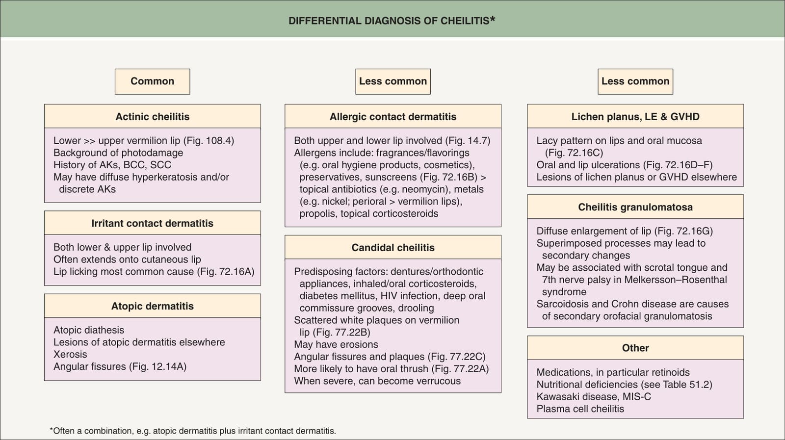
Fig. 72.15 Differential diagnosis of cheilitis. Uncommon causes include exfoliative cheilitis, cheilitis glandularis, and associations with other cutaneous disorders (e.g. cutaneous lupus erythematosus, actinic prurigo, lichen sclerosus), systemic disorders (e.g. Crohn disease, sarcoidosis, syphilis), and genetic disorders (e.g. Down syndrome, KID syndrome). AKs, actinic keratoses; BCC, basal cell carcinoma; GVHD, graft-versus-host disease; MIS-C, multisystem inflammatory syndrome in children; SCC, squamous cell carcinoma. Courtesy Jean L. Bolognia, MD.
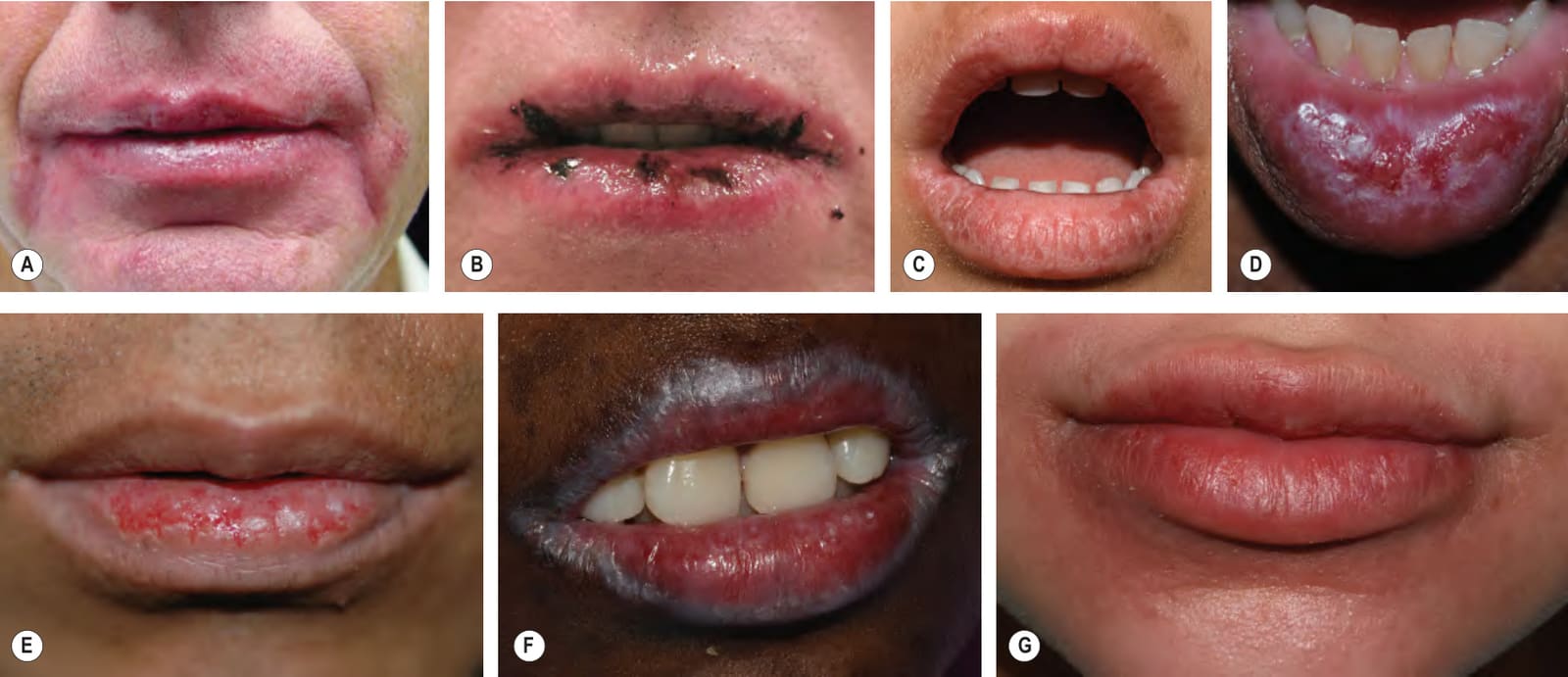
Fig. 72.16 Different causes of cheilitis.AIrritant contact dermatitis due to lip licking with involvement of the vermilion and cutaneous lips as well as the perioral region. BAllergic contact dermatitis to oxybenzone with involvement of the upper and lower vermilion lips. CLichen planus with a white lacy pattern of the lower lip and central upper lip. D Erosive lichen planus of the vermilion and mucosal lower lip. E Multiple jagged erosions limited to the lower vermilion lip in a patient with cutaneous lupus erythematosus. F Chronic graft-versus-host disease in which gray-white plaques are at the periphery of broad erosions. G Secondary orofacial granulomatosis characterized by diffuse enlargement of the lower > upper lip in an adolescent girl with Crohn disease. Note the coalescing tiny pink papules of the lower cutaneous lip and upper chin. A, Courtesy Jeffrey P. Callen, MD; B, D, Courtesy Kalman Watsky, MD; C, F, G, Courtesy Julie V. Schaffer, MD; E, Courtesy Luis Requena, MD.
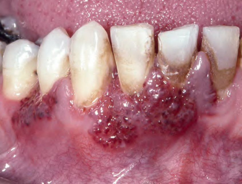
Fig. 72.17 Granulomatosis with polyangiitis – strawberry gums. The affected areas of the gingiva are red–purple, micropapular and friable, with a resemblance to ripe strawberries. Courtesy Carl M. Allen, DDS, MSD and Charles Camisa, MD.