EPITHELIAL PATHOLOGY
Oral Leukoplakia and Proliferative Verrucous Leukoplakia
Key features
Leukoplakia and proliferative verrucous leukoplakia represent clinical diagnoses
Leukoplakia is the most common potentially malignant disorder of the oral cavity
Biopsy is mandatory in order to assess the degree of dysplasia and to exclude invasive SCC
Introduction
The World Health Organization (WHO) defines oral potentially malignant disorders (OPMDs) as “clinical presentations that carry a risk of cancer development in the oral cavity, whether in a clinically definable precursor lesion or in clinically normal oral mucosa”. The major OPMDs are leukoplakia (including proliferative verrucous leukoplakia), erythroplakia, erythroleukoplakia, oral lichen planus and discoid lupus erythematous, oral submucous fibrosis, smokeless tobacco keratosis, palatal lesions associated with reverse smoking, chronic candidiasis, and actinic cheilitis.
Oral leukoplakia is defined as “a white plaque of questionable risk having excluded (other) known diseases or disorders that carry no increased risk for cancer”. In addition to smoking, excessive alcohol intake, betel nut use, older age, and male sex represent risk factors. Individuals who are immunocompromised or have genetic syndromes such as dyskeratosis congenita or Fanconi anemia also have an increased risk of developing oral leukoplakia. Proliferative verrucous leukoplakia (PVL) was first described in 1985 as a subset of leukoplakia. While sharing some features with leukoplakia, the transformation rate of PVL is far higher such that many consider PVL a distinct entity warranting a separate classification.
Epidemiology
Oral leukoplakia is the most common OPMD. It has a male predominance with a peak incidence after 50 years of age. In contrast, PVL is more common in older women and the majority have few to none of the traditional risk factors for leukoplakia and OSCC.
Clinical features
Oral leukoplakia presents as a white plaque or patch and is usually asymptomatic. Although leukoplakia can involve any site within the oral cavity, the tongue, floor of the mouth, gingivae, and buccal mucosa are the most commonly affected sites (Fig. 72.18). Based upon clinical presentation, oral leukoplakia is classified as: (1) homogenous leukoplakia – well-demarcated white plaque or patch that is mainly smooth-surfaced with varying degrees of surface fissuring; and (2) non-homogenous leukoplakia. The latter has three subtypes: (a) erythroleukoplakia – varying amounts of erythema within a well-demarcated, mostly white plaque and a patchy or speckled pattern; (b) verrucous leukoplakia – a rough surface that at times appears leathery and most commonly occurs on the gingivae; and (c) nodular leukoplakia – nodular surface, at least focally.
PVL is characterized by the progressive development of white, often verrucous and fissured, patches and plaques in multiple non-contiguous sites or as a single large lesion. Although the seventh decade represents the average age at the time of diagnosis, many patients have had the condition for years. Dysplasia is regarded as the best predictor of transformation, but a meta-analysis found that only 12% of dysplastic lesions transformed to OSCC. That said, as many as 70% of patients with PVL eventually develop OSCC despite their lack of traditional risk factors and benign histopathologic features in early stages.
Pathology
The diagnosis of both leukoplakia and PVL is clinically based, i.e. independent of the presence or absence of dysplasia. Nonetheless, biopsies are routinely obtained due to concern regarding OSCC. Histopathologic features vary from only hyperkeratosis to dysplasia to frank OSCC. Major features of dysplasia include irregular, altered epithelial architecture with loss of basal cell polarity, as well as nuclear pleomorphism, dyskeratosis, hyperchromasia, and an increased number of mitoses with superficial and atypical mitotic figures. Dysplasia is graded from mild to severe, depending on the portion of the involved epithelium (lower third or more). A band-like inflammatory infiltrate can be seen in the upper lamina propria (lichenoid dysplasia) and should be differentiated from oral lichen planus.
Differential diagnosis
Entities in the differential diagnosis include leukoedema, in which the white to gray–white discoloration disappears with stretching (Fig. 72.19), white sponge nevus due to mutations in KRT4 and KRT13, the leukokeratosis seen in patients with pachyonychia congenita (see Ch. 58), and oral lichen planus. Oral white plaques can also be seen in hereditary benign intraepithelial dyskeratosis, a disorder that occurs primarily in Native Americans.
Treatment
A baseline incisional biopsy is considered the gold standard for management of leukoplakia and PVL. Although complete excision of the lesion (via scalpel, CO laser, or cryosurgery) has been proposed as first-line therapy by some authors, removal of OPMDs has not been proven to prevent the onset of OSCC. When possible, surgical removal of the entire lesion is performed to avoid sampling error and to exclude OSCC. Habit cessation (in particular tobacco use), longitudinal evaluation by an oral medicine specialist, and repeat biopsies as indicated are recommended for all patients.
Erythroplakia
Key features
Erythroplakia is less common than leukoplakia but is also a clinical diagnosis
High likelihood of dysplasia or invasive SCC at the time of diagnosis
Introduction
The WHO defines erythroplakia as “a fiery red patch that cannot be characterized clinically or pathologically as any other definable disease”. Similar to leukoplakia, erythroplakia is an OPMD that is strongly associated with traditional risk factors for OSCC. A diagnosis of erythroplakia is only established after exclusion of other red lesions of the oral cavity.
Epidemiology
A rare condition that most commonly affects middle-aged and elderly men with traditional OSCC risk factors.
Clinical features
Erythroplakia typically presents as an asymptomatic, rather sharply circumscribed, velvety red patch or thin plaque. The most commonly involved sites are the floor of the mouth, tongue, soft palate, and buccal mucosa.
Pathology
Advanced dysplasia (see leukoplakia above) or invasive OSCC is found in >90% of lesional biopsies. In addition to a lack of keratinization and atrophy of the epithelium, varying degrees of cellular and architectural findings consistent with OSCC (see below) or dysplasia (see leukoplakia above) are seen in these cases.
Treatment
A baseline biopsy should be performed, with treatment based upon the histopathologic diagnosis. Patients with OSCC are referred to head and neck surgeons or oncologists. When possible, complete surgical removal of dysplastic lesions is performed to avoid sampling error and to exclude OSCC. Habit cessation (in particular tobacco use), longitudinal evaluation by an oral medicine specialist, and repeat biopsies as indicated are recommended.
Nicotinic Stomatitis
Synonyms: Nicotine stomatitis Stomatitis nicotina
Key features
Gray–white mucosa with umbilicated papules
Caused by exposure of the palatal mucosa to intense heat from smoking, not nicotine
Introduction
Nicotinic stomatitis is most commonly observed in pipe, cigar, and reverse smokers. However, the resultant inflammation of the palatal mucosa is more closely linked to exposure to the intense heat from smoking rather than nicotine, as the name has erroneously implied. For example, identical lesions are observed in hot beverage drinkers. The exact etiology in e-cigarette users is less clear and may be related to nicotine or other chemical compounds (e.g. strawberry flavoring) added to the liquid.
Epidemiology
Nicotinic stomatitis most frequently affects men in the fifth decade of life.
Clinical features
The palatal mucosa is thickened and has a diffuse gray or white discoloration. In addition, there are scattered 1–3 mm umbilicated papules with central red puncta which represent inflamed salivary gland orifices (Fig. 72.20). These changes are more prominent toward the posterior hard palate and anterior soft palate.
Pathology
The histopathologic features include hyperkeratosis, parakeratosis, acanthosis, and mild chronic sialodochitis, but the diagnosis is usually made on clinical grounds.
Treatment
Since habit cessation typically leads to resolution, it is necessary to exclude true leukoplakia or erythroplakia if any white or red plaque persists after habit cessation.
Actinic Cheilitis
Synonyms: Actinic cheilosis Solar cheilosis
Key features
Primarily affects the vermilion zone of the lower lip in fair-skinned individuals
Indicator of photodamage from cumulative sun exposure
Introduction
Actinic cheilitis is a common OPMD that has the same etiology as actinic keratoses (see Ch. 108). Given its relationship to chronic UV exposure, the vermilion zone of the lower lip is the primary mucosal site of involvement.
Epidemiology
Actinic cheilitis is most common in men over 40 years of age with skin phototypes I–II. It reflects significant photodamage from cumulative exposure to UV irradiation.
Clinical features
Early features include loss of normal dermatoglyphics, atrophy, and blurring of the margin between the vermilion zone and the skin. Signs of progression to precancerous actinic cheilitis include scaling and roughness, sometimes in conjunction with leukoplakia (see Fig. 108.4). Although often localized initially, over time multifocal involvement is characteristic. Fissuring and erosions may also be present as the condition worsens. Some patients have concomitant cheilitis glandularis (see above).
Pathology
There is hyperkeratosis and variable thickness of the spinous layer as well as dysplasia of the lower part of the epithelium. Actinic cheilitis may be observed in contiguity with SCC in situ or invasive SCC.
Treatment
Actinic cheilitis frequently represents a “field change”, and while worrisome clinical features may appear focal in nature, the photodamage and epithelial dysplasia are more extensive. Treatment options include topical 5-fluorouracil or imiquimod, photodynamic therapy, and cryosurgery. When severe or treatment-resistant, vermilionectomy with histopathologic examination is a therapeutic option. Any site suspicious for SCC should be biopsied, including prior to CO laser ablation.
Oral Squamous Cell Carcinoma
Key features
The most common malignancy of the oral cavity
Develops primarily in middle-aged or elderly men
Significant association with the use of tobacco and alcohol
In contrast to oropharyngeal SCC, only a small percentage of oral
SCCs are associated with HPV infection, particularly types 16 and 18
Most common intraoral sites are the lateral and ventral surfaces of the tongue, floor of the mouth, and soft palate
Introduction
SCC is the most common malignancy of the oral cavity, and may or may not be preceded by an OPMD. OSCC risk factors include tobacco use, excessive alcohol consumption, betel nut chewing (primarily in the Indian subcontinent and Southeast Asia), chronic immunosuppression, and genetic syndromes such as dyskeratosis congenita and Fanconi anemia. Human papillomavirus (HPV) infection, especially types 16 and 18, has been implicated in the etiopathogenesis of oropharyngeal SCC, with HPV-positive tumors associated with a more favorable outcome.
Epidemiology
OSCC accounts for >90% of all malignant neoplasms of the mouth. It is typically a disease of middle-aged or elderly men; however, an increasing incidence of oral SCC among women and younger patients has been noted. Globally, the overall age-adjusted incidence is 4 per 100 000 individuals, but there is marked variation depending upon geographic location. For example, the Indian subcontinent has recorded an incidence as high as 22 per 100 000 and worldwide accounts for one-third of all OSCCs.
Clinical features
While any intraoral site may be affected, the most common locations are the lateral and ventral surfaces of the tongue, floor of the mouth, and soft palate. OSCC may present as an ulcer, a white plaque, an exophytic mass, or an endophytic process with varying degrees of induration (Fig. 72.21). The surface is typically irregular, rough, or granular. Early on lesions are often painless, but with disease progression and local invasion, patients may experience pain, paresthesia, dysphagia, dysphonia, or difficulty moving the tongue.
In comparison to cutaneous SCC, OSCC is significantly more aggressive and has a less favorable prognosis. The five-year survival
rates for stage I or II disease approach 80%, while the 5-year survival rate for stages III and IV is <40%.
Pathology
OSCCs are histologically similar to those occurring elsewhere (see Ch. 108).
Treatment
Because treatment depends upon the clinical stage, computed tomography (CT), positron emission tomography (PET), and/or magnetic resonance imaging (MRI) may be required to determine the extent of local and distant disease. In general, smaller tumors (stages I and II) are surgically excised +/− sentinel lymph node biopsy or cervical lymph node dissection. For stages III and IV, various combinations of surgery, radiotherapy, chemotherapy, and immune checkpoint inhibitors are employed. Patients have a 3%–7% annual risk of developing a second primary SCC of the upper aerodigestive tract after treatment of their original tumor.
Verrucous Carcinoma
Subtype/synonyms: Oral verrucous carcinoma Oral florid papillomatosis Ackerman tumor
Key features
Uncommon, low-grade variant of SCC
Exophytic mass with a papillary or verrucous topography
Characterized by slow, persistent growth
Introduction
Verrucous carcinoma is a rare, low-grade variant of SCC that may involve the oral, laryngeal, sinonasal, esophageal, or genital mucosa as well as the plantar surface of the foot (see Ch. 108). Oral verrucous carcinoma has been associated with the use of snuff and chewing tobacco. Although HPV-6, -11, -16, and -18 have sometimes been isolated from these tumors, the precise etiologic role of HPV remains unclear.
Epidemiology
Verrucous carcinoma typically affects men over 55 years of age.
Clinical features
The tumor location often corresponds to the site where intraoral tobacco has been chronically placed. Therefore, tumors most commonly develop in the mandibular vestibule, buccal mucosa, tongue, and gingiva. In populations where tobacco is placed under the tongue or in the maxillary vestibule, the floor of the mouth and hard palate are commonly affected.
Verrucous carcinoma presents as a slowly growing, exophytic, papillomatous, or warty proliferation with well-demarcated margins. The color depends on associated inflammation and degree of hyperkeratosis but is most often white. If left untreated, local destruction of underlying bone, muscle, and salivary glands may occur; metastases are rare.
Pathology
Histologically, there is hyperkeratosis, acanthosis, and a papillary or verrucous surface. The epithelium is well differentiated, with minimal atypia and rare mitotic figures. A dense infiltrate of chronic inflammatory cells is frequently present in the superficial connective tissue. Multiple sections are recommended because up to 20% of verrucous carcinomas show foci of conventional OSCC (hybrid carcinoma).
Treatment
Compared to conventional OSCC, verrucous carcinoma has a better prognosis. The 5-year survival rate is 85%–90% with prognosis based upon stage at diagnosis. The treatment of choice is surgical excision. Although cervical lymph node metastases are rare, sentinel lymph node biopsy can be performed. In contrast, hybrid carcinomas may metastasize and should be managed as conventional OSCC. Treatment failure has been attributed to extensive involvement, delayed diagnosis, comorbidities, and failure to identify hybrid carcinomas.
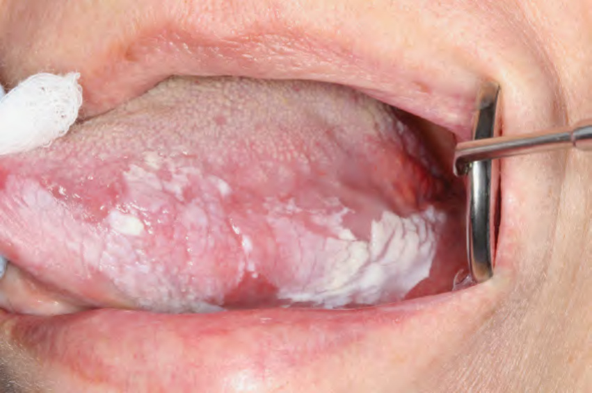
Fig 72.18 Leukoplakia. Non-homogenous, sharply demarcated white plaque involving the left ventrolateral tongue.
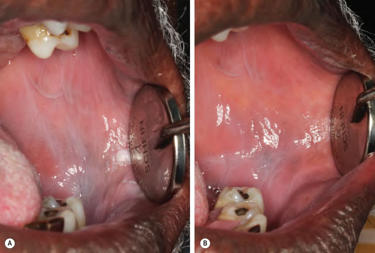
Fig 72.19 Leukoedema.A Diffuse grayishwhite and wrinkled appearance of the left buccal mucosa. B These findings disappear when the cheek is stretched.
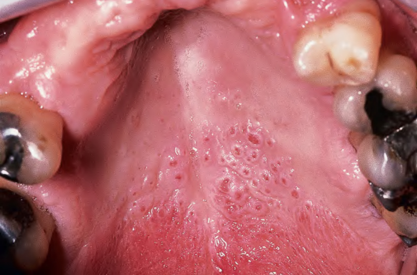
Fig. 72.20 Nicotinic stomatitis. Gray–white palatal mucosa with numerous umbilicated papules representing inflamed palatal salivary glands. Courtesy Carl M. Allen, DDS, MSD and Charles Camisa, MD.
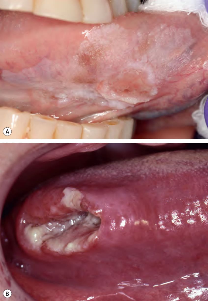
Fig. 72.21 Squamous cell carcinoma.A Ulcerated leukoplakia of the ventrolateral tongue. B Ulcerated, indurated, exophytic mass involving the right lateral border of the tongue. Both are typical presentations for this tumor. Courtesy Carl M. Allen, DDS, MSD, Charles Camisa, MD, and Kristin K. McNamara, DDS, MS.