SALIVARY GLAND DISEASES
Mucocele
Synonyms: Mucous cyst of oral mucosa Mucous extravasation phenomenon
Key features
Disruption of a minor salivary gland duct with resultant leakage of mucus into the submucosal tissue
Fluid-filled, dome-shaped mucosal swelling ranging from a few millimeters to 1–2 cm
Most commonly seen on the lower labial mucosa
May require surgical excision
Introduction
Mucocele is a common disorder that results from disruption of a salivary gland duct and escape of mucus into the adjacent submucosal connective tissue (see Ch. 110). Although the term mucocele is sometimes used to describe both retention and extravasation mucoceles, these two conditions differ in location, etiology, and histopathology. The preferred term for a retention mucocele is salivary duct cyst. Herein, the term mucocele refers exclusively to extravasation mucocele.
Epidemiology
While the majority of mucoceles are detected in younger individuals (second through fourth decades), they can present at any age and are
usually due to trauma. Patients with chronic oral GVHD can develop superficial mucoceles, primarily on the palate.
Clinical features
Mucoceles present as dome-shaped, mucosal swellings ranging from 1–2 mm to a few centimeters in size. Their color depends on the depth of the swelling relative to the surface mucosa, with very superficial lesions often appearing vesicular and slightly deeper mucoceles having a bluish tinge (Fig. 72.22); the deepest lesions have no discernible color change. In general, mucoceles are asymptomatic and undergo multiple cycles of swelling, rupturing, and then refilling. While the lower labial mucosa is the most common site of involvement, any oral mucosal area that contains minor salivary glands can potentially be affected. In particular, mucoceles may develop on the ventral tongue, buccal mucosa, and posterior hard palate. A mucocele on the floor of the mouth and confined to sublingual tissues superior to the mylohyoid muscle is referred to as a ranula.
Pathology (see Ch. 110)
Treatment
While some mucoceles may resolve spontaneously, others require surgical excision. Other therapeutic modalities are outlined in Chapter 110.
Necrotizing Sialometaplasia
Key features
Uncommon ulcerative process of the hard palate thought to be due to ischemia
Frequently misdiagnosed as a malignancy, both clinically and histologically
Introduction
Necrotizing sialometaplasia is an uncommon, locally destructive disease that is most likely due to an interruption of the blood supply to salivary gland tissue, leading to ischemic necrosis. While such a disruption in blood supply can follow local trauma, in most patients the cause of the ischemic injury is unknown. Very infrequently, a submucosal malignancy may occlude the palatal blood vessels.
Epidemiology
The condition is almost always seen in adults, with a slight male predominance.
Clinical features
The patient will typically report a sudden onset of swelling of the hard palate, in addition to localized pain, paresthesia, or anesthesia. Within 1–2 weeks, the ischemic tissue sloughs and the patient will often state that
“part of the roof of my mouth fell off”. This leads to a well-demarcated ulcer with a smooth or minimally elevated border. By palpation, the ulcer has no underlying induration. A malignancy is often suspected clinically and therefore a biopsy is performed to exclude OSCC or a salivary gland tumor.
Pathology
Early on, acinar necrosis is seen, followed by squamous metaplasia of the salivary ducts, spillage of mucin into the surrounding connective tissue, and an associated inflammatory infiltrate. When present, preservation of the lobular architecture of the gland helps to distinguish necrotizing sialometaplasia from mucoepidermoid carcinoma and squamous cell carcinoma.
Treatment
Once a diagnosis of necrotizing sialometaplasia is established, additional treatment is not required. The condition resolves spontaneously within 5–6 weeks.
Sjögren Syndrome
Key features
Autoimmune disorder characterized by keratoconjunctivitis sicca and xerostomia
Primarily affects women
Introduction
Sjögren syndrome (SjS) is a chronic autoimmune connective tissue disease in which lacrimal and salivary gland dysfunction leads to dry eyes (keratoconjunctivitis sicca) and mouth (xerostomia) (see Ch. 45). While the etiology and pathogenesis of SjS remain unclear, patients do produce several autoantibodies, in particular anti-SSA/Ro and -SSB/La (see Ch. 40). Patients with SjS have a 40-fold increased risk of lymphoma, predominantly extranodal B cell non-Hodgkin type. Diagnostic criteria for SjS are outlined in Table 40.4.
Epidemiology
SjS primarily affects women in their 50s and 60s.
Clinical features
Both the major and minor salivary glands may be involved, leading to reduced salivary production. Major gland involvement is clinically evident in fewer than one-half of affected patients, presenting as firm, diffuse, non-tender or slightly tender glandular enlargement that is usually bilateral.
Patients have variable degrees of ocular and oral dryness, usually with a gradual onset of symptoms. There may be changes in either quality (frothy) or quantity (reduced) of saliva and most patients have a lack of the usual pooling in the floor of the mouth. Affected patients may present with difficulty swallowing, dysgeusia, halitosis, dental caries along the gum line, difficulty wearing dentures, coated or hairy tongue, and oral candidiasis.
Pathology
An SjS diagnostic criterion is one or more foci of inflammatory cells (≥50 lymphocytes) in 4 mm2 of minor salivary gland tissue (see Table 40.4). In major salivary glands, there are intraglandular infiltrates of lymphocytes as well as hyperplasia and metaplasia of salivary duct epithelia, forming the characteristic epimyoepithelial islands.
Treatment (see Table 40.5)
Salivary Gland Tumors
Key features
Most commonly occur in the parotid gland
Benign salivary gland tumors are more common than malignant tumors
Introduction
Salivary gland tumors (SGTs) can occur in both minor and major salivary glands. They are considered primary epithelial neoplasms and there exists a wide array of distinct benign and malignant salivary gland tumors.
Epidemiology
The low incidence, ever-evolving classification, and disparities between old and new studies are some of the obstacles in estimating the true incidence of SGTs. Pleomorphic adenoma followed by lymphomatous papillary cystadenoma (Warthin tumor) are the most common benign SGTs and mucoepidermoid carcinoma followed by acinic cell carcinoma are the most common malignant SGTs (Table 72.5). SGTs account for <10% of head and neck tumors, with radiation, smoking, viral infections, and environmental exposures (e.g. nickel compounds) implicated in their development.
Clinical features
The parotid gland is the most frequent site of SGTs, and the majority (>70%) are benign. SGTs involving major glands present as a painless, rubbery to firm, slowly growing swelling. Less frequently, SGTs develop within submandibular, sublingual, or minor salivary glands. They can present as a painless submucosal mass or mucosal ulceration, most commonly of the posterior hard palate/anterior soft palate followed by the upper lip or buccal mucosa. When malignant, SGTs occur most often in the sublingual gland followed by minor salivary glands.
Pathology
For a definitive diagnosis of SGT, histopathologic examination is required which demonstrates different features, depending upon the type of tumor. Ultrasound-guided fine needle biopsy may be performed prior to surgical intervention. For staging information, see https:// cancer.net/cancer-types/salivary-gland-cancer/stages-and-grades.
Treatment
The treatment of benign SGTs consists of conservative surgical excision. Malignant tumors usually require more extensive surgery and postoperative radiation therapy.
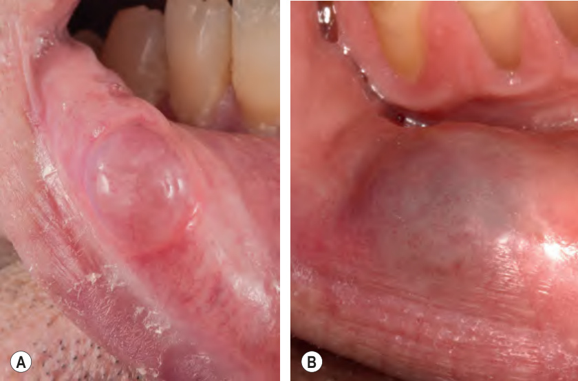
Fig. 72.22 Mucoceles of the mucosal lower lip.A Superficial mucocele that is translucent and has a vesicular appearance. B Slightly deeper mucocele presenting as a soft nodule with a bluish hue. This lateral location is commonly observed. A, Courtesy Edward Cowen, MD.
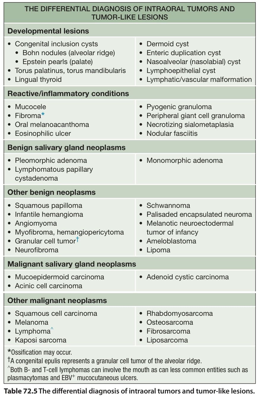
Table 72.5 The differential diagnosis of intraoral tumors and tumor-like lesions.