LICHEN SCLEROSUS
Synonyms: Lichen sclerosus et atrophicus Balanitis xerotica obliterans (men) Kraurosis vulvae (women)
Key features
Chronic inflammatory disease with a predilection for the anogenital region
Pruritus is the most frequent symptom
Major clinical signs are pallor, atrophy, fissures, and foci of hyperkeratosis
Scarring may cause loss of the normal architecture of the vulva in women and phimosis in men
Introduction
Lichen sclerosus is a chronic inflammatory disease that preferentially affects the anogenital region, although any cutaneous site may be affected; 15%–20% of patients with genital lichen sclerosus will have extragenital disease. Symptoms may be longstanding as there is frequently a delay in diagnosis. Patients require long-term evaluation due to the risk of scarring and the risk of developing squamous cell carcinoma (SCC).
Epidemiology
Lichen sclerosus is 6–10 times more prevalent in women than in men. The disorder may occur at any age, but the two peaks of onset are childhood and after menopause (or in the 4th or 5th decade in men). Although most studies have centered on White European patients, there are reports of lichen sclerosus occurring in Africans and Asians. Powell et al. calculated a prevalence of 1 in 660 women and an annual incidence of 52/100 000 in postmenopausal women. The incidence of lichen sclerosus in boys may be underestimated; when foreskins of circumcised boys were examined, 14% were found to have the characteristic features of lichen sclerosus.
Etiology
The cause of lichen sclerosus is unknown. In women, there is evidence to support an association with autoimmunity and heritability. Autoantibodies to extracellular matrix protein 1 (ECM1) are found in the majority of women with lichen sclerosus, but their role in pathogenesis is uncertain. A heritable risk is suggested by the occurrence of familial cases, as well as an association with HLA-DRB112 (and the related haplotype DRB112/DQB1*0301/04/09/010) and autoimmune disorders such as thyroid disease. However, in male patients, familial cases are uncommon, and HLA and autoimmune associations are rarely observed.
An infectious etiology has been proposed, but Borrelia or other spirochetal infection has not been confirmed in large studies. Local friction or rubbing may induce lesions of lichen sclerosus via the Koebner phenomenon, and some cases of early-onset lichen sclerosus may be associated with the antiandrogenic oral contraceptive pill. In men, chronic exposure of susceptible epithelium to urine due to naviculomeatal dysfunction (microurinary incontinence) has been proposed as causative. Of note, lichen sclerosus of the penis does not occur in men circumcised at birth. Whether urinary dysfunction plays a role in women and children is not known.
Clinical features
Both male and female patients may present with pruritus or soreness, but rarely are entirely asymptomatic. Dyspareunia is frequently reported. Children with lichen sclerosus are more likely to present with urinary or bowel symptoms (e.g. constipation), purpura, or even hemorrhagic bullae, which may lead to a misdiagnosis of child abuse.
In women and girls, the characteristic clinical findings are vulvar hypopigmentation and thin, wrinkled, atrophic skin in a figure-of-eight distribution encircling the vulvar and perianal region (Fig. 73.2). Focal areas of hyperkeratosis, erosions, and fissures are frequently seen, with the perineum a common location for fissures. Additional cutaneous features that are helpful in identification are purpura (Fig. 73.3), telangiectasias, and rarely, hemorrhagic bullae. The hemorrhagic component can occasionally be so pronounced that it mimics a vascular lesion. Because lichen sclerosus is a scarring disease, architectural change is common (Fig. 73.4). Burying of the clitoris secondary to midline fusion or fusion of the labia minora to the labia majora may occur. In untreated or severe disease, there may be total loss of the labia minora
Hypopigmented, shiny skin in a figure-of-eight configuration with purpura, multiple erosions, and foci of hyperkeratosis. Courtesy Julie V. Schaffer, MD.
and narrowing of the introitus. Darkly pigmented lentigines with an irregular outline may arise within areas of involvement.
Lichen sclerosus affects the glans penis and foreskin of men and, in contrast to women, involvement of the perianal region is uncommon. The disease usually presents with pruritus, pain, and dyspareunia. Affected boys may have recurrent balanitis and a poor urinary stream, and circumcision is sometimes required. Men may report painful erections or erectile dysfunction as well as dysuria and spraying, dribbling, or poor flow of urine. Clinically, gray–white discoloration, purpura, and scarring can be seen (Fig. 73.5), leading to phimosis, paraphimosis, or constrictive posthitis. Additional architectural changes include coronal adhesions, reduced coronal sulcus and rim, and narrowing of the urethral meatus.
Extragenital lichen sclerosus favors the submammary region, as well as the shoulders, neck, and wrists, presenting as asymptomatic, hypopigmented, wrinkled patches with follicular plugging (see Ch. 44); occasionally, there are multiple guttate lesions. The diagnosis is established by a combination of the characteristic clinical and histologic findings.
Pathology
The principal histologic findings are compact orthokeratosis, follicular plugging (in non-mucosal sites), a thinned epidermis, vacuolization of the basal layer, and hyalinization of the papillary dermis. A dense band-like lymphocytic infiltrate is often observed. Squamous hyperplasia may be present and may be associated with an increased risk of SCC. In bullous lichen sclerosus, there is extensive vacuolar degeneration of the basal layer with clefting and hemorrhage. Early on, the papillary dermis may be edematous and homogenous, but, with time, hyalinization and sclerosis are observed.
Histologic examination of the skin is recommended as it can be difficult to distinguish lichen sclerosus from mucous membrane (cicatricial) pemphigoid or erosive lichen planus on the basis of clinical appearance alone. Occasionally, despite marked clinical changes, the disease cannot be confirmed histologically and repeat biopsies over time may be needed to establish the diagnosis. Of note, in genital lesions, a dense band-like lymphoid infiltrate with several intraepithelial lymphocytes may be the only histopathologic finding, with hyalinization of the superficial collagen absent or only present focally. In lesions that have been treated with potent topical corticosteroids, ascertaining the diagnosis histologically may also prove challenging.
Differential diagnosis
Very mild or early disease may be confused with dermatitis. The presence of genital scarring necessitates the exclusion of other scarring disorders such as erosive lichen planus and mucous membrane pemphigoid. The absence of oral or vaginal mucosal involvement helps to distinguish lichen sclerosus from erosive lichen planus. In children, sexual abuse must be considered, as lichen sclerosus and sexual abuse are not mutually exclusive. Associated pruritus and topical therapies can lead to superimposed lichen simplex chronicus as well as allergic or irritant contact dermatitis. Histologically, the presence of a dense lymphoid infiltrate may be misdiagnosed as mycosis fungoides; the latter diagnosis, especially in the genital region, requires compelling evidence, particularly in children.
Treatment
The main aim of therapy is to bring the disease under control as quickly as possible with the fewest side effects. For induction treatment (after the diagnosis is established), a superpotent topical corticosteroid such as clobetasol propionate 0.05% ointment is prescribed until signs and symptoms improve, typically 1 to 3 months. This alleviates symptoms in the majority of patients, often within weeks. Subsequently, the corticosteroid is tapered over 2–3 weeks and clinical remission maintained by limited (typically twice weekly) application of a higher potency corticosteroid or daily application of a lower potency topical corticosteroid. In some women, there is complete resolution of signs as well as symptoms, with the exception of scarring, which is irreversible. Beyond preserving clinical remission, maintenance therapy has been associated with a reduced occurrence of SCC.
In the management of vulvar lichen sclerosus without secondary SCC, there is no longer a role for vulvectomy. For longstanding disease in which narrowing of the introitus has resulted in dyspareunia, surgical
refashioning with division of adhesions may be helpful. Vaginal dilators may also be needed, in addition to psychosexual counseling to address the psychological distress that frequently accompanies lichen sclerosus.
In male patients, first-line therapy consists of potent topical corticosteroids, with cotton swab/bud application for meatal involvement. Those with urinary symptoms should be evaluated by a urologist. If treatment does not lead to improvement or a tight phimosis is present, circumcision is often necessary.
Longitudinal evaluation is essential for patients with lichen sclerosus because both men and women are at increased risk of developing genital SCCs (estimates of the risk are 2%–6%). Women with childhoodonset lichen sclerosus may be particularly at risk of developing genital SCC, and notably the myth that childhood lichen sclerosus resolves at puberty has been debunked. Women with lichen sclerosus may develop differentiated vulvar intraepithelial neoplasia (VIN), which has a higher risk of developing into invasive SCC when compared to high-grade squamous intraepithelial lesion (HSIL); the latter is an HPV-associated disease and was previously known as undifferentiated VIN. Men are at risk of penile intraepithelial neoplasia. Non-healing fissures, ulcers, or nodules must be examined histologically. Written educational material and patient support groups are very helpful. It is essential that patients who notice any features of malignancy seek prompt medical assessment.
The first- and second-line therapies for anogenital lichen sclerosus are outlined in Table 73.1.
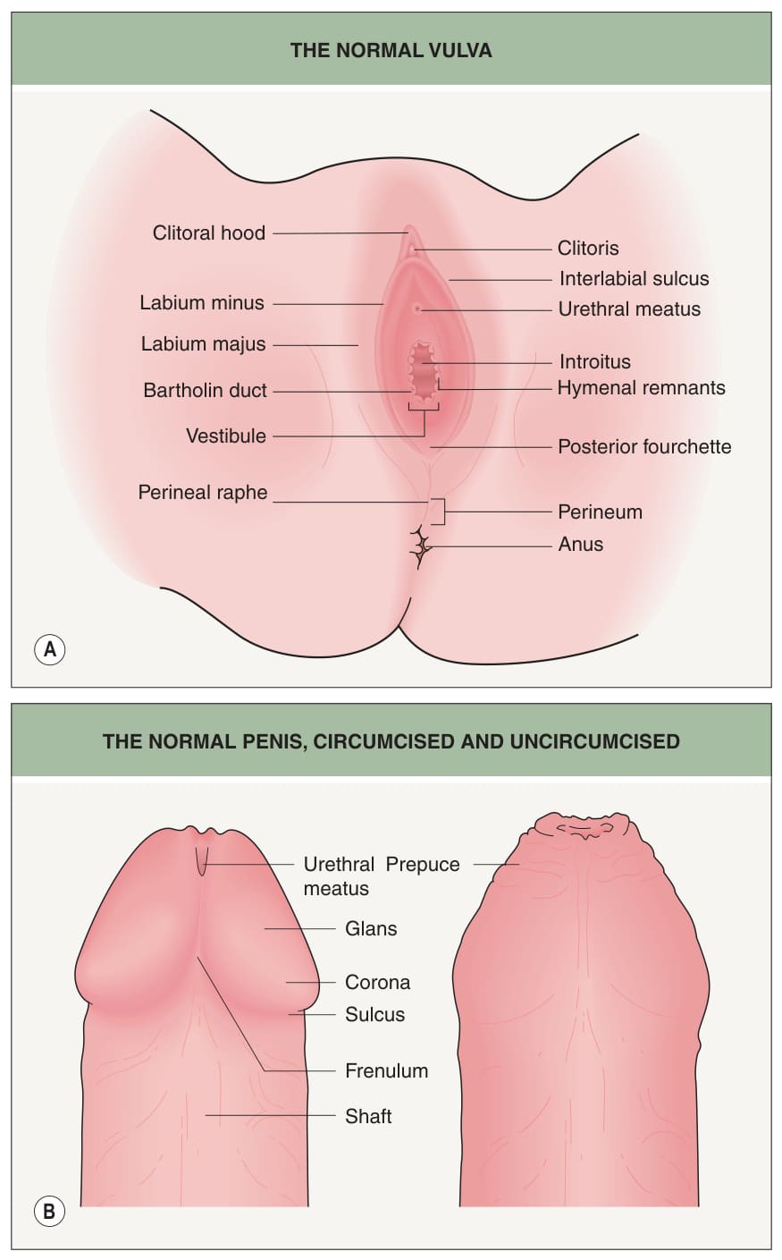
Fig. 73.1 Genital anatomy.A The normal vulva. B The normal penis, circumcised and uncircumcised.
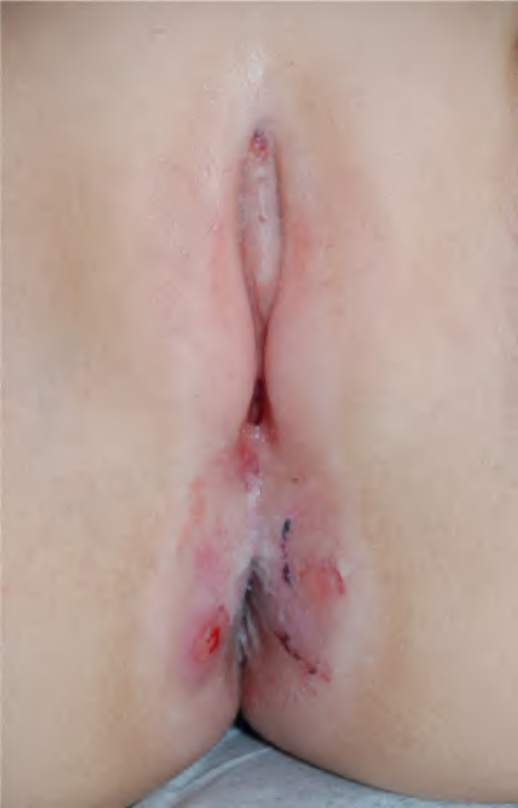
Fig. 73.2 Anogenital lichen sclerosus in a girl.
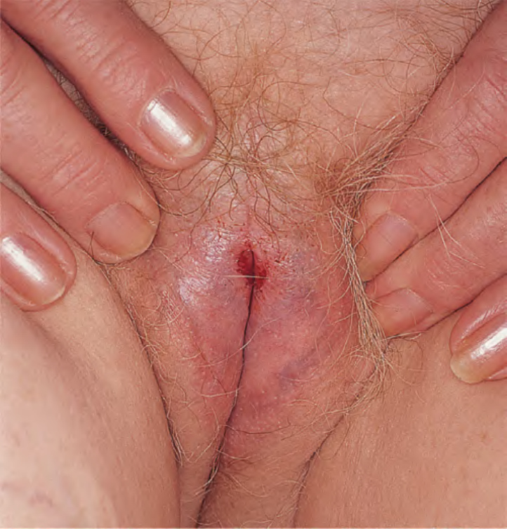
Fig. 73.3 Lichen sclerosus of the vulva with characteristic purpura.Courtesy Kalman Watsky, MD.
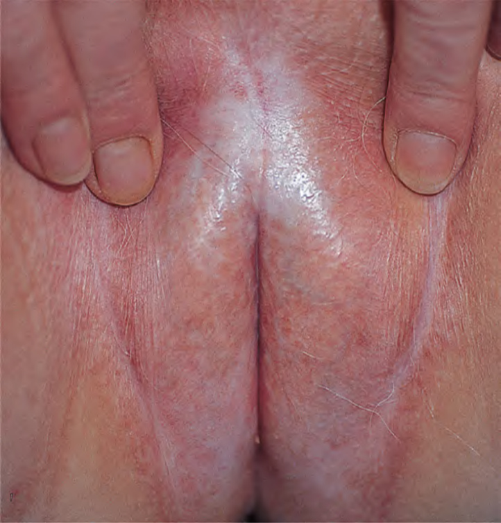
Fig. 73.4 Lichen sclerosus of the vulva. There is marked architectural change with loss of the labia minora and marked midline fusion.
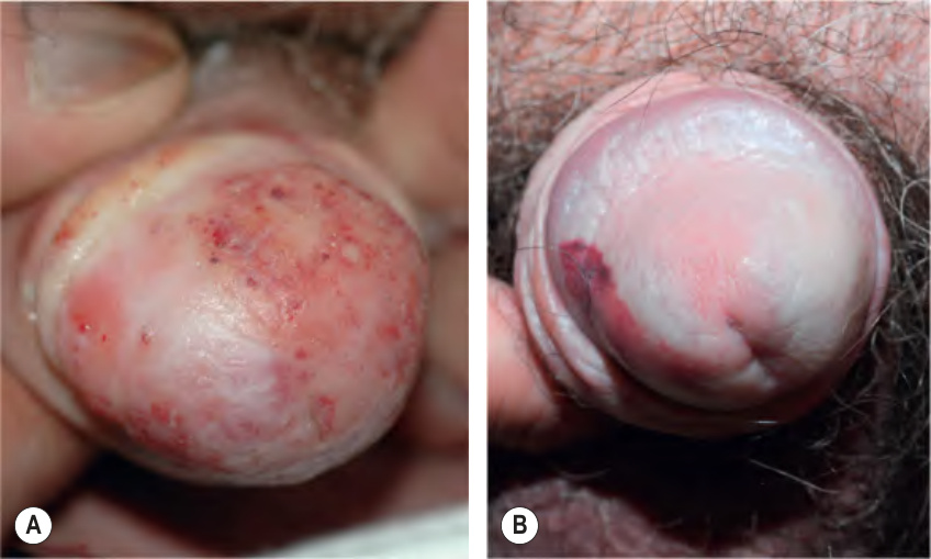
Fig. 73.5 Lichen sclerosus of the penis.A Erythematous and white plaque on the glans. B Shiny hypopigmented plaques of the glans in addition to purpura. A, Courtesy Luis Requena, MD; B, Courtesy Kalman Watsky, MD.
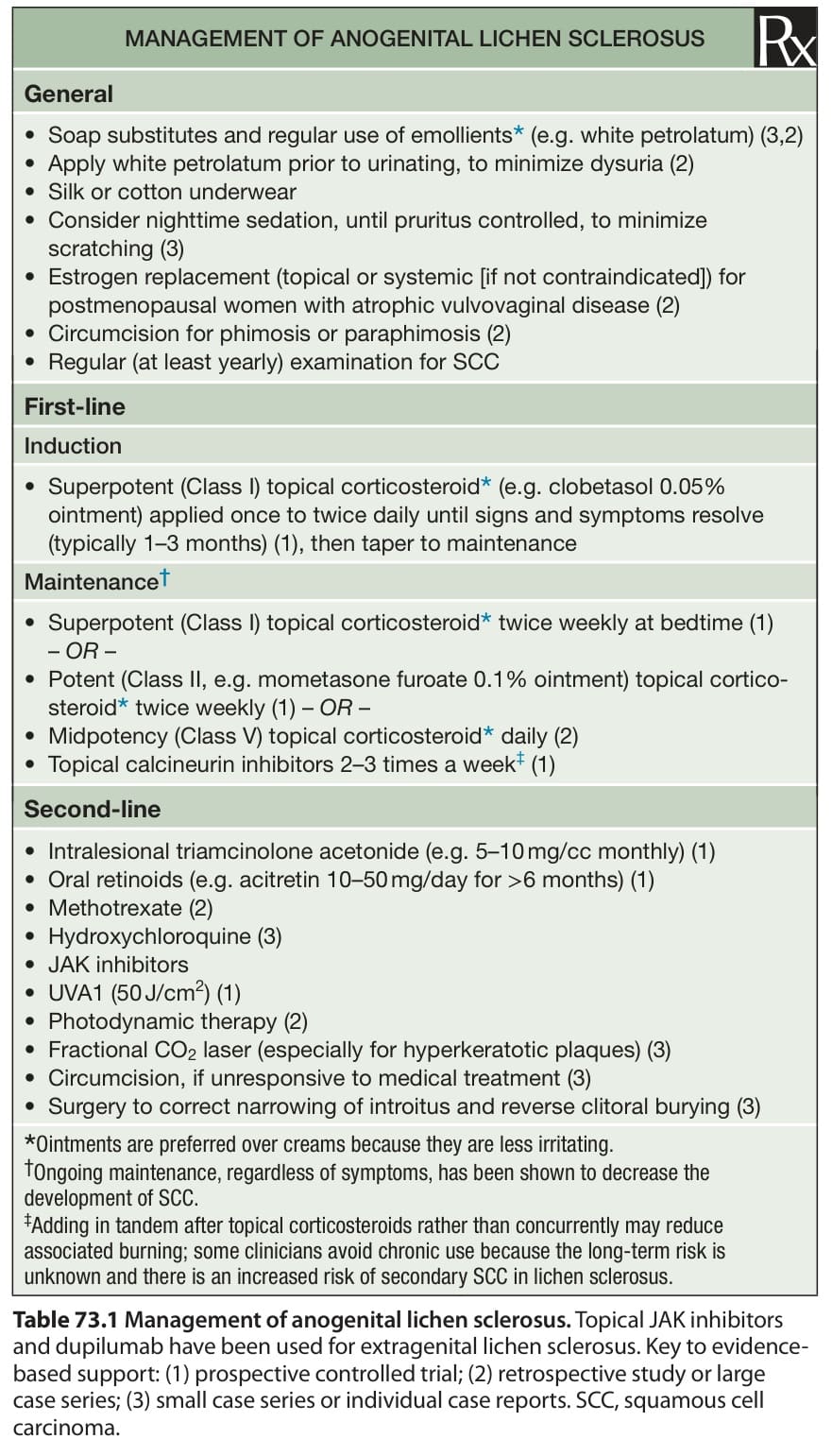
Table 73.1 Management of anogenital lichen sclerosus. Topical JAK inhibitors and dupilumab have been used for extragenital lichen sclerosus. Key to evidencebased support: (1) prospective controlled trial; (2) retrospective study or large case series; (3) small case series or individual case reports. SCC, squamous cell carcinoma.