LICHEN PLANUS
Synonyms: Vulvovaginal gingival syndrome/vulvo-vaginal-gingival syndrome (erosive lichen planus in women) Peno-gingival syndrome in men Mucosal lichen planus
Key features
Four distinct forms of genital lichen planus are observed: (1)
papules or plaques in the setting of generalized cutaneous lichen planus; (2) erosive disease; (3) hypertrophic disease; and (4) lichen planopilaris
Although cutaneous disease is often self-limiting, mucous membrane disease is more persistent
The classic findings in cutaneous disease are violaceous, flat-topped papules and plaques; lacy white streaks typically occur on the genitalia as well as the oral mucosa
Erosive lichen planus often involves multiple mucous membrane sites – in particular, the vulva, vagina, and oral mucosa
Introduction
Approximately 50% of women and 25% of men with cutaneous lichen planus have genital involvement. The classic violaceous papules and plaques typically affect the labia minora and majora or the mons pubis in women and the glans or shaft of the penis in men. Erosive lichen planus is a distinct subtype of the disease, characterized by severe, scarring erosive disease of the vestibule, introitus, vagina, and oral cavity. Hypertrophic genital lichen planus and lichen planopilaris are the least frequent forms.
Epidemiology
Lichen planus is more common in women, generally presenting in the sixth decade. The exact prevalence is unknown, but it is likely that some cases of genital involvement in cutaneous lichen planus may be missed.
Etiology
Lichen planus probably represents T cell-mediated damage to epidermal cells that express altered self or foreign antigens on their surface, but the exact etiology is unknown (see Ch. 11). There is an autoimmune phenotype (see Lichen sclerosus above) in female patients with vulvar lichen planus and an association with autoimmune disease. While some cases of lichen planus may be drug-induced, there are no definite associations with genital disease. Infectious triggers, particularly hepatitis C viral infection, may be associated with mucosal lichen planus in certain parts of the world such as the Mediterranean basin and Japan.
Clinical features
There are four distinct presentations of genital lichen planus, as follows:
●In classic lichen planus, the genital lesions are similar to those in other cutaneous sites, but lacy reticulation may be very prominent. Genital involvement is usually limited to the mons pubis and the labia majora and minora in women or the glans penis and shaft in men (see Fig. 11.4B). Penile plaques are frequently annular (Fig. 73.6A). Scarring is not a feature of classic lichen planus. Severe cutaneous pruritus is the usual presenting symptom, but genital lesions may be asymptomatic. Lesions are usually self-limited.
●Erosive lichen planus occurs much more frequently in women than in men. Pain and dyspareunia are the most common complaints. In female patients, extensive erosions occur around the vaginal orifice, edged by lacy white reticulations; similar lesions may also occur on the labia (Fig. 73.6B). Erosive lichen planus frequently leads to scarring, resulting in distortion of the vulvar architecture. In severe cases, there may be complete obliteration of the vaginal canal and urinary retention. Vaginal involvement can present with a discharge, erythema and contact bleeding of the vaginal mucosa, and/or scarring with adhesions. Oral involvement is frequently observed, usually in the form of a desquamative gingivitis, but lacy
white reticulations and erosions may also occur. Erosions limited to the perianal area alone have been described.
●In the hypertrophic form of lichen planus, hyperkeratotic white plaques on the vulva or shaft of the penis may be seen.
●In lichen planopilaris, follicular keratotic papules are limited to the hair-bearing labia majora and the mons pubis, but may also be seen on the scalp and trunk and in the axillae.
Pathology
The typical histologic findings are irregular epidermal acanthosis, accentuation of the granular layer, liquefactive degeneration of the basal layer, and a band-like dermal infiltrate of lymphocytes. It is more difficult to interpret biopsy specimens of mucous membranes, where these findings are more variable. Because there is loss of the epithelium in erosive disease, the specimen should include the edge of the erosion as well as the surrounding lacy reticulated skin. Despite this, it is sometimes difficult to unequivocally confirm the disease, but histologic evaluation can aid in excluding other disorders in the differential diagnosis.
Differential diagnosis
The principal entities in the differential diagnosis are lichen sclerosus; autoimmune bullous disease, including mucous membrane pemphigoid; Zoon (plasma cell) vulvitis; Behçet disease; graft-versus-host disease; and high-grade squamous intraepithelial lesion (HSIL; previously referred to as undifferentiated vulvar intraepithelial neoplasia) or SCC. Direct immunofluorescence may be necessary to exclude an autoimmune bullous disease. Orogenital ulceration raises the possibility of Behçet disease (see Ch. 26). For oral lesions, lupus erythematosus is in the differential diagnosis, but this would be unusual in the anogenital region. Early lichen planus may be confused with vulvar eczema and there may be superimposed lichen simplex chronicus. For hypertrophic disease, HSIL, SCC, and lichen simplex chronicus are the major entities in the differential diagnosis.
Treatment
The treatment of genital lichen planus is outlined in Table 73.2. Potent or moderately potent corticosteroid ointments will control most symptoms of classic genital lichen planus, as the disease is usually self-limiting. Hypertrophic disease may require intralesional corticosteroids.
The management of erosive lichen planus is more difficult, as the disease is often more persistent. The majority of patients will respond to a 3-month course of topical clobetasol propionate 0.05% ointment (as described for lichen sclerosus) and thereafter can be maintained on moderately potent corticosteroids or twice weekly application of the potent corticosteroid. A combination of a moderately potent corticosteroid with a topical antifungal cream (e.g. econazole) may be as effective as more potent corticosteroids, particularly for maintenance therapy. For refractory disease, anecdotal case reports describe benefit from intralesional corticosteroid injections, topical calcineurin inhibitors, oral hydroxychloroquine, systemic immunosuppressants, and acitretin (see Table 73.2). Tricyclic antidepressants can be used to treat associated vulvodynia. Erosive vaginal disease requires
intravaginal corticosteroids and regular use of vaginal dilators to prevent stenosis; surgery to lyse adhesions may be required.
Long-term evaluation of these patients is advised because of the risk of developing SCC; the incidence of the latter has been estimated to be ~2.4%. For further details on the treatment of lichen planus, including systemic agents for severe, refractory disease, see Chapter 11.
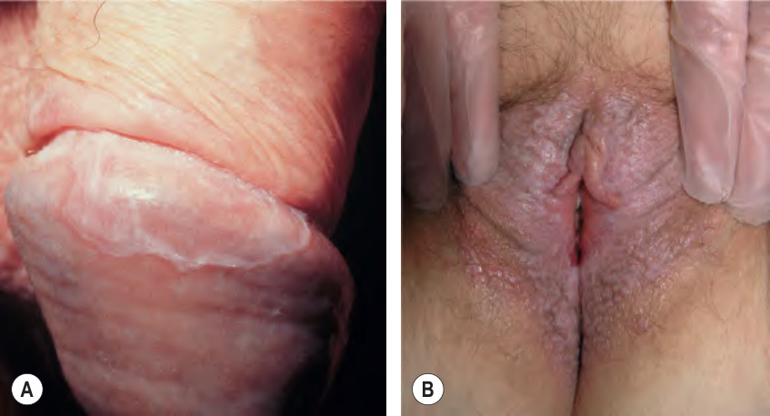
Fig. 73.6 Lichen planus.A An annular band is seen on the glans of the penis. This is a typical finding. B Erosive lichen planus of the vulva with fissures and an extensive white lacy pattern. A, Courtesy R. Turner, MD.
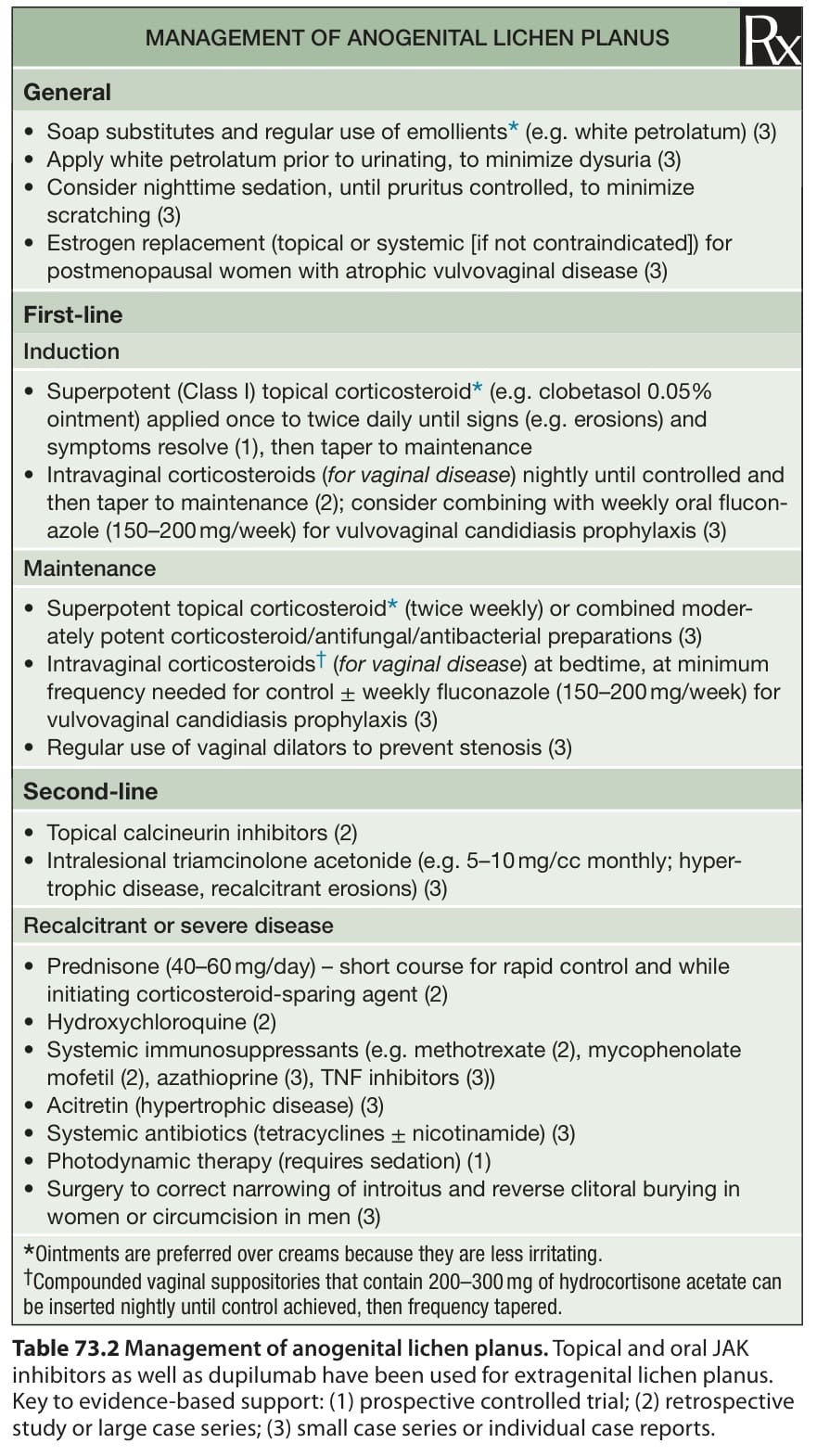
Table 73.2 Management of anogenital lichen planus. Topical and oral JAK inhibitors as well as dupilumab have been used for extragenital lichen planus. Key to evidence-based support: (1) prospective controlled trial; (2) retrospective study or large case series; (3) small case series or individual case reports.