ZOON BALANITIS/VULVITIS
Synonyms: Plasma cell balanitis/vulvitis Vulvitis/balanitis circumscripta plasmacellularis Zoon’s erythroplasia
Key features
Male predominance
Erythematous, moist, speckled, discrete thin plaques
Involvement of adjacent surfaces produces “kissing” lesions in men
In men, cured by circumcision
Introduction
While most authorities believe this condition occurs in men, its existence as a separate entity in women has been questioned. This is because many of the clinical features in women are indistinguishable from those of erosive lichen planus, mucous membrane pemphigoid, or lupus erythematosus, and the lichenoid features histologically overlap with erosive lichen planus and lichen sclerosus.
Epidemiology
Zoon balanitis/vulvitis may occur at any age but is usually seen from the third decade onwards in uncircumcised men and in postmenopausal women.
Etiology
The etiology is unknown, but poor penile hygiene or chronic irritation from warmth or rubbing has been suggested. Zoon balanitis has not been observed in circumcised men. In women, the vulvitis may represent a reactive process secondary to trauma or reflect a postmenopausal hypoestrogenic state.
Clinical features
In men, the lesions on the glans penis may be asymptomatic or give rise to pruritus or dysuria. The lesions are erythematous, discrete, moist thin plaques with a “cayenne pepper” speckled appearance and a deep red to reddish-brown hue. “Kissing” lesions with involvement of adjacent, touching areas, e.g. around the urethral meatus, are a characteristic finding (Fig. 73.7A). Shallow erosions may resolve slowly, leaving a rusty color. In women, thin erythematous plaques similar to those in men may be seen anywhere on the vulva (Fig. 73.7B). Lesions may be asymptomatic or cause burning, dyspareunia, dysuria, pruritus, or pain.
Pathology
The typical histologic picture is a dense lichenoid infiltrate in the upper and mid dermis with a predominance of plasma cells. Additional features include “lozenge-shaped” keratinocytes, vascular proliferation, hemosiderin deposition, and erythrocyte extravasation.
Differential diagnosis
In order to establish the diagnosis, a biopsy is usually performed. The major disorder in the differential diagnosis is erosive lichen planus, but the possibility of cutaneous malignancy (e.g. SCC in situ, extra mammary Paget disease) must be considered.
Treatment
In men, a circumcision will almost always cure the disease. Potent topical corticosteroids or topical calcineurin inhibitors may provide some symptomatic relief. Vaginal estrogen may be helpful for some women.
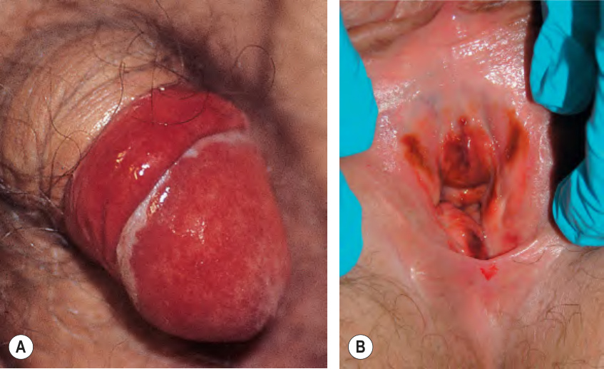
Fig. 73.7 Zoon balanitis and vulvitis.A There are moist red “kissing” lesions on adjacent surfaces of the glans and prepuce. B Light to dark red patches of the clitoris, vestibule, introitus, and labia.