EROSIVE GENITAL DISEASE
Erosions and ulcerations are seen in a wide variety of conditions (Table 73.6). Genital aphthae and HSV infections are common. Erosions may occur after scratching or rubbing or as a result of blister formation. To determine the cause of the erosions, it is essential to take a careful history and perform a full examination of the skin, including other mucosal surfaces. For example, a white lacy pattern on the buccal mucosa may suggest a diagnosis of lichen planus. Further investigations will be determined by the clinical appearance, but swabs for bacterial and fungal culture are often necessary. Biopsies of lesional skin for light microscopy and perilesional skin for direct immunofluorescence are indicated if an autoimmune blistering disorder is suspected.
Primarily in adolescent girls, reactive non-sexually related acute genital ulcers, sometimes referred to as Lipschutz ulcers, may be seen (Fig. 73.18). These ulcers can be large and deep and are triggered by a variety of viral or bacterial infections, including Epstein–Barr virus, Mycoplasma, cytomegalovirus, influenza, and streptococci. However, a cause is not always identified.
Acquired Autoimmune Bullous Diseases
Immunofluorescence (direct and indirect) is an essential investigation in suspected autoimmune bullous diseases as it may be very difficult to distinguish between the different blistering disorders on clinical grounds alone (Table 73.7). The most common autoimmune bullous diseases seen in the genital region are bullous pemphigoid, including the childhood variant, and mucous membrane pemphigoid. Unless blisters are seen or described, anogenital mucous membrane pemphigoid is often indistinguishable from erosive lichen planus; thus, histology and immunofluorescence are essential (Fig. 73.19). The extent and severity of the disease determine the treatment, which usually consists of a combination of topical and systemic anti-inflammatory and immunosuppressive agents. Great symptomatic relief can be gained from the use of potent topical corticosteroids, either as sole treatment in localized disease or as an adjunct to systemic treatment in widespread or more severe disease.
Acquired Inflammatory Bullous Diseases
Erythema multiforme and Stevens–Johnson syndrome
Erythema multiforme (EM, primarily the major form) and Stevens– Johnson syndrome (SJS) may present as a bullous eruption in the genital region. In recurrent EM, HSV infection should be sought, remembering that there may be a delay of up to 2 weeks from infection to appearance of the lesions. In recurrent cases, a trial of prophylactic oral anti-HSV therapy may be indicated. Confluent dusky macular lesions point to SJS, in which drugs are the most common etiology. Prominent mucosal involvement also occurs in Mycoplasma-induced rash and mucositis (MIRM)/reactive infectious mucocutaneous eruption (RIME); see Ch. 20).
Fixed drug eruption
Recurrent erythematous or eroded plaques may occur on both skin and mucous membranes (see Ch. 21). Often, cutaneous lesions are strikingly round, but this may not hold true in mucous membrane sites. The lesions recur in exactly the same site with each exposure to the drug. Bullous genital lesions frequently present as erosions. Common culprits are acetaminophen (paracetamol), tetracyclines, trimethoprim–sulfamethoxazole, and nonsteroidal anti-inflammatory drugs.
Inherited Bullous Disease
Hailey–Hailey disease
Patients with autosomal dominant Hailey–Hailey disease have recurrent involvement of flexural areas (see Ch. 59). Expanding erythematous plaques with superimposed erosions and sometimes vesicles and pustules are seen. In the inguinal creases, there is a particular tendency to form hypertrophic plaques. Secondary infection with bacteria, fungi, and HSV is a frequent problem and must be excluded in any exacerbation; for recurrent disease, suppressive therapy may be considered. Topical corticosteroids in combination with antifungal/antibacterial agents can ease symptoms. Low-dose naltrexone or CO laser ablation may also prove helpful.
Additional figures and table on the 2015 International Society for the Study of Vulvovaginal Disease (ISSVD) terminology of vulvar squamous intraepithelial lesions, available in our eBook (see inside front cover for access code).
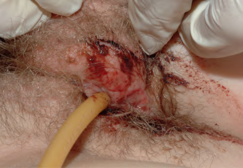
Fig. 73.18 Reactive non-sexually related acute genital ulcers associated with primary EBV infection. Extreme pain with urination required placement of a Foley catheter in this 13-year-old girl. EBV-related genital ulcers are often misdiagnosed as a genital herpes simplex viral infection. Courtesy Julie V. Schaffer, MD.
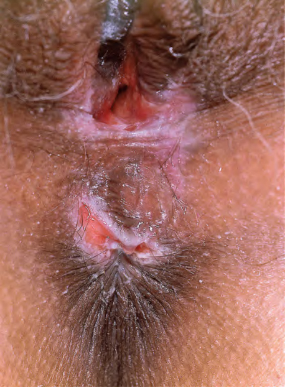
Fig. 73.19 Mucous membrane pemphigoid of the vulva. Ulcerations and scarring of the perianal area and perineum (positive direct immunofluorescence distinguished this from erosive lichen planus).
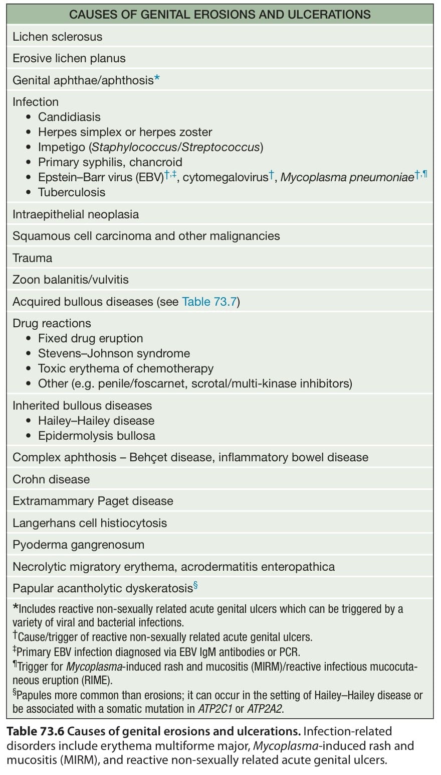
Table 73.6 Causes of genital erosions and ulcerations. Infection-related disorders include erythema multiforme major, Mycoplasma-induced rash and mucositis (MIRM), and reactive non-sexually related acute genital ulcers.
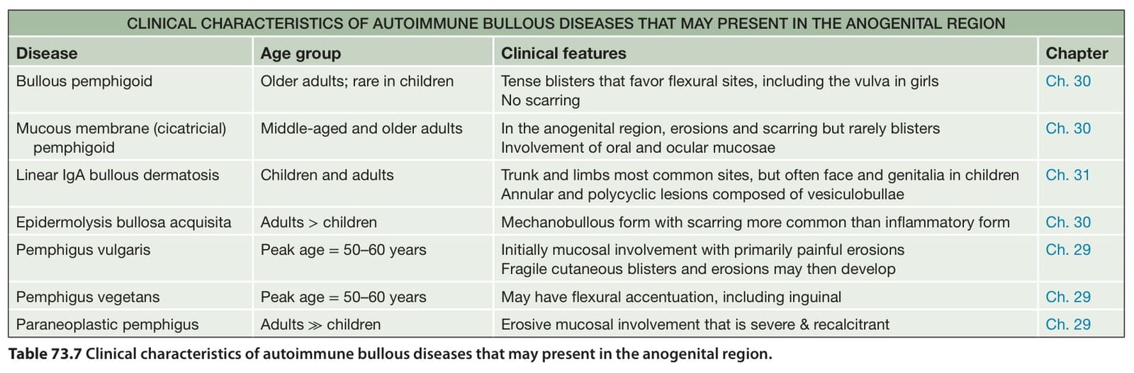
Table 73.7 Clinical characteristics of autoimmune bullous diseases that may present in the anogenital region.