OTHER GRAM-POSITIVE SKIN INFECTIONS
Cutaneous Anthrax
Introduction and epidemiology
The name Bacillus anthracis is derived from the Greek word for coal, anthrakis, reflecting the black, coal-like eschars. B. anthracis is an aerobic, sporulating, Gram-positive rod measuring 1–1.5 × 3–5 microns. The bacterium causes disease in humans via three routes: inhalation, ingestion, and cutaneous inoculation. While the latter produces the least severe form of anthrax, it accounts for ~95% of infections. Anthrax is primarily a disease of animals (e.g. sheep, cows, horses, goats) that is endemic in parts of Africa and the Middle East. Most human cases are due to occupational exposure to infected animals or their carcasses, including hides and wool (“woolsorter’s disease”). Two thousand cases are reported annually worldwide, with <10 per year in the US.
History
The fifth and sixth plagues of Egypt that are described in the book of Exodus are thought to have been anthrax. B. anthracis was the first bacterium proven to be the etiologic agent of a specific disease, independently demonstrated by Dr. Robert Koch and Dr. Louis Pasteur. Anthrax was first used as a biologic weapon during World War I. Accidental release of anthrax from a biologic weapons complex in the Soviet Union in 1979 resulted in 68 fatalities. In 2001, anthrax spores were sent through the US mail as a terrorist weapon, resulting in 22 cases of inhalational or cutaneous anthrax and 5 fatalities.
Pathogenesis
Anthrax spores are environmentally hardy and can survive for decades in the soil. Full virulence requires the presence of an antiphagocytic capsule and three toxin components (protective antigen, lethal factor, and edema factor) that combine to form both lethal toxin and edema toxin. Edema toxin impairs neutrophil function and affects water homeostasis, leading to edema; lethal toxin causes release of TNF and IL-1β.
Clinical features
The clinical features of cutaneous anthrax are outlined in Table 74.12. Additional key points include the following:
●The lack of pain despite a necrotic eschar helps to differentiate cutaneous anthrax from the bite of a brown recluse spider.
●A pustular primary lesion is unlikely to be cutaneous anthrax, although “cloudy” vesicles may form a “pearly wreath” around the eschar. The term “malignant pustule”, which is often attributed to anthrax, is therefore a misnomer.
●Vesicles may discharge serosanguineous fluid containing numerous organisms, which are evident on Gram stain.
●Lymphangitis and painful lymphadenopathy with systemic symptoms (fever, malaise, headache) occur rarely.
●Antibiotic therapy does not alter the progression from vesicle to ulcer to eschar, as the process is toxin-mediated.
●Multiple lesions, when present, tend to cluster in the same area of the body.
●Palpebral anthrax manifests with eyelid swelling and necrosis, often resulting in cicatricial ectropion.
Pathology
Spongiosis, papillary dermal edema, and a superficial and deep inflammatory infiltrate with abundant neutrophils are observed. Substantial hemorrhage is often evident, and the inflammatory infiltrate may involve nerves. Extensive ulceration and coagulative necrosis can be seen in older lesions. Elongated Gram-positive bacilli are present, often in small clusters. Spores grow readily on all routine culture media at 37°C, with a “jointed bamboo-rod” cellular appearance and a unique “curled-hair” colonial morphology.
Diagnosis and differential diagnosis
Gram staining of exudate or touch preparations can aid in rapid diagnosis. The organism grows in culture within 6–24 hours; however, antibiotic administration for more than 24 hours prevents the recovery of pathogens in culture.
Laboratory personnel must be alerted to the possibility of anthrax to ensure that the Bacillus isolate is not assumed to be B. cereus and that further speciation is performed. Definitive diagnosis of anthrax requires specific testing such as detection of lethal factor, PCR-based analysis,
and immunohistochemical staining. Serologic assays can become positive as early as 10 days after onset of symptoms, with peak titers at 40 days.
The differential diagnosis includes cutaneous infection with Bacillus megaterium, a brown recluse spider bite, the eschar of several rickettsial diseases, cellulitis, ecthyma gangrenosum, orf, milker’s nodule, opportunistic bacterial and fungal infections, and entities in the ulceroglandular category (e.g. tularemia; see below).
Treatment
Without antibiotic treatment, mortality from cutaneous anthrax may be as high as 20% (usually from septicemia), but that number falls to almost zero with proper antimicrobial therapy. Although antibiotics do not alter the evolution of the cutaneous lesions (which are toxin-mediated), the likelihood of systemic illness is reduced. When cutaneous anthrax is associated with a concomitant inhalational exposure, the Centers for Disease Control and Prevention (CDC) recommend antibiotic therapy for at least 60 days, compared to a 7- to 10-day course for a purely cutaneous exposure to an animal-based source. Quinolones and doxycycline are first-line agents, with clindamycin and (if susceptible) penicillin or amoxicillin as alternatives.
An anthrax vaccine, consisting of an inactivated cell-free filtrate of a non-encapsulated, attenuated strain of B. anthracis, is available. The recommended five-dose pre-exposure series includes administration at 0, 1, 6, 12, and 18 months, followed by yearly boosters. For post-exposure prophylaxis, three vaccine doses can be given at 0, 2, and 4 weeks together with antimicrobial treatment. Raxibacumab and obiltoxaximab are recombinant human immunoglobulin monoclonal antibodies targeting the B. anthracis protective antigen. Either of these antibodies or polyclonal anthrax immune globulin may be administered as a single intravenous infusion (in conjunction with antibiotics) for the treatment and post-exposure prophylaxis of inhalational anthrax.
Bacillus cereus Infection
Bacillus cereus is a large Gram-positive rod best known for causing food poisoning. However, it can produce primary cutaneous disease in immunocompetent and immunosuppressed patients as well as pneumonia, meningitis, and necrotizing fasciitis in immunocompromised individuals. An outbreak of skin infections in neonates due to contamination of equipment used during delivery has been reported. Skin infection typically presents as a single necrotic bulla, which may occur in association with high fevers and negative blood cultures in neutropenic individuals. Vancomycin is considered the drug of choice, but quinolones and imipenem may also be used.
Erysipeloid
Synonyms: Diamond skin disease Erysipeloid of Rosenbach (localized cutaneous form) Pseudoerysipelas
Erysipeloid is an acute cutaneous infection caused by Erysipelothrix rhusiopathiae (formerly insidiosa), a Gram-positive, non-motile, smooth or curved bacillus. It is caused by traumatic inoculation of the organism into the skin, which most often occurs in fishermen or persons who prepare meat, poultry, or fish. It has also been reported following animal bites. There are two types of erysipeloid, localized and generalized, and they usually occur within 1 week of inoculation. The localized form is characterized by an erythematous to violaceous area of non-suppurative cellulitis (Fig. 74.24), which can be pruritic or painful. Hemorrhagic vesicles may develop. Although the classic finding is involvement of the finger webs with sparing of the terminal phalanges, other sites may be affected. Constitutional symptoms are unusual in localized erysipeloid, which is typically a self-limited condition. However, the generalized form presents with fever and arthralgias in addition to widespread cutaneous lesions. The latter range from perifollicular papules to erythematous plaques to macular purpura and necrosis. Complications can include bacteremia, endocarditis, septic arthritis, and cerebral and other visceral abscesses. The organism is often difficult to culture, and PCR-based assays can help to establish the diagnosis. Erysipelas, cellulitis, spider bites, and fixed drug eruptions may simulate localized erysipeloid (see
Table 74.10). Most patients with untreated localized disease recover spontaneously. Penicillin is the drug of choice; erythromycin, cephalosporins, clindamycin, linezolid, and quinolones are alternatives. The best means of prevention is using gloves when preparing fish or meat.
Listeriosis
Listeria monocytogenes is a motile Gram-positive bacillus that represents a common veterinary (e.g. cattle, sheep) and zoonotic pathogen. Infection is usually acquired by eating contaminated food (e.g. soft cheeses, ice cream). Listeriosis primarily affects the elderly, pregnant women, and immunocompromised individuals. Manifestations include meningitis and gastroenteritis accompanied by fever, myalgias, and bacteremia. Pregnant women are at risk of miscarriage, stillbirth, and premature delivery. Neonatal septicemia and meningitis due to vertical transmission can occur and may be associated with disseminated vesiculopustules, purpura, or granulomatous papules and nodules (“granulomatosis infantiseptica”). Primary cutaneous listeriosis can present as grouped papulopustules, an abscess, or cellulitis. Transmission may
occur via skin exposure to bovine products of conception in veterinarians and farmers, but this has decreased in recent years due to safer occupational practices. In otherwise healthy individuals, primary cutaneous listeriosis may be self-limited. The first-line treatment for listeriosis is ampicillin, and TMP-SMX represents an alternative; L. monocytogenes is resistant to cephalosporins.
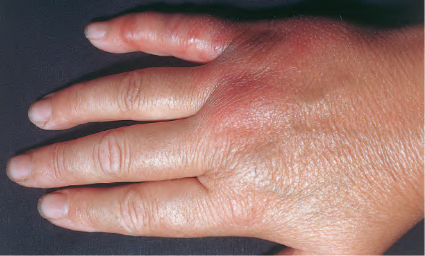
Fig. 74.24 Erysipeloid. Erythema and edema with vesicle formation on the hand and fifth digit.
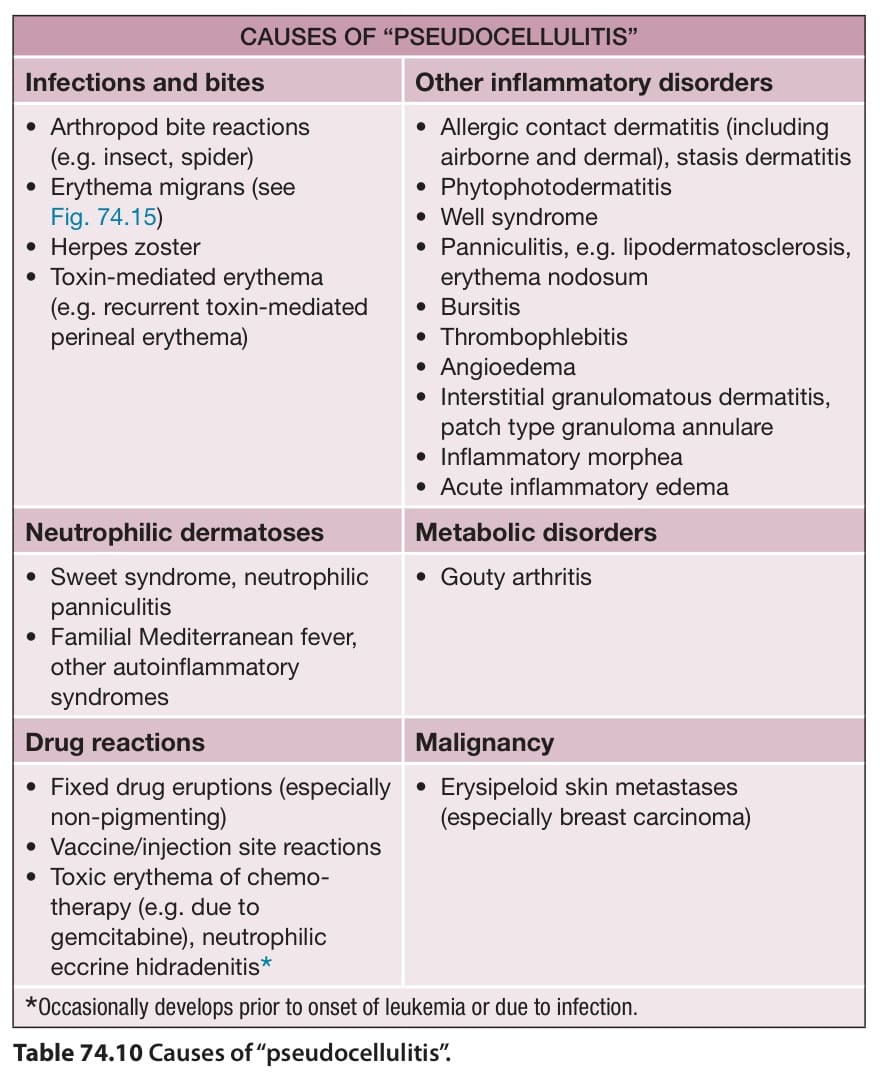
Table 74.10 Causes of “pseudocellulitis”.
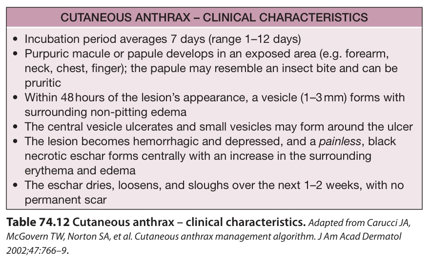
Table 74.12 Cutaneous anthrax – clinical characteristics.Adapted from Carucci JA, McGovern TW, Norton SA, et al. Cutaneous anthrax management algorithm. J Am Acad Dermatol 2002;47:766–9.