NEISSERIA MENINGITIDIS
Acute and Chronic Meningococcemia
Introduction
Meningococcemia is characterized by fever and a petechial eruption, and it has acute and chronic forms. The causative organism, Neisseria meningitidis, is an aerobic Gram-negative diplococcus.
Epidemiology and pathogenesis
Meningococcal infections occur worldwide and affect primarily infants (6 months to 1 year), adolescents, and young adults. There is a male predominance, with a male : female ratio of 3–4 : 1. Most cases develop in the winter and spring. Risk factors include HIV infection and asplenia; deficiencies in complement component C3 (which may be associated with partial lipodystrophy), terminal complement components (C5–C9), properdin, factor D or H, and immunoglobulins (see Ch. 60); and treatment with terminal complement inhibitors (e.g. eculizumab, ravulizumab) and potentially TNF inhibitors. In addition, there is evidence that patients with low TNF production, high IL-10 or plasminogen activator inhibitor 1 production, or homozygosity of certain alleles at the IL-1 locus are at increased risk of a fatal outcome.
Thirteen clinically significant N. meningitidis serogroups are distinguished by their capsular polysaccharide virulence factors. Serogroups A, B, C, X, Y, and W-135 are most often implicated in human disease. Proliferation of N. meningitidis in contact with host cells increases the production of a bacterial transferase that adds phosphoglycerol onto its type IV pili; this modification results in bacterial detachment and
migration across the epithelium, a prerequisite for dissemination and invasive disease. The bacteria elaborate an endotoxin that triggers inflammatory processes leading to shock, multi-organ failure, and purpura fulminans.
The only known reservoir is the human nasopharynx. Rates of asymptomatic carriage in the US range from 5% to 15%. Transmission is by the respiratory route, with an incubation period of 2–10 days. Active or passive exposure to tobacco smoke, as well as concurrent viral infection of the upper respiratory tract, disrupts the integrity of the respiratory mucosa, thereby increasing the risk of invasive meningococcal disease.
Clinical features
Infection with N. meningitidis most often results in an asymptomatic carrier state, which leads to lifelong immunity against that subgroup. Upper respiratory symptoms as well as bacteremia without sepsis may also occur. When acute meningococcemia develops, one-third to one-half of patients present with a petechial eruption, typically in association with a fever, chills, myalgias, and headache. Retiform purpura and ischemic necrosis may follow (Fig. 74.25). Bullous hemorrhagic lesions occasionally arise, most commonly on the trunk and lower extremities. In a minority of patients, only a transient, blanchable morbilliform eruption is seen. Hypotension, meningitis, meningoencephalitis, pneumonia, arthritis, pericarditis, myocarditis, and progression to disseminated intravascular coagulation (DIC) can develop.
Chronic meningococcemia comprises less than 5% of invasive meningococcal disease cases and is defined by blood cultures positive for N. meningitidis and symptom duration >7 days, without either meningitis or shock. Typical symptoms include recurrent fevers, migratory arthralgias, and the development of polymorphous erythematous macules and papules 12–24 hours after the onset of fevers. The skin lesions may develop into tender nodules with indistinct borders. Petechiae, retiform purpura, pustules, and erythema multiforme-like lesions have also been described. The fever and rash of chronic meningococcemia tend to resolve concurrently and then recur within 2–10 days.
Pathology
Biopsy specimens from patients with acute meningococcemia show leukocytoclastic vasculitis and thrombosis; organisms can be visualized with Gram stain in 70% of cases. In chronic meningococcemia, a perivascular infiltrate composed of lymphocytes and a few neutrophils can be appreciated; leukocytoclastic vasculitis may be seen in petechial lesions, and Gram or silver staining is often negative.
Diagnosis and differential diagnosis
The initial diagnosis is often clinical. Time is of the essence in diagnosing patients with meningococcemia and initiating treatment, as rapid decompensation occurs in acute infections. Culture of N. meningitidis from the skin or a normally sterile body fluid (e.g. blood, cerebrospinal fluid [CSF], synovial fluid) can establish the diagnosis; however, the sensitivity may be low, especially after administration of antibiotics. Latex agglutination tests that detect group A, B, C, Y, and W-135 antigens in the CSF and urine have good specificity but low sensitivity.
PCR-based assays are also specific and have better sensitivity, which may be higher in skin biopsy specimens than blood or CSF samples, but bacterial culture is still necessary for susceptibility testing.
Acute meningococcemia must be differentiated from enteroviral infections (which can present with meningitis and a petechial exanthem), Rocky Mountain spotted fever, septic vasculitis due to acute bacteremia and endocarditis, TSS, purpura fulminans, leptospirosis, and various non-infectious vasculitides. The differential diagnosis of chronic meningococcemia includes bacterial endocarditis, Sweet syndrome, Henoch–Schönlein purpura, rat-bite fever, erythema multiforme, and chronic gonococcemia. The rash and arthralgias associated with chronic meningococcemia must not be confused with inflammatory diseases, as administration of corticosteroids or other immunosuppressive medications could provoke progression of the infection.
Treatment
While early institution of appropriate therapy for acute meningococcemia is essential, if possible, blood and CSF samples should be obtained prior to antibiotic administration. A third-generation cephalosporin is the recommended treatment for suspected (e.g. Gram stain with Gram-negative diplococci) or culture-proven acute meningococcemia prior to obtaining susceptibility results. Chloramphenicol and quinolones are alternatives in patients with a history of immediate-type hypersensitivity to penicillins, although quinolone-resistant strains have been reported. Chronic meningococcemia is treated with similar antibiotics. All close contacts should receive prophylactic treatment with ciprofloxacin, rifampin, or ceftriaxone. Experimental therapies that have been tried in small numbers of patients include activated protein C, anti-endotoxin monoclonal antibodies, tissue plasminogen activator, antithrombin III infusion, topical nitroglycerin, plasmapheresis, and extracorporeal membrane oxygenation. To date, these interventions have not proven to be effective in decreasing mortality.
Several meningococcal vaccines are approved for use in the US: three quadrivalent vaccines effective against serogroups A, C, W, and Y; a bivalent vaccine effective against serogroups C and Y plus H. influenzae type b; and two monovalent vaccines effective against serogroup B. The duration of immunity for these vaccines is ~3–5 years. A quadrivalent conjugated vaccine is routinely given to children at age 11–12 years with a booster at age 16 years; this vaccine is also administered to other individuals at high risk for infection, such as college students living in dormitories and military recruits, as well as patients ≥2 months of age at increased risk of severe meningococcal infection due to conditions such as complement deficiencies and asplenia. The monovalent serogroup B vaccine is recommended for patients ≥10 years of age in the latter high-risk group; in addition, it is offered to teens, preferably at age 16–18 years.
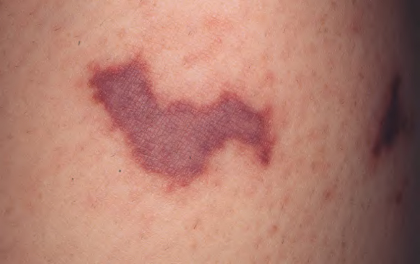
Fig. 74.25 Acute meningococcemia. Purpura with irregular outline and central gunmetal gray color. Courtesy Kalman Watsky, MD.
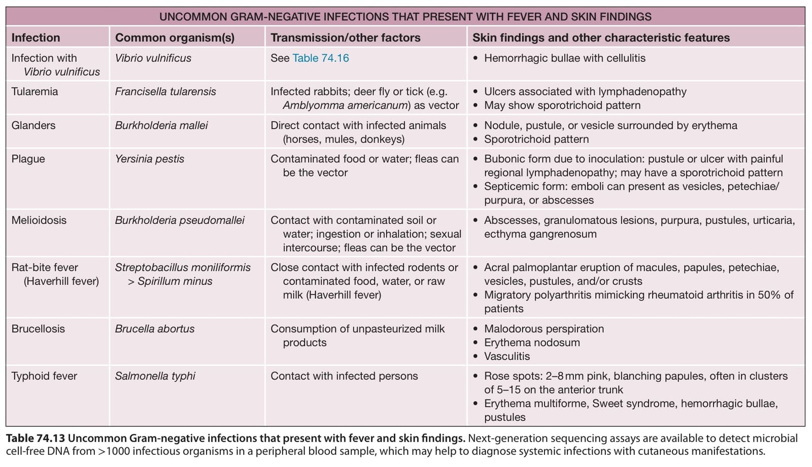
Table 74.13 Uncommon Gram-negative infections that present with fever and skin findings. Next-generation sequencing assays are available to detect microbial cell-free DNA from >1000 infectious organisms in a peripheral blood sample, which may help to diagnose systemic infections with cutaneous manifestations.