PSEUDOMONAS AERUGINOSA
Pseudomonas aeruginosa is a ubiquitous Gram-negative, strictly aerobic, motile bacillus. It is widely distributed in the soil and on plants, growing especially well in aqueous environments. This organism has relatively low virulence and usually requires a local anatomic or immunologic defect to cause disease. Primary cutaneous infection with P. aeruginosa can occur in otherwise healthy individuals in areas exposed to high levels of moisture in conjunction with skin barrier disruption; these infections have a very good prognosis. In contrast, cutaneous manifestations of Pseudomonas sepsis in immunocompromised individuals are associated with a grave prognosis.
Green Nail Syndrome
In green nail syndrome (chloronychia), green–blue to green–black discoloration of the nail develops due to pyocyanin, a blue–green pigment produced by P. aeruginosa (Fig. 74.26). There is typically associated distal onycholysis and sometimes proximal chronic paronychia. Predisposing factors include frequent or prolonged exposure to water, excessive use of detergents and soaps, nail trauma, onychomycosis, and other causes of onycholysis (see Ch. 71). The diagnosis of green nail syndrome is usually clinical; if necessary, it can be confirmed by Gram stain and culture of exudate and nail fragments. The differential diagnosis includes a subungual hematoma, melanocytic nevus, melanoma, and Aspergillus infection. Treatment involves avoidance of
predisposing factors, clipping the nail, and topical application of 2% sodium hypochlorite (household bleach diluted 1 : 4; see Ch. 71), a quinolone, or aminoglycoside (e.g. tobramycin) solution for 1–4 months. Treatment of coexistent onychomycosis may help prevent recurrence. In refractory cases, removal of the affected nail may be required.
Pseudomonal Pyoderma and Blastomycosis-Like Pyoderma
Pseudomonal pyoderma is a superficial infection of the skin with P. aeruginosa. Features include a blue–green purulence, a “grape juice-like” or “mousy” odor, and a moth-eaten appearance of the skin. The borders are often macerated and eroded (Fig. 74.27). Pseudomonal pyoderma can complicate burns, decubitus ulcers, and other chronic cutaneous ulcers. Pseudomonal pyoderma often plays a role in Gram-negative toe web infections and in “infectious eczematoid dermatitis” on the hands and feet or in the anogenital region.
Blastomycosis-like pyoderma is a rare reactive condition that occurs in the setting of a long-standing localized infection, presenting as large verrucous plaques with multiple pustules and elevated borders. Histologic examination reveals pseudoepitheliomatous hyperplasia with intraepidermal abscess formation; fungi are not seen, and fungal cultures are negative. This condition usually occurs in immunocompromised patients, and other bacteria (e.g. S. aureus) have also been implicated.
Treatment of these conditions includes systemic antipseudomonal antibiotics, topical antimicrobial and drying agents, and debridement of the hyperkeratotic rim in web space infections. Additional treatment options for blastomycosis-like pyoderma include acitretin, saturated solution of potassium iodide, surgical excision, electrodesiccation and curettage, and ablative laser therapy.
Otitis Externa and Necrotizing (Malignant) Otitis Externa
P. aeruginosa colonizes 1%–2% of normal ear canals. However, pseudomonal infection can result in otitis externa (“swimmer’s ear”). Predisposing factors include eczema and psoriasis of the ear, swimming, mechanical trauma, hearing-aid use, and ear irrigation. In this condition, the external auditory canal is swollen and macerated with a green, purulent discharge. The tympanic membrane is uninvolved, and manipulation of the pinna results in extreme pain. An exudative dermatitis of the external ear and retroauricular area may also be present. Treatment includes removal of canal debris, antibiotic eardrops (preferably those that cover S. aureus co-infection), and possibly the instillation of a cotton wick. Oral therapy is only occasionally required, but recurrences can occur.
Necrotizing (malignant) otitis externa refers to infection that progresses to affect the temporal bone and adjacent structures. It is seen most often in elderly diabetic patients and those with HIV infection or other immunodeficiencies. Manifestations include severe pain, persistent drainage, and granulation tissue within the ear canal. Local lymphadenopathy and parotid swelling may be present. Deeper invasion can result in osteomyelitis of the skull, nerve palsies, mastoiditis, sepsis, and sigmoid sinus thrombosis. Treatment includes extended courses of intravenous antipseudomonal penicillins or cephalosporins as well as oral ciprofloxacin; surgical intervention may be required.
Auricular chondritis secondary to Pseudomonas spp. has been associated with piercing of the cartilaginous portion of the pinna.
Pseudomonal Folliculitis
Pseudomonal folliculitis (“hot tub folliculitis”) is associated with the use of whirlpools, hot tubs, and (less often) swimming pools with low chlorine levels; transmission from fomites such as nylon bath towels and rubber gloves has also been reported. Erythematous, edematous perifollicular papules and papulopustules arise 8–48 hours after exposure and resolve spontaneously in 7–14 days (see Ch. 38). The lesions frequently occur in sites covered by bathing suits, and the face and neck are usually spared. Associated symptoms (which do not reflect systemic spread of P. aeruginosa) may include pruritus, painful eyes, earaches, a sore throat, headache, fever, painful swollen breasts, malaise, rhinorrhea, nausea, vomiting, and abdominal pain.
The diagnosis can be confirmed by isolation of P. aeruginosa, especially serotype O-11, from lesions. The differential diagnosis includes other forms of folliculitis (e.g. due to S. aureus) and papular urticaria. Treatment is generally not indicated in immunocompetent hosts, as it is usually a self-limited process. Warm compresses with 2% acetic acid and application of topical polymyxin B or gentamicin may be of benefit. In the case of widespread eruptions, recurrences, an immunosuppressed host, or associated systemic symptoms, an oral quinolone can be used.
Pseudomonas Hot-Foot Syndrome
Pseudomonas hot-foot syndrome occurs after swimming or wading in pool water that contains high concentrations of P. aeruginosa. The soles may be diffusely erythematous, and extremely painful, red-to-purple, 1–2 cm nodules develop on weight-bearing surfaces (Fig. 74.28); palmar involvement occasionally occurs. Systemic symptoms are uncommon.
Histologic examination of the nodules shows perivascular and perieccrine neutrophilic infiltrates with microabscess formation. The condition is self-limiting and requires only symptomatic treatment. Idiopathic palmoplantar hidradenitis (see Ch. 39) has identical clinical features and may be pathogenically related to pseudomonas hot-foot syndrome. Additional diagnostic considerations may include pernio, symmetric lividities, and erythema nodosum.
Ecthyma Gangrenosum
Ecthyma gangrenosum represents an infectious vasculitis that causes skin necrosis. The most common clinical scenario is Pseudomonas bacteremia in a neutropenic patient. However, it may also occur in previously healthy individuals, sometimes as the initial presentation of immunodeficiency (e.g. X-linked agammaglobulinemia in young boys; see Ch. 60). Clinically identical lesions can result from other bacteria (e.g. E. coli, Klebsiella pneumoniae, Aeromonas, Stenotrophomonas, Citrobacter, S. aureus) or fungi (Candida, Mucor, Fusarium, Scytalidium, Aspergillus). Although ecthyma gangrenosum usually occurs in the setting of bacteremia (or fungemia), a localized anogenital form without identifiable bacteremia has also been described.
Lesions are usually few in number and begin as erythematous or purpuric macules, most commonly located in the anogenital area or on the lower extremities. They then evolve into hemorrhagic vesicles or bullae, which rupture and become necrotic ulcers with a central black eschar. The surrounding tissue is erythematous and tender (Fig. 74.29).
Histologic examination shows a necrotizing hemorrhagic vasculitis, and Gram-negative rods or other causative organisms can be seen in the medial and adventitial walls of deeper vessels. The intima is characteristically spared. Multiple lesions, prolonged neutropenia, and a delay in antibiotic treatment portend a poor prognosis. Once the diagnosis is suspected, biopsy and tissue culture of a skin lesion (as well as blood and urine cultures) should be performed, followed by prompt initiation of intravenous therapy (e.g. with an aminoglycoside and an antipseudomonal penicillin). Surgical debridement is often necessary. Ecthyma gangrenosum in a previously healthy patient should prompt a search for an underlying immunocompromised state.
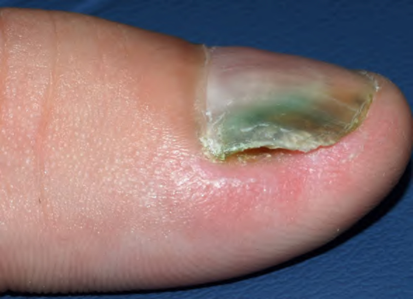
Fig. 74.26 Green nail syndrome. Blue–green discoloration of the nail due to pyocyanin produced by Pseudomonas aeruginosa. Note the associated onycholysis. Courtesy Julie V. Schaffer, MD.
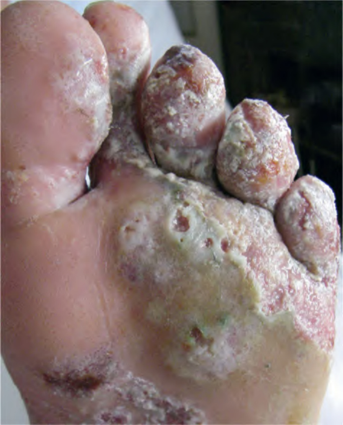
Fig. 74.27 Superficial infection of the skin with Pseudomonas. Note the maceration, erosions, and moth-eaten appearance of the skin. Courtesy Kalman Watsky, MD.
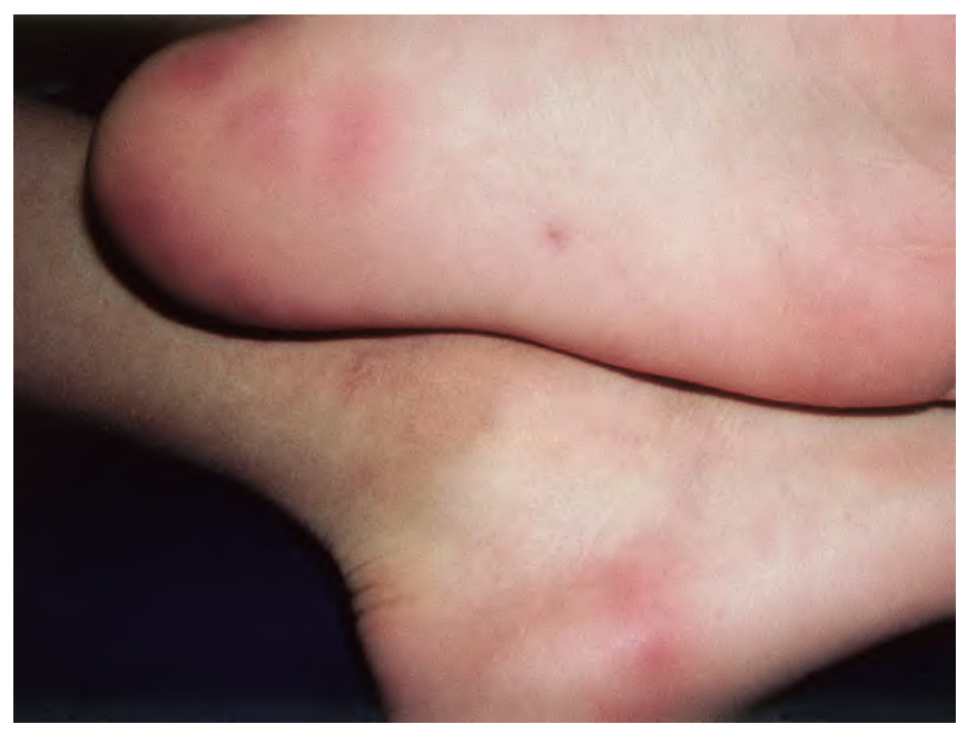
Fig. 74.28 Pseudomonas hot-foot syndrome. Tender erythematous nodules on the heel. Courtesy Justin J. Green, MD.
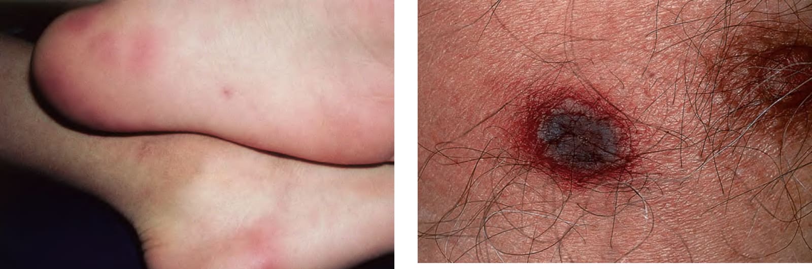
Fig. 74.29 Ecthyma gangrenosum. Embolic lesion of Pseudomonas aeruginosa on the chest. Note the necrotic center and inflammatory border.