BARTONELLA
Bartonella are small, pleomorphic, facultative intracellular Gram-negative bacilli (closely related to Brucella) that are capable of infecting erythrocytes and endothelial cells. Although there are >30 known species of Bartonella, B. henselae (cat scratch disease), B. quintana (trench fever), and B. bacilliformis (Carrion disease) represent the three major human pathogens. Both B. henselae and B. quintana also cause bacillary angiomatosis and endocarditis. Transmission occurs via close contact between animals and humans, arthropod vectors, and
possibly blood transfusions. Depending on factors such as the immune status of the infected individual, a particular Bartonella species can cause either acute or chronic infection and manifestations ranging from vascular proliferation to suppuration. There is no therapeutic regimen that guarantees eradication of Bartonella.
Bartonellosis
Synonyms: Carrion disease Oroya fever Verruga peruana
Introduction
Bartonellosis is a potentially life-threatening biphasic infection caused by Bartonella bacilliformis. Two distinct clinical forms may occur independently or sequentially: (1) Oroya fever, an acute febrile disease with associated hemolytic anemia and transient immunodeficiency; and (2) verruga peruana (Peruvian wart), a chronic disease characterized by cutaneous vasoproliferative lesions.
History
The biphasic nature of bartonellosis was first described in 1540 by Miguel de Steta. In 1885, a Peruvian medical student, Daniel Carrion, died of complications of Oroya fever after inoculating himself with blood derived from a lesion of verruga peruana, proving a common causality for the two disease states.
Epidemiology and pathogenesis
The disease is transmitted by the bite of female Phlebotomine sand flies (Lutzomyia verrucarum). It occurs in mountain valleys of Peru, Ecuador, and southwestern Colombia, where the vector resides. There is no racial, sex, or age predilection, but the disease tends to be milder in children than in adults. The incubation period varies between 3 and 14 weeks.
Clinical features
The onset of Oroya fever is often sudden, with dyspnea, weakness, pallor, tachycardia, fever, thirst, anorexia, arthralgias, and headache. A precipitous drop in hemoglobin may occur secondary to invasion of erythrocytes, and hyperbilirubinemia and hemoglobinuria can result. After the initial septicemia, a variable degree of immunodeficiency ensues. During this phase, a third of patients develop secondary bacterial infections; when death occurs, it is often secondary to an enteropathic bacterial infection, especially Salmonella enterica. Lymphadenopathy, splenomegaly, and megaloblastic hyperplasia of the bone marrow are seen in severe cases. Patients with Oroya fever usually require 8–10 weeks for full recovery.
Most patients who recover from Oroya fever develop cutaneous nodules during or after their convalescent period, but verruga peruana can also appear in previously asymptomatic individuals and in those treated with antibiotics for acute infection. Bright red papules and nodules measuring 2 mm to several centimeters in diameter appear within small erythematous patches. Lesions may be sessile or pedunculated and are usually located on the head and extremities. Hemorrhage, ulceration, and secondary bacterial infection can occur. In the absence of complications, lesions usually heal without scarring but may recur and serve as a reservoir for further transmission.
Pathology
The histologic features of verruga peruana vary and can resemble those of a pyogenic granuloma or Kaposi sarcoma. Masses of intracytoplasmic Bartonella organisms (Rocha–Lima inclusions) are present within swollen endothelial cells as well as in red blood cells; the bacilli are also found extracellularly. Thrombosis and occlusion of vessels occurs.
Diagnosis and differential diagnosis
The presence of organisms within red blood cells or within the cytoplasm of endothelial cells is diagnostic. B. bacilliformis can be isolated from blood cultures; immunologic testing (e.g. ELISA, immunoblot) and PCR-based assays for blood or tissue samples are also available. The differential diagnosis of verruga peruana includes multiple pyogenic granulomas, bacillary angiomatosis, warts, molluscum contagiosum, and yaws.
Treatment
Table 74.14 outlines the treatment recommendations for bartonellosis.
Cat Scratch Disease
Synonyms: Cat scratch fever Subacute regional lymphadenitis Inoculation lymphoreticulosis English–Wear infection
Introduction
Cat scratch disease (CSD) is caused by infection with Bartonella henselae. In immunocompetent hosts, it is a benign, self-limited illness characterized by tender regional lymphadenitis that lasts for weeks to months. Over 90% of patients report a recent scratch and/or bite from a cat.
Epidemiology and pathogenesis
CSD occurs worldwide, and ~12 500 cases are diagnosed annually in the US. Exposure to cats is the primary factor for CSD. Although fleavectored transmission to humans has not been documented, it could explain some cases in which there is no history of exposure to cats.
Clinical features
CSD is a common cause of persistent lymphadenopathy (>3–4 weeks) in children and adolescents. Lymphadenopathy usually presents 2–4 weeks after a scratch and can last 2–6 months, or even longer if fibrosis occurs. A single, large (1–10 cm), tender, mobile lymph node, with erythema and swelling of the overlying skin, is most often noted in the axilla. Suppuration occurs in only 10%–25% of patients. A skin lesion (e.g. a red or crusted pustule) develops at the inoculation site in most patients. Other skin findings can include a morbilliform eruption, erythema multiforme, granuloma annulare, and erythema nodosum.
Although most patients are otherwise well, the lymphadenopathy can be accompanied by fevers, malaise, fatigue, weakness, and headaches. Additional manifestations such as encephalopathy, hepatic granulomas, osteomyelitis, and pulmonary involvement develop in up to 15% of patients. The oculoglandular syndrome of Parinaud – unilateral conjunctivitis and ipsilateral preauricular lymphadenopathy – occurs in ~5% of individuals with CSD. Immunosuppressed patients (e.g. those with HIV infection) are prone to a more severe and complicated disease course.
Pathology
Affected lymph nodes display central necrosis surrounded by histiocytic and epithelioid cells with an inner palisading layer. Giant cells with a peripheral zone of lymphoid cells are also evident. The primary skin lesion can show similar changes, albeit less well developed. The Warthin–Starry silver stain demonstrates bacilli within areas of necrosis.
Diagnosis and differential diagnosis
For patients who present with regional adenopathy and a history of a recent cat scratch, the diagnosis is usually made clinically. A sensitive and specific assay for B. henselae antibodies is available, and antibody titers are usually high during the first few weeks after the onset of lymphadenopathy; PCR-based testing can also be performed. Culture is difficult and not routinely recommended. Other causes of lymphadenopathy such as infections (bacterial, fungal, or viral), reactive hyperplasia, drug reactions, or malignancy can be excluded by fine-needle aspiration histology or biopsy and culture of lymph node tissue.
Treatment
Table 74.14 outlines current recommendations for the treatment of CSD.
Bacillary Angiomatosis
Synonyms: Epithelioid angiomatosis Disseminated cat scratch disease
Introduction
Bacillary angiomatosis is caused by infection with B. henselae or B. quintana. Vascular proliferation is the predominant feature of this condition, which most often affects HIV-infected individuals.
History
The disorder was first described in 1983 as subcutaneous vascular proliferations in individuals with AIDS. Electron microscopy and Warthin–Starry-stained sections of the lesions revealed small bacilli. In 1989, LeBoit and colleagues coined the name “bacillary angiomatosis” and proposed histologic criteria for the disorder. In the early 1990s, Rochalimaea quintana and R. henselae (subsequently reclassified in the genus Bartonella) were recognized as the causes of bacillary angiomatosis (see Table 74.14).
Epidemiology and pathogenesis
Bacillary angiomatosis is an uncommon disease that is most often seen in HIV-infected patients when their CD4+ count is <200 cells/mcl (see Ch. 78). Fortunately, with the advent of effective antiretroviral therapy (ART), the prevalence of bacillary angiomatosis has decreased substantially. The associated vascular proliferation may reflect aberrant vascular endothelial growth factor (VEGF) signaling in endothelial cells. Only 20% of patients with bacillary angiomatosis due to B. henselae report a preceding cat scratch or bite, compared to 90% of those with CSD.
Clinical features
Cutaneous lesions of bacillary angiomatosis may present as superficial angiomatous papules and nodules, violaceous lichenoid plaques, or deep subcutaneous nodules. The papules and nodules resemble pyogenic granulomas (Fig. 74.30A) and may be smooth-surfaced, pedunculated, or surrounded by a collarette of scale. These lesions may display central umbilication, crusting, or ulceration. Subcutaneous nodules are often several centimeters in diameter and skin-colored; overlying erosions or ulcerations may develop. Less commonly, bacillary angiomatosis presents as a cellulitic erythematous plaque that can involve the under-lying bone. The host immune status largely determines the number and distribution of the lesions. Immunocompetent patients may have a single lesion at the site of inoculation, whereas the entire cutaneous surface can be affected in those who are severely immunocompromised.
Extracutaneous bacillary angiomatosis may occur with or without cutaneous disease and can affect any organ. Bacillary peliosis hepatis caused by B. henselae (but not B. quintana) presents with nausea, vomiting, diarrhea, abdominal pain, hepatosplenomegaly, and elevated liver enzymes. Bacillary peliosis splenis may lead to pancytopenia and require splenectomy. Patients can also present with malaise and night sweats.
Pathology
Biopsy specimens reveal lobular proliferations of capillaries and venules, plump and protuberant endothelial cells, and a marked neutrophilic infiltrate with evidence of leukocytoclasis throughout the interstitium
(Fig. 74.30B). In H&E-stained sections, interstitial clumps of bacilli appear as purplish granular material. Bacteria can be readily identified with a Warthin–Starry stain (see Fig. 74.30B, inset).
Diagnosis and differential diagnosis
PCR-based assays are a rapid and sensitive method to identify Bartonella from tissue specimens. Culture of B. henselae or B. quintana on chocolate or supplemented agar may take 20 to 40 days. Testing to detect antibodies against B. henselae (but not B. quintana) is sensitive and specific.
The clinical differential diagnosis includes pyogenic granulomas, Kaposi sarcoma, cherry angiomas, angiokeratomas, verruga peruana, and a disseminated non-tuberculous mycobacterial infection. Histologically, bacillary angiomatosis can be confused with verruga peruana, Kaposi sarcoma, pyogenic granuloma, and angiosarcoma.
Treatment
Cutaneous lesions begin to improve within 1 week of appropriate antibiotic therapy and resolve completely within 4 weeks. At least 3 months of treatment is recommended, as therapeutic failures and relapses can occur with shorter courses. Immunosuppressed patients with disease relapses may require maintenance therapy. Surgical excision of solitary cutaneous lesions, although effective, is usually not necessary. Table 74.14 outlines treatment recommendations for bacillary angiomatosis.
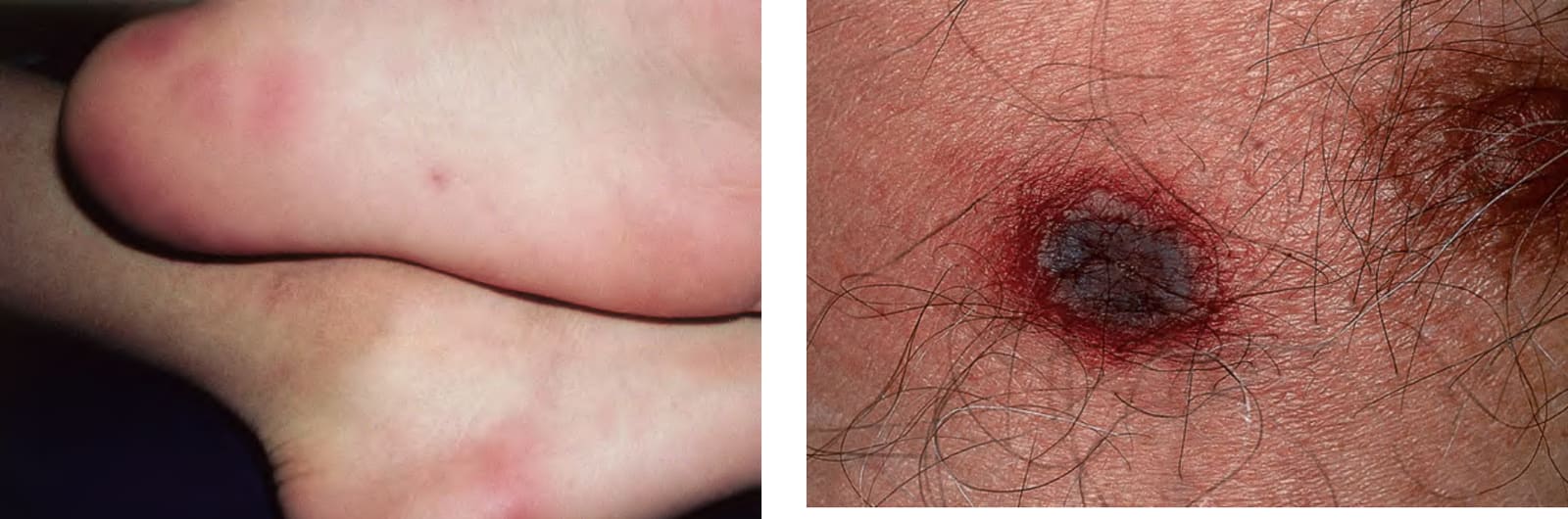
Fig. 74.29 Ecthyma gangrenosum. Embolic lesion of Pseudomonas aeruginosa on the chest. Note the necrotic center and inflammatory border.
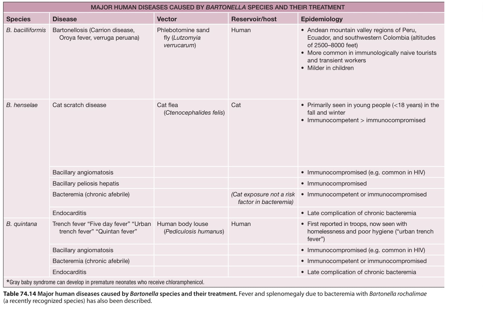
Table 74.14 Major human diseases caused by Bartonella species and their treatment. Fever and splenomegaly due to bacteremia with Bartonella rochalimae (a recently recognized species) has also been described.