OTHER GRAM-NEGATIVE SKIN INFECTIONS
Brucellosis
Synonyms: Undulant fever Malta fever Bang disease
Brucellosis is a chronic granulomatous disease caused by Brucella, a genus of Gram-negative coccobacilli. It is the most common zoonosis worldwide, with >500 000 cases annually. Endemic regions include the Middle East, Mediterranean basin, Central Asia, and Indian subcontinent. Transmission occurs through the consumption of contaminated unpasteurized milk products (commonly raw goat’s milk and cheese), direct contact with infected animal parts, or inhalation of aerosolized particles. Infection is rare in the US (~100 cases per year) and is primarily an occupational disease of farmers, laboratory personnel, butchers, and veterinarians. Children tend to have a more benign course, with fewer complications and better response to treatment, whereas pregnant women with brucellosis have a substantial risk of miscarriage.
Brucellosis demonstrates substantial clinical variability (Fig. 74.31). Acute disease is characterized by fevers, chills, headache, arthralgias, myalgias, back pain, and hepatosplenomegaly. Additional manifestations can include peripheral arthritis, spondylitis, epididymo-orchitis, endocarditis, and depression. Malodorous perspiration is highly characteristic. Skin lesions occur in <10% of patients, most often presenting as a disseminated eruption of violaceous papulonodules. Other cutaneous manifestations of brucellosis include erythema nodosum-like lesions, extensive purpura, and morbilliform eruptions. Endocarditis represents the major cause of death.
Acute brucellosis may mimic mononucleosis, influenza, malaria, typhoid fever, typhus, tularemia, tuberculosis, or sarcoidosis. The chronic form can be confused with rheumatologic, neurologic, or psychosomatic conditions. Multidrug regimens are recommended for brucellosis, with six weeks of doxycycline plus streptomycin, gentamicin, or rifampin representing a first-line treatment regimen; quinolones and TMP-SMX are other possible components of combination therapy. There is some concern regarding the use of rifampin in areas with a high prevalence of tuberculosis, as resistance may develop.
Glanders
Glanders is principally a disease of horses, mules, and donkeys that is caused by Burkholderia mallei (formerly Pseudomonas mallei), a non-motile, strictly aerobic, facultative intracellular, Gram-negative bacillus. Human disease occurs chiefly in those with direct contact with infected animals and laboratory personnel. A naturally occurring human case has not been reported in the US since 1934, but infections still occur in East Asia, South America, Eastern Europe, North Africa, and the Middle East. Use of B. mallei as a biological weapon has been documented. Tissue damage results from production of exotoxins, including pyocyanin (interferes with electron transfer), lecithinase (causes cell lysis), collagenase, lipase, and hemolysin.
Glanders has four clinical forms – septicemic, localized, pulmonary, and chronic. Acute septicemic glanders is characterized by malaise, anorexia, chills, fever, diarrhea, and joint pains. Patients may have flushing, cyanosis, a disseminated pustular eruption, cellulitis, lymphangitis, erythroderma, and/or jaundice. Multi-organ failure ensues, and the mortality rate is ~50% (>90% without treatment).
In the localized form, a nodule, pustule, or vesicle surrounded by hemorrhagic edema appears at the inoculation site after approximately 1 to 5 days. The superficial portion of the lesion sloughs, producing an ulcer with a gray–brown base. Glanders may also involve the nose and cause ulceration or perforation of the nasal septum or palate.
Chronic glanders features malaise, myalgias, and often fevers. Painful subcutaneous and intramuscular abscesses may appear at any site and can ulcerate, drain, or extend into joints, periosteum, and bone. Multiple subcutaneous and intramuscular nodules (“farcy buds”) appear along the draining lymphatics, and the term “farcy pipes” refers to the firm cords that result from lymphatic involvement.
Glanders should be considered when localized cutaneous abscesses and secondary lesions appear in the path of lymphatic drainage (see Table 77.17). Definitive diagnosis requires isolation of B. mallei, but serological, PCR-based, and indirect hemagglutination assays are also available. For localized disease, a 60- to 150-day course of oral amoxicillin/clavulanate, doxycycline, or TMP-SMX can be prescribed; the regimens recommended for melioidosis represent another option (see below). Alternatives include ciprofloxacin, streptomycin, and gentamicin.
Melioidosis
Synonym: Whitmore disease
Melioidosis is caused by Burkholderia pseudomallei, a mobile, obligately aerobic, intracellular, non-spore-forming bacillus found in soil and water. The name melioidosis is derived from the Greek words melis, meaning “donkey distemper”, and eidos, meaning “resembles glanders”. Melioidosis is endemic in Southeast Asia and northern Australia, but it also occurs in Africa, the Middle East, and Central and South America. B. pseudomallei has been identified in the environment along the Gulf Coast of the US, with two reports of local transmission of melioidosis in Texas; contaminated imported products (e.g. aromatherapy spray from India) have also been implicated in melioidosis outbreaks in the US. Infection develops via direct contact of abraded or lacerated skin with contaminated soil or water as well as through ingestion, inhalation, or sexual intercourse. Cases peak during the rainy season, likely due to aerosolization of bacteria. It is more common in adults with underlying medical conditions, especially diabetes mellitus, alcohol use, chronic lung disease, and chronic kidney disease.
Acute melioidosis can produce localized cutaneous infections, genitourinary infections, pneumonia, or septicemia. Subacute melioidosis, primarily a pulmonary disease, is frequently confused with tuberculosis. Chronic melioidosis is characterized by abscesses and granulomas in
multiple sites. Cutaneous manifestations may include cellulitis, subcutaneous abscesses, granulomatous lesions, ecthyma gangrenosum, purpura, pustules, Sweet syndrome, and urticaria.
Bacterial culture represents the standard for diagnosis of melioidosis, but the sensitivity is as low as 60%. Multiplex PCR-based assays are more sensitive as well as species-specific. Both complement-fixing and agglutinating antibodies appear within 4 to 6 weeks after infection, but serologic testing alone is inadequate in endemic regions.
Treatment of melioidosis is difficult, and susceptibility testing should be performed. Current guidelines recommend an initial intensive phase with intravenous administration of ceftazidime or a carbapenem for at least 10–14 days, followed by an eradication phase with oral TMP-SMX or amoxicillin/clavulanate for ≥3 months. Of note, B. pseudomallei has shown resistance to quinolones, macrolides, and aminoglycosides. Melioidosis-associated sepsis has a mortality rate of 50%–90%; however, intensive supportive care lowers this to ~20%. Melioidosis has a relapse rate of ~10%, and B. pseudomallei can remain latent in the body for decades.
Because B. mallei and B. pseudomallei represent potential agents of bioterrorism, attempts are underway to develop vaccines for these infections.
Cutaneous Malacoplakia (Malakoplakia)
Cutaneous malacoplakia, from the Greek malakos (“soft”) and plakia (“plaque”), is a chronic infectious condition that is characterized by
granulomatous inflammation with distinctive macrophages that are unable to kill bacteria effectively. It occurs primarily in immunocompromised hosts, including solid organ (especially kidney) transplant recipients, people living with HIV, and children with primary immunodeficiencies. Malacoplakia is most commonly due to E. coli, although Pseudomonas, Proteus, Klebsiella, Staphylococcus, Shigella, Enterococcus, Rhodococcus, and Mycobacterium spp. have also been implicated.
Malacoplakia most often affects the genitourinary tract but rarely involves the skin. Cutaneous lesions usually develop in the perianal region and can present as ulcerations, abscesses with multiple draining sinuses, yellow-to-pink soft papules, and erythematous indurated nodules.
Histologic examination of involved tissue shows highly characteristic findings. Michaelis–Gutmann bodies are intracytoplasmic laminated concretions that represent accumulations of calcified, iron-containing phagolysosomes; they stain with PAS, von Kossa, Perls’, and Giemsa stains. The presence of such concretions is noted in other conditions by different names (e.g. Schaumann bodies in sarcoidosis) and thus not pathognomonic for malacoplakia. Von Hansemann cells are large macrophages that contain Michaelis–Gutmann bodies and stain positively for CD68, lysozyme, and alpha-1 antitrypsin. The histopathologic differential diagnosis may include sarcoidosis, infectious granulomas, Langerhans cell histiocytosis, granular cell tumor, and fibrous histiocytoma.
The treatment of malacoplakia is difficult. Localized disease may be amenable to surgical excision. Prolonged courses of antibiotics such as quinolones, TMP-SMX, penicillins, and clofazimine may be of benefit. Ascorbic acid and bethanechol chloride can be employed to improve macrophage function. In severe cases, reducing the dosage of immunosuppressive therapy (if possible) may be of benefit. Lesions may spontaneously regress after ~6 months but can also be recalcitrant to therapy.
Tularemia
Synonyms: Deer fly fever Rabbit fever Pahvant Valley plague
Tularemia is a bacterial infection caused by Francisella tularensis, a Gram-negative, non-motile coccobacillus. First described in 1911 as a plague-like illness, there were ~2100 cases reported in the US from 2011 to 2020. Transmission to humans may occur via direct contact with mucous membranes or eroded skin, ingestion of contaminated food or water, inhalation of infectious particles, or bites from ticks or deer flies. Rodents and rabbits are the main reservoirs of disease, which may also affect domestic cats and sheep.
Tularemia occurs in six clinical forms according to the mode of transmission – ulceroglandular, glandular, oculoglandular, oropharyngeal/gastrointestinal, typhoidal/septicemic, and pneumonic (Fig. 74.32). Ulceroglandular tularemia is the most common form and is characterized by lymphadenopathy and an erythematous, indurated, punched-out ulcer at the site of cutaneous exposure that may last for weeks. Regional lymph nodes can become fluctuant and suppurate. Occasionally, ulceration or lymphadenopathy occurs in isolation. Other rare cutaneous manifestations include buboes, a morbilliform or vesicular eruption, erythema nodosum, and erythema multiforme.
Because of concern regarding the potential use of F. tularensis as a biologic weapon, tularemia is a reportable disease in the US. Diagnostic methods include direct fluorescent antibody testing (available in designated reference laboratories), immunohistochemical staining of tissue samples, and PCR-based assays. Attempts to culture F. tularensis are usually not made due to its high infectivity, risk to laboratory workers, and inability to grow on most media. The differential diagnosis of ulceroglandular tularemia includes other ulceroglandular entities (e.g. bubonic plague, tuberculous chancre, rat-bite fever, glanders), anthrax, soft tissue infections (i.e. Staphylococcus, Streptococcus, Pasteurella multocida), and infectious causes of a sporotrichoid pattern (see Ch. 77).
Treatment recommendations include a 10-day course of streptomycin (the classic first-line agent), gentamicin, or ciprofloxacin; a 14- to
21-day course of doxycycline is another option that results in a higher risk of relapse. A Jarisch–Herxheimer-like reaction may occur following the initiation of therapy. There are ongoing efforts to develop a safe and effective vaccine.
Haemophilus influenzae Cellulitis
Haemophilus influenzae is a Gram-negative coccobacillus that can cause facial cellulitis with a violaceous hue in infants and young children (usually 6–24 months of age) following an upper respiratory tract infection. It favors the buccal and periorbital areas and is often associated with high fevers, an increased leukocyte count with a left shift, and positive blood cultures. If diagnosis is delayed, systemic spread may lead to meningitis. Routine vaccination of children against H. influenzae type b has greatly reduced the incidence of this form of cellulitis. However, H. influenzae type f and non-typeable (unencapsulated) strains have been reported to cause cellulitis of the extremities in patients immunized to H. influenzae type b. First-line therapy for severe H. influenzae infections is a third-generation cephalosporin; selection of empiric antibiotic therapy for cellulitis is discussed above.
Rhinoscleroma
Rhinoscleroma is a slowly progressive, chronic granulomatous infection involving the nose and upper respiratory tract. It is caused by Klebsiella rhinoscleromatis, a short, immotile Gram-negative coccobacillus that is a subspecies of Klebsiella pneumoniae. Endemic foci exist in Central Europe, Egypt, India, Indonesia, Mexico, Central America, and tropical Africa. Rhinoscleroma is acquired via inhalation of contaminated droplets. Ineffective macrophage killing of K. rhinoscleromatis results in large, vacuolated, non-lipidized histiocytes with intracellular bacteria (Mikulicz cells). Clinical features, diagnostic techniques, the differential diagnosis, and treatment recommendations are outlined in Table 74.15. Recurrence after treatment is common. Histologic findings mimicking Rosai–Dorfman disease have been observed in cutaneous lesions and lymph nodes of patients with rhinoscleroma.
Salmonellosis
Salmonellosis refers to the spectrum of infections caused by Gram-negative aerobic bacilli in the genus Salmonella. Resultant forms of enteric fever include typhoid fever due to S. typhi and paratyphoid fever due to S. paratyphi. S. typhi is spread via direct contact with persons who have typhoid fever or are chronic carriers. In contrast, paratyphoid fever is most commonly acquired from inadequately cooked poultry or eggs as well as other contaminated food products or water, resulting in gastroenteritis.
Typhoid fever is characterized by fever, headache, malaise, myalgias, cough, sore throat, nausea, vomiting, and diarrhea or constipation. The characteristic cutaneous sign, “rose spots”, are pink, blanching, slightly elevated papules that measure 2–8 mm and are usually found on the anterior trunk in groups of 5–15 lesions. They occur in up to 30% of patients with typhoid fever and less often in non-typhoidal enteric fever. Rose spots often occur in crops during the second to fourth weeks of the illness, and Salmonella spp. can usually be cultured from these lesions. Other cutaneous manifestations of salmonellosis include erythema multiforme, Sweet syndrome, hemorrhagic bullae, pustular dermatitis, and a generalized erythematous eruption known as erythema typhosum. Hemophagocytic lymphohistiocytosis represents a rare complication of salmonellosis.
A history of travel to endemic areas (e.g. Southeast Asia, Africa), persistence of fevers, and positive cultures aid in distinguishing typhoid fever from influenza and other viral infections. Culture of Salmonella spp. from the bone marrow is the most sensitive diagnostic procedure but is rarely used in clinical practice. Blood cultures are less sensitive and PCR-based detection is limited by the low concentrations of microbial DNA in clinical samples. Since the late 1980s, S. typhi has developed resistance to the drugs that were previously used as first-line treatments (e.g. chloramphenicol, TMP-SMX, and ampicillin). Current treatment options include azithromycin, carbapenems, and (depending on resistance patterns) quinolones and third-generation cephalosporins. Typhoid conjugate vaccines are available and are recommended for travelers to endemic areas.
Rat-Bite Fever
Synonyms: Haverhill fever Sodoku Erythema arthriticum epidemicum
Rat-bite fever is most often due to infection with Streptobacillus moniliformis in North America and Spirillum minus in Asia, where it is known as sodoku. Infection results in an acute illness characterized by fever, arthritis, and a rash. While the disorder is usually caused by a bite (rat-bite fever), it can also occur from close contact with rodents or ingestion of contaminated food, water, or raw milk (Haverhill fever).
The incidence of rat-bite fever is highest in urban areas with poor sanitation resulting in a large rat population, but it can also occur from exposure to pet rats or laboratory rats. Approximately half of cases are in children. Erythema, edema, abscess formation, ulceration, and secondary infection may develop at the site of the bite. Regional lymphadenopathy is common. Paroxysms of fever interspersed with afebrile periods as well as headaches, nausea, vomiting, and myalgias may occur. In infants and children, weight loss and diarrhea can be significant. One of the hallmarks of the disease is a migratory polyarthritis that occurs in 50% of patients and may mimic rheumatoid arthritis. At 2 to 4 days following the onset of fever and arthritis, most patients with rat-bite fever develop an acrally distributed eruption involving the palms and soles. Erythematous macules and papules, petechiae, vesicles, pustules, and crusts may be seen.
Rat-bite fever can be diagnosed by culturing Streptobacillus moniliformis from blood, synovial fluid, or abscess aspirates, which requires an environment free of sodium polyanethol sulfonate; Spirillum minus does not grow in culture. PCR-based assays may also be utilized. The triad of fever, arthritis/arthralgias, and a rash can be caused by a variety of infectious diseases, including viral infections (e.g. enteroviruses, EBV, parvovirus B19), Rocky Mountain spotted fever, acute or chronic meningococcemia, acute rheumatic fever, and secondary syphilis. Non-infectious causes include Still disease, systemic lupus erythematosus, serum sickness-like drug reactions, and Sweet, Schnitzler, and periodic fever syndromes.
Penicillin is the drug of choice for rat-bite fever and is usually administered for a week, although patients with septicemia may require up to 6 weeks of parenteral therapy. Ceftriaxone, tetracyclines, and streptomycin represent additional options; erythromycin and clindamycin have also been used, but studies of their efficacy are lacking. Although most infections resolve spontaneously within 2 weeks, 10%–15% of untreated cases are fatal.
Plague
Plague is caused by Yersinia pestis, a Gram-negative, rounded, bipolar bacillus transmitted predominantly by fleas (Fig. 74.33). Peru, Democratic Republic of the Congo, and Madagascar are currently the most plague-endemic countries. There are typically <10 cases reported yearly in the US, with most occurring in two regions: (1) northern New Mexico, northern Arizona and southern Colorado; and (2) California, southern Oregon and far western Nevada.
Three clinical forms of plague are recognized – bubonic, septicemic, and pneumonic. The symptoms of bubonic plague are variable. Constitutional symptoms range from absent to severe. In approximately 10% of patients, the inoculation wound develops into a pustule or ulcer. This is followed by painful regional lymphadenopathy (bubo); suppuration and discharge from the lymph nodes may occur. In septicemic plague, vesicles, carbuncles, petechiae, and purpura may be seen. Necrotic lesions and hemorrhage from the nasopharynx or gastrointestinal tract have also been described. In pneumonic plague, the patient develops acute pneumonitis. Apathy, delirium, coma, and seizures occur in seriously ill patients. Bubonic plague may simulate tularemia, rat-bite fever, sporotrichosis, tuberculosis, streptococcal ecthyma, syphilis, or lymphogranuloma venereum. Septicemic plague must be differentiated from typhus, typhoid fever, tularemia, malaria, and other bacteremias. Y. pestis produces a virulence factor that reduces pyrin activity, explaining why individuals with familial Mediterranean fever (MEFV) gene variants that result in increased pyrin activity demonstrate resistance to plague.
Untreated bubonic plague has a 40%–60% fatality rate, while untreated septicemic or pneumonic plague is nearly always fatal. Streptomycin and other aminoglycosides (e.g. gentamicin) are first-line treatments for plague. Doxycycline and quinolones may be used when oral therapy is acceptable. Chloramphenicol (which attains high concentrations in the cerebrospinal fluid) is the drug of choice for plague meningitis or endophthalmitis. Postexposure prophylaxis with doxycycline or ciprofloxacin, if taken within 6 days, is effective in preventing disease. Live vaccines are utilized for prevention of plague in some parts of the world, but they have poor safety profiles. There are ongoing efforts to develop a safe and effective Y. pestis vaccine.
Vibrio vulnificus Infection
The genus Vibrio constitutes a group of Gram-negative anaerobic bacteria, among which V. vulnificus is the most common cause of cutaneous disease. V. vulnificus infections occur most commonly in men over 40 years of age who have chronic liver disease, diabetes mellitus, or immunosuppression plus a history of exposure to warm seawater or raw/undercooked seafood. “Vibrio season” traditionally lasts from May to October in the US, but climate change has elongated this to the later fall and expanded the bacterium’s geographic range. Additional risk factors are listed in Table 74.16.
The clinical presentation of V. vulnificus septicemia includes fever, chills, nausea, vomiting, diarrhea, abdominal cramps, and hypotension. Skin lesions develop in 75% of these patients, with progression from erythematous and purpuric macules to vesicles and hemorrhagic bullae (Fig. 74.34) and then to necrotic ulcers in a manner reminiscent of purpura fulminans. Wounds acquired in or contaminated by seawater can also lead to Vibrio cellulitis that is erythematous, edematous, and painful, typically with rapid evolution to hemorrhagic bullae. Progression to necrotizing fasciitis or myositis occasionally occurs. First-line treatment is doxycycline (oral or intravenous) plus a thirdgeneration cephalosporin (intravenous or intramuscular). Necrotic lesions may require surgical debridement.
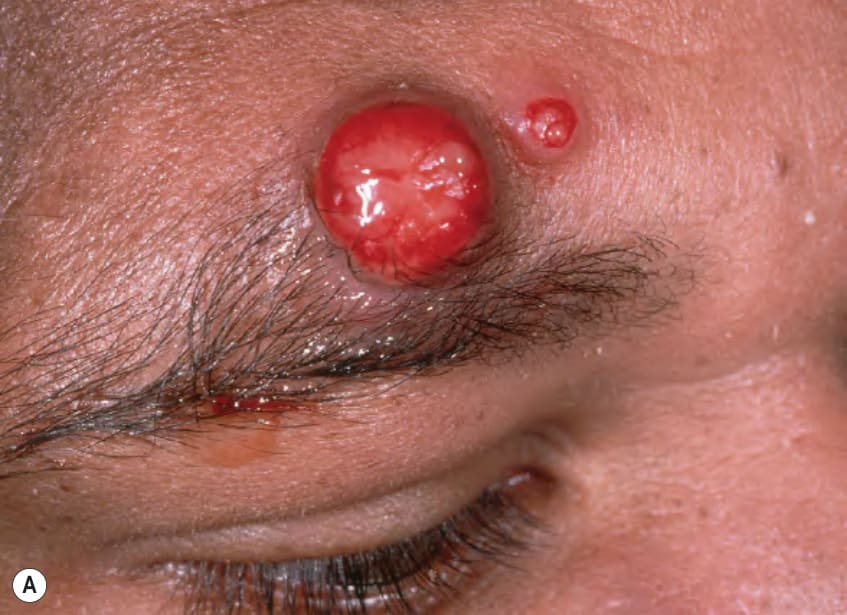
Fig. 74.30 Bacillary angiomatosis.A Bright red nodule and papule on the forehead. B Histologically, a dermal proliferation of vessels with plump endothelial cells is evident and scattered neutrophils are present in the accompanying infiltrate. Bacilli (in this case Bartonella henselae) are identified with the Warthin–Starry stain (inset). B, Courtesy James Patterson, MD.
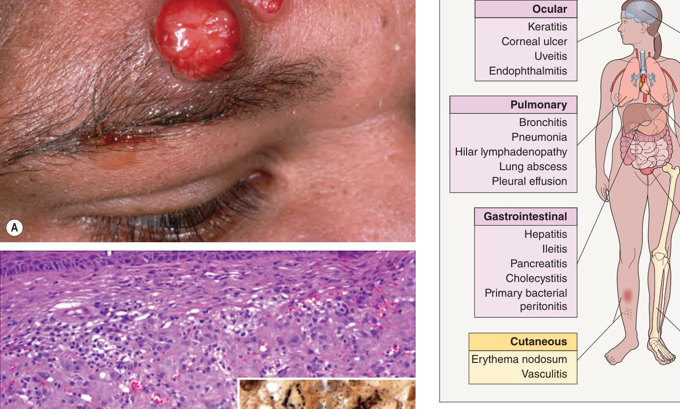
Fig. 74.31 Clinical manifestations of brucellosis.With permission from Slack MPE. Gram-negative coccobacilli. In: Armstrong D, Powderly WG (eds). Infectious Diseases. Edinburgh: Mosby, 2004.
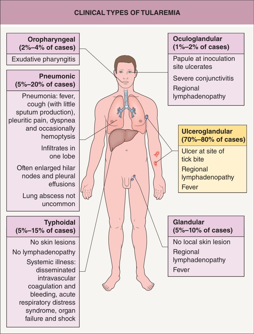
Fig. 74.32 Clinical types of tularemia.Adapted with permission from Slack MPE. Gram-negative coccobacilli. In: Armstrong D, Powderly WG (eds). Infectious Diseases. Edinburgh: Mosby, 2004.
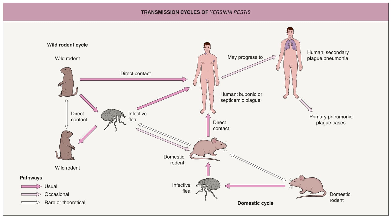
Fig. 74.33 Transmission cycles of Yersinia pestis.With permission from Dennis DT, Gage KL. Plague. In: Armstrong D, Powderly WG (eds). Infectious Diseases. Edinburgh: Mosby, 2004.
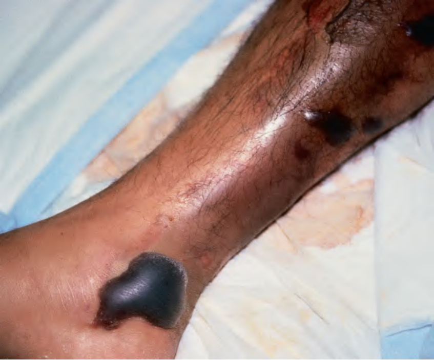
Fig. 74.34 Hemorrhagic bullae of the leg secondary to Vibrio vulnificus infection.
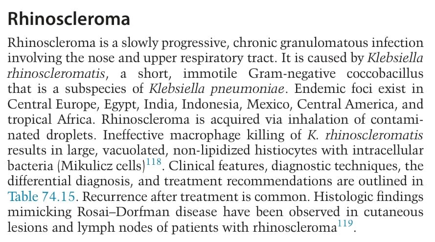
Table 74.15 Rhinoscleroma – clinical features, differential diagnosis, and treatment.
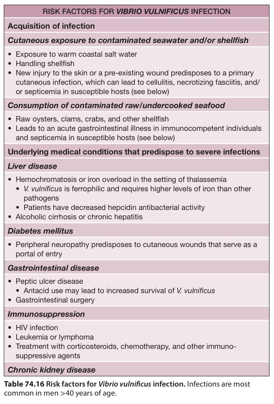
Table 74.16 Risk factors for Vibrio vulnificus infection. Infections are most common in men >40 years of age.