BORRELIA BURGDORFERI
Lyme Disease
Synonym: Lyme borreliosis
Lyme disease is a multisystem disorder with prominent skin findings that is caused by Borrelia species of spirochetes. Although found
worldwide, it has a particularly high incidence in North America and Europe, representing the most common tick-borne disease in the US. Lyme disease can occur year-round, but most cases present in the summer. If diagnosed in its early stages, it is a completely curable illness.
The species of Ixodes tick vector differs depending on the geographic region, e.g. I. pacificus in the western US, I. scapularis (also called I. dammini) in the eastern US and Great Lakes region, I. ricinus in Europe, and I. persulcatus in Asia. B. burgdorferi is the predominant etiologic organism in the US, whereas B. garinii and B. afzelii are the predominant causes of Lyme disease in Europe. This may explain the occurrence of borrelial lymphocytoma and acrodermatitis chronica atrophicans in Europe, but not the US. Once the spirochete is in the midgut of the tick vector after feeding on an infected host (e.g. whitetailed deer), it produces outer surface protein C, which enables it to traverse the midgut epithelium, enter the hemocoel, and make its way to the salivary glands. B. burgdorferi is ultimately transmitted to humans via the tick’s saliva. The rate of transmission is extremely low during the first 48 hours of tick attachment.
Chapter 19 describes the clinical features and treatment of Lyme disease, in particular erythema migrans (see Fig. 74.15), and Table 74.17 outlines the extracutaneous manifestations of Lyme disease. The next sections discuss borrelial lymphocytoma and acrodermatitis chronica atrophicans.
Borrelial Lymphocytoma
Synonyms: Lymphadenosis benigna cutis Lymphocytoma cutis Cutaneous lymphoid hyperplasia Spiegler–Fendt lymphoid hyperplasia, pseudolymphoma of Spiegler and Fendt, sarcoidosis of Spiegler and Fendt
Lymphocytoma is a benign, reactive form of lymphoid hyperplasia that can be attributed to various stimuli, including Borrelia infection. Borrelial lymphocytoma usually appears during the early disseminated stage of Lyme disease (see Ch. 19). Because it is caused by B. afzelii and B. garinii, neither of which is found in North America, borrelial lymphocytoma is virtually never seen in those who have not traveled outside of the US. However, it occurs in approximately 1% of patients with Lyme disease in Europe, with a predilection for children.
The formation of a lymphocytoma following a tick bite was first reported in 1950, but it was not until 1986 that Borrelia was cultured from such a lesion. A firm, bluish-red, occasionally tender nodule or plaque appears most commonly on the earlobes in children and the nipple/areola in adults, less often developing on the genitalia, trunk,
or extremities. It may be associated with regional lymphadenopathy or concomitant erythema migrans.
Histologically, the epidermis is normal and separated from the dermal infiltrate by a Grenz zone. The dense dermal infiltrate of lymphocytes forms a pattern that closely resembles the architecture of a lymphoid follicle. The histologic differential diagnosis includes an arthropod bite reaction and cutaneous lymphoma, with the presence of Bcl-2 protein within follicular cells and monoclonality by PCR favoring the latter (see Ch. 119). Of note, B. burgdorferi-associated B cell lymphomas have rarely been described. Borrelial lymphocytoma responds to the antibiotics used for Lyme disease (see Ch. 19).
Acrodermatitis Chronica Atrophicans
Synonym: Herxheimer disease
Acrodermatitis chronica atrophicans (ACA) is a cutaneous manifestation of chronic Lyme disease. First described in Europe in 1883 by Buchwald as “idiopathic atrophy”, it is most commonly due to an infection with B. afzelii, although it has also been associated with B. burgdorferi and B. garinii. As a result, it is extremely rare in the Americas but is seen in up to 10% of patients with Lyme disease in Europe.
ACA occurs months to years after the initial infection, predominantly in women 40–70 years of age, and appears to be associated with persistence of Borrelia organisms in the skin. It is a biphasic disorder consisting of an early, easily treatable, inflammatory stage and a late, treatment-resistant, atrophic stage. Initially, erythematous to violaceous plaques and nodules develop on the acral portion of the extremities, often insidiously. The skin is frequently doughy and swollen. This early stage follows a waxing and waning course for weeks to years. In the late stage, the skin has a glistening (“cigarette-paper”) appearance with prominent blood vessels (Fig. 74.35). Fibrous nodules may form on extensor surfaces (ulnar or tibial bands). Hypopigmentation, hyperpigmentation, pain, pruritus, hyperesthesia, paresthesias, and scaling may also be present. Rarely, the condition may be complicated by the development of basal cell or squamous cell carcinoma.
In biopsy specimens of early lesions, a dermal perivascular lymphocytic infiltrate with plasma cells, telangiectatic endothelial-lined spaces, and mild epidermal atrophy are seen. Histologic examination of late lesions shows an atrophic epidermis and an interstitial lymphocytic infiltrate with plasma cells and occasional histiocytes and mast cells. The dermis may be attenuated, with periadnexal fibrosis. Borrelia DNA may be detected by PCR in skin biopsy specimens. The differential diagnosis may include eczematous dermatitis (e.g. due to stasis), cold injury, atrophy secondary to chronic use of potent topical corticosteroids, severe photodamage, and, in the case of fibrotic lesions, scars, morphea, and fibromatoses.
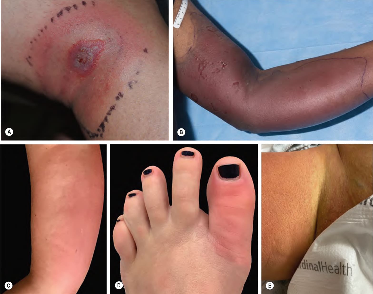
Fig. 74.15 Pseudocellulitis – clinical examples.A Erythema migrans with erosion following rupture of a central bulla. Inking of the skin reflects initial diagnosis of cellulitis. B Extravasation reaction following infusion of docetaxel. C Excessive limb swelling (ELS) following injection of multiple vaccines. This reaction does not preclude future immunizations. D Acute gout with abrupt onset of severe pain, warmth and erythema of the left great toe with difficulty in weight bearing. E Acute inflammatory edema with erythematous, edematous plaque sparing the inguinal fold. A, Courtesy Kalman Watsky, MD; B, Courtesy Edward Cowen, MD; C, D, Courtesy Karynne O. Duncan, MD.
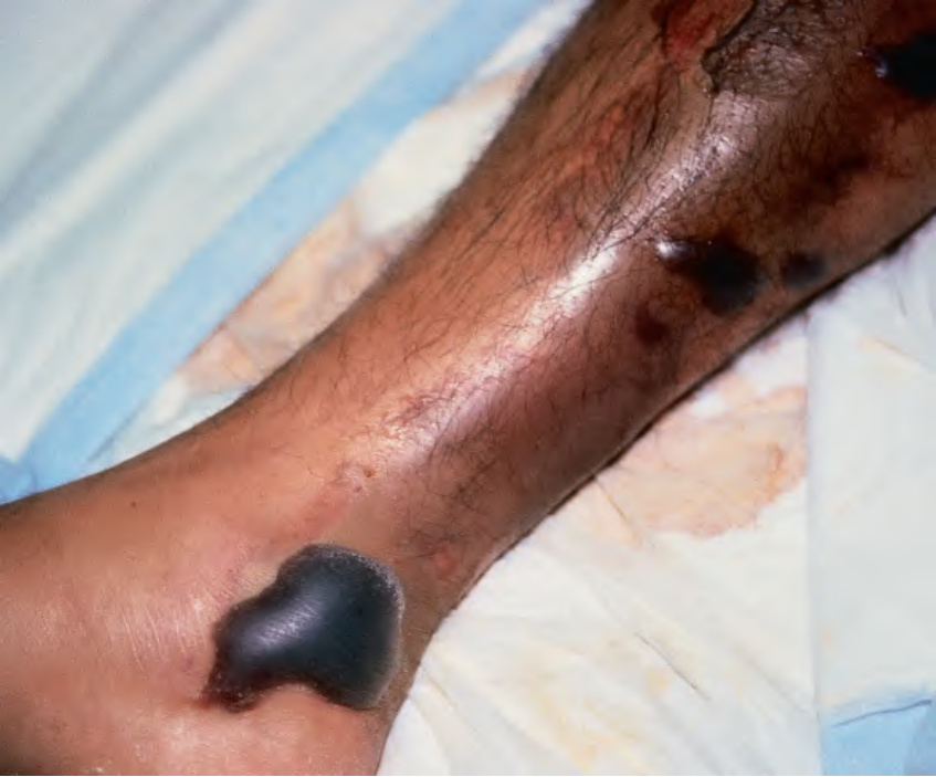
Fig. 74.34 Hemorrhagic bullae of the leg secondary to Vibrio vulnificus infection.
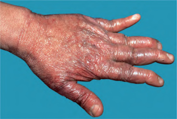
Fig. 74.35 Acrodermatitis chronica atrophicans. The acral skin is atrophic, shiny, and wrinkled, with prominent superficial veins.
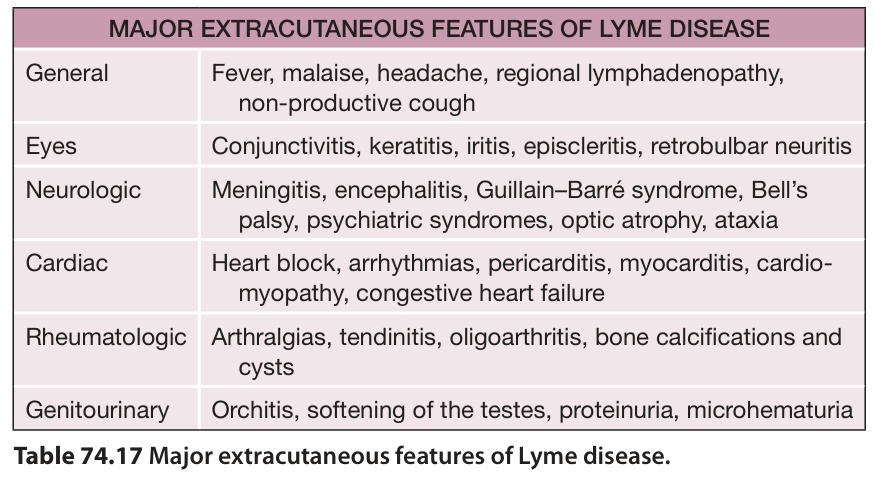
Table 74.17 Major extracutaneous features of Lyme disease.