NON-VENEREAL (ENDEMIC) TREPONEMATOSES
Yaws, pinta, and bejel (endemic syphilis) are caused by organisms that are morphologically and antigenically identical to the causative organism of venereal syphilis, Treponema pallidum (see Ch. 82). All three diseases have chronic relapsing courses with major dermatologic
manifestations. The primary route of transmission is person-to-person via skin, mucous membrane, or possibly fomite contact. With the exception of pinta, children are most often affected.
The diagnosis of yaws, pinta, and bejel is based primarily upon clinical features. The same serologic assays used for venereal syphilis can be used to diagnose the endemic treponematoses, but a positive test does not differentiate among the four diseases. Treponemal tests (e.g. TPHA, FTA-ABS, MHA-TP) are specific for treponemal infections (see Ch. 82) and may remain positive for life, regardless of treatment. The non-treponemal tests (e.g. VDRL, RPR) may indicate a current or recent infection, or a biologic false-positive result (in 1%–3% of the population). The vast majority of false-positive sera have antibody titers of <1 : 4. The quantitative non-treponemal tests are particularly useful in the evaluation of patients following therapy; a fourfold decrease in antibody titers indicates successful treatment, while a fourfold rise indicates reinfection or relapse. Diagnosis can also be made through darkfield examination of exudates from lesions.
For over 50 years the primary treatment for patients with endemic treponematoses was benzathine penicillin, given as a single intramuscular dose of 1.2 million units in individuals ≥10 years of age and 0.6 million units in children <10 years of age. A single high dose of oral azithromycin (30 mg/kg, maximum 2 g) was shown to be non-inferior to benzathine penicillin for the treatment of active and latent yaws. Due to the ease of administration and safe use in patients with penicillin allergy, azithromycin is now recommended as the first-line treatment for yaws by the WHO, with goals of mass treatment and disease eradication (see below). To date, no formal trials have studied azithromycin efficacy for pinta and bejel.
Yaws
Synonyms: Pian (French) Frambösie (German) Buba (Spanish) Parangi (Malay)
Yaws is a three-stage infection caused by T. pallidum subspecies pertenue and represents the most common and severe endemic treponematosis. It occurs in tropical climates, most often in the Pacific islands, Southeast Asia, and Africa. Yaws most commonly involves the lower extremities of children <15 years of age (Fig. 74.36). A “mother yaw”, the main lesion of the primary stage, occurs at the site of inoculation within 10 days to 3 months. It begins as an erythematous, infiltrated, painless papule that over time enlarges peripherally to become 1–5 cm in diameter, then ulcerates and develops an amber-yellow crust. The lesion is rich in treponemes and eventually heals spontaneously over 3 to 6 months.
“Daughter yaws”, the lesions of secondary yaws, are smaller and more widespread than the primary lesion. They usually occur adjacent to body orifices, such as the nose and mouth, and can expand or ulcerate. Both types of lesions are highly infectious. Patients may then enter a latent phase with reactive serology but absent clinical findings. Only 10% of patients progress to the final stage, during which abscesses form,
Indonesia.Courtesy Peter Ehrnstrom, MD.
become necrotic, and ulcerate. The ulcers may coalesce into serpiginous tracts, which heal with significant scarring and produce crippling deformities. Yaws can also cause periostitis, dactylitis, and osteitis, with the latter potentially leading to curvature of the tibia (“saber shins”).
Histologic examination of early yaws lesions demonstrates spongiosis, acanthosis, and papillomatosis. A moderate to dense dermal inflammatory infiltrate composed mostly of plasma cells and lymphocytes can be seen. Silver stains easily identify treponemes in specimens. Clinically, the skin lesions of yaws can resemble venereal syphilis, chancroid, eczema, psoriasis, verrucae, calluses, scabies, tungiasis, sarcoidosis, and vitamin deficiencies.
Renewed eradication efforts against yaws began in 2012, and in 2016 India was the first country to be declared free of yaws by the WHO. However, the emergence of azithromycin-resistant yaws in Papua New Guinea threatens eradication efforts.
Pinta
Pinta affects the skin exclusively and is caused by an infection with T. carateum. The disease is found only in the western hemisphere (Central and South America) in semiarid, warm climates. All ages are equally affected. The primary lesions occur 7 days to 2 months after inoculation, most often on the lower extremities. They begin as tiny macules or papules surrounded by an erythematous halo. Over a period of months, they develop into poorly defined, erythematous, infiltrated plaques that measure 10–12 cm in diameter. Secondary lesions (“pintids”) start as small, scaly papules, subsequently enlarging and coalescing into psoriasiform plaques. They are initially red but later become slate-blue, brown, gray, and black. The primary and secondary lesions are highly infectious. Tertiary pinta is characterized by symmetric depigmented, vitiligo-like lesions, which may be atrophic or hyperkeratotic and are not considered to be infectious.
Biopsy specimens of primary and secondary lesions reveal moderate acanthosis, slight spongiosis, and a superficial dermal inflammatory infiltrate consisting of lymphocytes, plasma cells, and neutrophils around dilated blood vessels. Some lesions show lichenoid changes, with hyperkeratosis, hypergranulosis, and vacuolar degeneration of the basal layer. The depigmented lesions of late pinta have epidermal atrophy and a complete absence of melanin. Except for longstanding, late depigmented lesions, treponemes can be visualized in biopsy specimens with silver stains. Early pinta is difficult to differentiate from venereal syphilis, yaws, and bejel. The early lesions can also be confused with eczema, psoriasis, leprosy, lichen planus, lupus erythematosus, and tinea corporis. The lesions of late pinta are frequently mistaken for vitiligo.
Bejel
Synonym: Endemic syphilis
Bejel is caused by an infection with T. pallidum subspecies endemicum. Most cases are seen in the arid, warm climates of North and West Africa and the Arabian Peninsula. Children <15 years of age are most commonly affected. Bejel is usually acquired via skin-to-skin or oral contact but can be transmitted sexually. In contrast to yaws and pinta, a primary lesion is rarely noticed. It usually consists of an inconspicuous small papule or ulcer in the oropharynx or on the nipples of breastfeeding women. Secondary bejel may present with findings similar to venereal syphilis, such as patches on mucous membranes, split papules, angular stomatitis, non-pruritic papular skin eruptions, and generalized lymphadenopathy. Condylomata lata are also frequently present. During the secondary stage, some patients experience osteoperiostitis of the long bones, which can cause nocturnal leg pain.
Six months to several years after inoculation, some patients develop the tertiary stage of bejel. Gumma formation may lead to gross mutilation of the skin, mucous membranes, muscle, cartilage, and bone. Without treatment, disfiguring lesions of the palate and nasal septum may occur, leading to difficulty in articulation and swallowing. The eyes and bones can also be severely affected in late-stage bejel.
The histologic picture closely resembles that of venereal syphilis. In the early stages, biopsy specimens demonstrate a perivascular dermal infiltrate of mostly plasma cells and lymphocytes. Oral lesions may resemble venereal syphilis, aphthosis, perlèche, vitamin deficiencies, and primary herpes simplex viral infection. The mutilating nasopharyngeal lesions of bejel can be mistaken for tertiary venereal syphilis, leprosy, rhinoscleroma, mucocutaneous leishmaniasis, paracoccidioidomycosis, and tuberculosis (see Tables 45.3 & 74.15).
Fig. 74.35 Acrodermatitis chronica atrophicans. The acral skin is atrophic, shiny, and wrinkled, with prominent superficial veins.
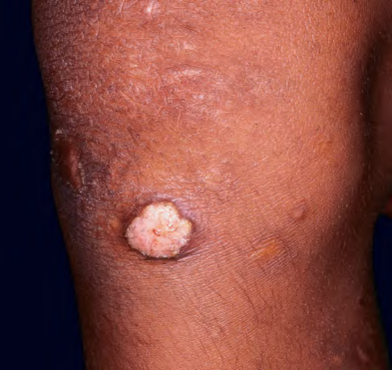
Fig. 74.36 Cutaneous yaws on the knee of an adolescent from