ACTINOMYCOSIS
Introduction
Actinomycosis is a subacute or chronic bacterial infection characterized by suppurating abscesses, granulomatous inflammation, and sinus formation. Actinomyces israelii, an anaerobic or microaerophilic Gram-positive, non-acid-fast actinomycete, is the most common causative organism. Cervicofacial (accounting for two-thirds of infections), pulmonary/thoracic, gastrointestinal, and pelvic forms exist.
Epidemiology and pathogenesis
Actinomycosis is seen worldwide, and men are affected more often than women. Humans are the only known reservoir for Actinomyces spp. A. israelii is part of the normal flora of the oral cavity and is also found in the gastrointestinal and female genital tracts. Actinomyces spp. are not considered particularly pathogenic, and their virulence is enhanced by co-infection with other organisms. In actinomycotic mycetoma,
species of Actinomyces other than A. israelii are usually responsible for the formation of sulfur granules (see Ch. 77).
Clinical features
In patients with cervicofacial actinomycosis (“lumpy jaw”), there is usually a history of poor dental hygiene, dental disease, or a previous orofacial injury or dental procedure. Additional risk factors include chronic tonsillitis and radiation therapy. An initial bluish swelling in the mandibular area progresses to brawny erythematous nodules, which become indurated (Fig. 74.37). Further progression leads to development of sinus tracts which drain purulent material with characteristic yellow “sulfur granules” that represent clumps of bacteria. Fever, pain, and leukocytosis may develop. Lymphadenopathy is usually absent. Once actinomycosis may spread via direct extension or hematogenous dissemination. Destruction of surrounding bony structures can result in facial deformity, and central nervous system involvement may lead to brain abscesses.
Pulmonary actinomycosis (15%–20% of patients) occurs after bacteria gain access to the lung through the aspiration of infected oral material. Pulmonary cavities are usually seen at the bases of the lungs. Extension to the pleura and chest wall may occur, and pleurocutaneous fistulas can develop. Gastrointestinal actinomycosis is usually preceded by trauma or inflammatory disease but may occur spontaneously. Granulomatous lesions involving the bowel can eventually extend to the abdominal wall, producing an erythematous mass with draining sinus tracts. Pelvic actinomycosis in women is often associated with intrauterine contraceptive devices. Actinomycosis may also involve the musculoskeletal system, gastrointestinal tract, and heart.
Pathology
An intense neutrophilic infiltrate is followed by the development of granulomatous inflammation with giant cells. Histiocytes, plasma cells, and epithelioid cells can be seen at the periphery of the abscess, while granules representing microcolonies of Actinomyces are seen centrally. Histologically these “sulfur granules” have a basophilic center and acidophilic periphery; they measure up to 30 microns in diameter.
Diagnosis and differential diagnosis
Microscopic examination of purulent material should be performed to detect the sulfur granules that are characteristic of actinomycosis (or actinomycotic mycetoma). They are composed of Gram-positive, branching filaments that fragment into diphtheroid forms and coccobacilli; acid-fast staining is negative. Actinomyces grows slowly, so cultures should be held for ≥2 weeks. Cervicofacial actinomycosis may resemble symbiotic anaerobic (e.g. Eikenella corrodens) and aerobic bacterial infections, scrofuloderma, dimorphic fungal infections, dental sinus tracts, and even neoplasms.
Treatment
The drug of choice for actinomycosis is penicillin G or ampicillin. Deep-seated, chronic infections should be treated with 2–6 weeks of intravenous therapy followed by 3–12 months of oral penicillin. For acute infections, 2–3 weeks of oral penicillin plus incision and drainage and surgical excision of sinus tracts is sufficient. Doxycycline, minocycline, and clindamycin are acceptable alternatives for penicillin-allergic patients. Imipenem has been shown to be effective in treatmentresistant cases. In order to avoid relapse, therapy should be extended beyond the resolution of symptoms.
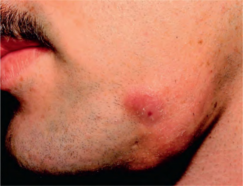
Fig. 74.37 Cervicofacial actinomycosis or “lumpy jaw”. Soft tissue swelling and draining erythematous nodules are seen. The discharge contained sulfur granules. Courtesy Joyce Rico, MD.