NOCARDIOSIS
Introduction
Nocardiosis is caused by various species of Nocardia, a filamentous, Gram-positive, acid-fast organism. Nocardia is recognized as an opportunistic pathogen in immunocompromised individuals, causing disseminated or systemic disease; in contrast, cutaneous nocardiosis commonly occurs in immunocompetent hosts.
Epidemiology and pathogenesis
Nocardiosis has a worldwide distribution and affects all age groups. Men are affected more often than women, and children may be more prone to developing lymphocutaneous disease. Nocardia spp. are ubiquitous in the soil. Localized trauma (e.g. puncture wounds), occupational exposures (e.g. farmers, gardeners), and immunodeficiency are risk factors for contracting nocardiosis.
Clinical features
The three major forms of primary cutaneous nocardiosis are: (1) mycetoma (see Ch. 77); (2) lymphocutaneous nocardiosis; (3) superficial cutaneous nocardiosis. Secondary skin infection may also develop after hematogenous dissemination, often from a pulmonary source. Table 74.18 details the various clinical presentations of Nocardia infection.
Pathology
Histologically, an intense neutrophilic infiltrate with abscess formation is seen. Sulfur granules are observed in Nocardia mycetoma but are absent in other forms of cutaneous nocardiosis. Organisms are not visible in routinely stained sections but appear as branching filaments on Gram stain. Nocardia also stains with methenamine silver and acid-fast stains.
Diagnosis and differential diagnosis
Nocardia grows easily on all common laboratory media. However, growth is slow, so cultures should be held for ≥2 weeks. Nocardiosis should be suspected in patients with sporotrichoid lesions or
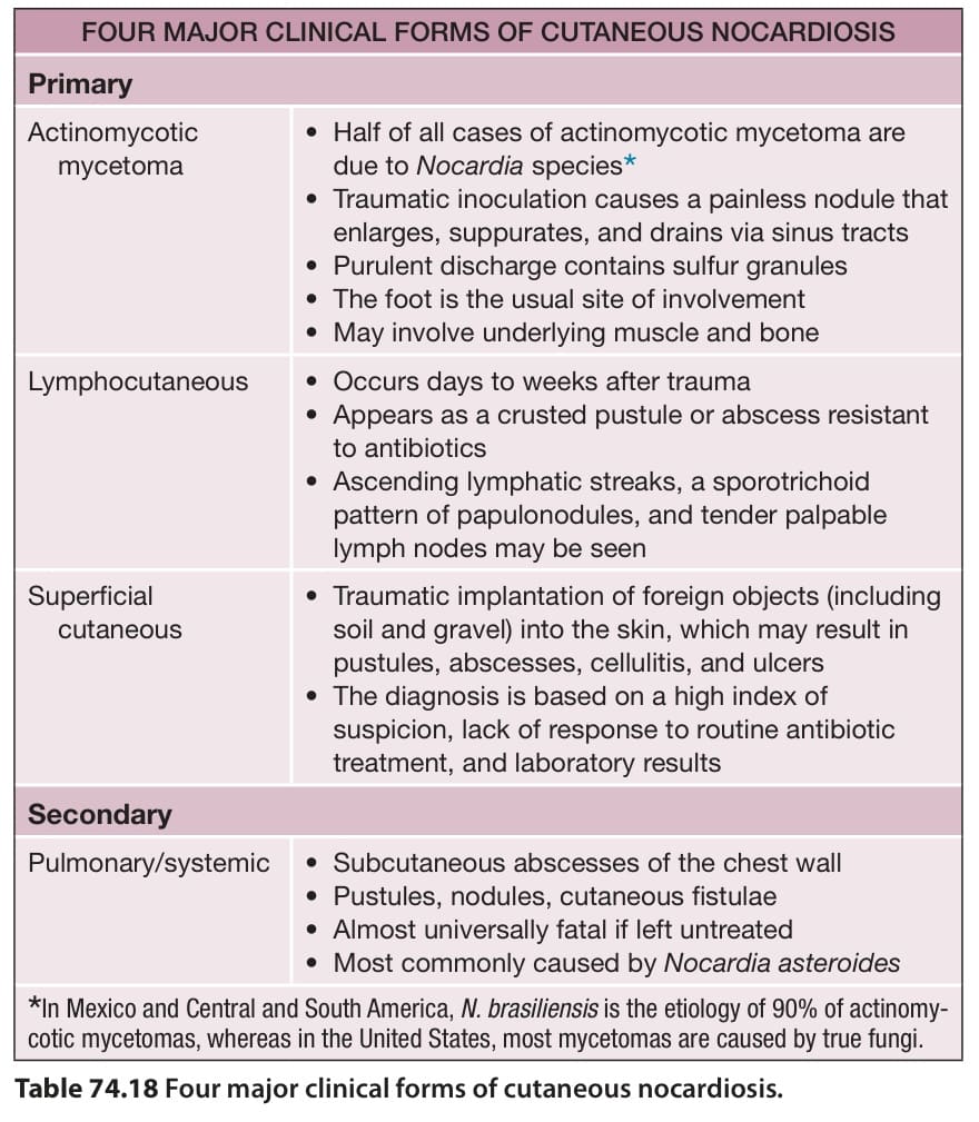
Table 74.18 Four major clinical forms of cutaneous nocardiosis.
trauma-associated superficial cutaneous infections that do not respond to routine treatment. It should also be considered in patients at risk for opportunistic infections.
Sulfonamides are the drugs of choice for nocardiosis. Minocycline is an effective alternative for sulfonamide-allergic patients, and linezolid may be added in resistant cases. For subcutaneous abscesses, surgical treatment is often required. Local forms of nocardiosis should be treated for 6–12 weeks, whereas immunocompromised patients and those with disseminated disease require 3–12 months of antimicrobial therapy.
Additional tables and figures available in our eBook (see inside front cover for access code).
Table 74.18 Four major clinical forms of cutaneous nocardiosis.