HANSEN DISEASE (LEPROSY)
Key features
Chronic infectious disease caused by Mycobacterium leprae or
M. lepromatosis, intracytoplasmic bacteria that parasitize macrophages and Schwann cells
Slowly progressive condition characterized by granulomas and neurotropism, with a predilection for the skin as well as peripheral nerves
The primary skin lesion is erythematous or hypopigmented, and it is often anesthetic
Based upon the clinicopathologic findings, which reflect the degree/type of immunity, Hansen disease is divided into two major forms – lepromatous with a predominantly Th2 response and tuberculoid with a predominantly Th1 response
In the lepromatous form of Hansen disease, multiple organisms are present in the dermis, whereas in the tuberculoid form, there are only a few organisms
Introduction
Hansen disease is a chronic infectious disease that is caused by Mycobacterium leprae or M. lepromatosis and has a predilection for the skin and nerves. It is divided into two major forms depending upon the degree and type of immunity – lepromatous with a predominantly
Th2 response and tuberculoid with a predominantly Th1 response. Hansen disease is classified by the World Health Organization (WHO) as a “neglected tropical disease” that represents a public health concern in a number of low-income countries. Once worldwide in distribution, Hansen disease is now seen primarily in tropical and subtropical regions of Asia, Africa, and Central and South America (Fig. 75.1). Early diagnosis and prompt therapy are key components in the strategy to control this chronic infectious disease.
History
The first written descriptions of leprosy may date back to as early as 600 BCE in India, and to 200 BCE in China and Japan. Many of the cases mentioned in the Bible may actually have been psoriasis, vitiligo, or other skin disorders rather than leprosy. The earliest scientific evidence of leprosy has come from an Egyptian skeleton and two Coptic mummies from the second century BCE and the fifth century CE, respectively. One theory was that leprosy was brought from India to the Mediterranean basin by Alexander the Great’s soldiers between 327 and 326 BCE and then spread throughout the Greek and Roman Empires. Recent studies utilizing comparative genomics have shown that all extant cases of leprosy are attributable to a single clone, and the disease appears to have originated in the Near East or eastern Africa. During the Middle Ages, leprosy reached epidemic proportions in Europe and then spread to the New World via the wave of exploration in the late fifteenth and sixteenth centuries. Independent of geographic location, the afflicted have frequently been ostracized by their communities and families.
During the nineteenth century, Danielssen and Boeck provided the first modern description of leprosy, and in the 1870s, a third Norwegian, Gerhard Henrik Armauer Hansen, identified M. leprae. The term Hansen disease is commonly used nowadays and may help to decrease the stigma associated with this infection. Mitsuda created the skin test for leprosy in 1919, and the value of sulfones in the treatment of leprosy was reported in 1942. In 1966, Ridley and Jopling suggested a classification of leprosy based on the immunologic status of the patient (see below).
Epidemiology
In the early 1980s, 11–15 million people worldwide were estimated to have Hansen disease. Introduction of multidrug therapy (MDT) has substantially reduced the number of affected individuals. The number of new cases per year decreased from >500 000 in 2003 to <150 000 in 2021, with only ~130 000 patients requiring treatment remaining worldwide. However, the incidence remains relatively high among patients <15 years of age. The goal of the WHO is a prevalence rate of <1 case per 10 000 persons, which has been achieved in all but a few countries. In 2021, Brazil (especially western Amazonia), India, and Indonesia accounted for ~75% of new cases, with other countries that have high infection rates located primarily in Southeast Asia and Africa (see Fig. 75.1). In the US, ~150–200 new cases are reported yearly. Recent evidence suggests that Hansen disease may be endemic in Florida17a.
Although men and women are equally affected, the lepromatous form of Hansen disease is seen more often in men than in women (~1.5 : 1). Hansen disease affects all races and ages, with the highest incidences in individuals 10–15 and 30–60 years of age. Key factors in the acquisition of Hansen disease are close contact of a susceptible person (e.g. based on genetic predisposition; see below) with a contagious individual (e.g. with multibacillary disease). The risk of acquiring the disease from household contacts is 25%, emphasizing the public health importance of domicile control. The incubation period varies widely, from months to >30 years, but it is usually 4–10 years. M. leprae is spread predominantly by patients with multibacillary disease via nasal and oral droplets and much less often from eroded skin.
Although Hansen disease primarily affects humans, it can be found in nine-banded armadillos (southern US from Texas to Florida, Brazil) and red squirrels (British Isles). In one study, ~40% of patients with Hansen disease from the southeastern US were infected with one of two M. leprae genotypes associated with wild armadillos, suggesting that Hansen disease represents a zoonosis in this region.
Pathogenesis
M. leprae and M. lepromatosis are very small, slightly curved rods that are characteristically acid-fast. They are obligate intracellular microorganisms with a predilection for macrophages and Schwann cells. The major sites of involvement are the peripheral nerves, skin, mucous membranes, bones, and viscera (e.g. testes, liver). In animal models, these bacilli grow best at a temperature of 27–33°C, and they thus prefer cooler regions of the body (e.g. nose, testicles, ear lobes) as well as regions where the peripheral nerves are close to the skin.
The fact that the majority of exposed individuals do not develop disease implies that there is variability in susceptibility/resistance, depending upon genetic and environmental factors. For example, susceptibility and the type of immune response appear to correlate with specific HLA types, such that individuals with HLA-DR2 and HLA-DR3 are more likely to develop the tuberculoid form and those with HLA-DQ1 the lepromatous form.
Polymorphisms in the genes that encode interleukin (IL)-6, 10, 12, and 17 A/F; tumor necrosis factor (TNF); lymphotoxin-α (a low-producing allele linked to early-onset Hansen disease); and the IL-23 receptor, vitamin D receptor (VDR), and Toll-like receptors (TLRs) 1 and 2 have been shown to be related to either overall Hansen disease susceptibility or the particular form of Hansen disease that develops. The genetic factors implicated vary in different populations. For example, in Vietnamese and Brazilian populations, variants in the shared regulatory region of the PRKN (encoding Parkin, an E3 ubiquitin protein ligase) and PACRG genes have been associated with susceptibility to Hansen disease. Genome-wide association studies in Chinese populations have linked variants in genes encoding proteins in the NOD2 (nucleotide-binding oligomerization domain containing 2) pathway with susceptibility to Hansen disease, especially the multibacillary form. The NOD2 pathway, along with the VDR, TLRs, and Parkin, play key roles in the innate immune response, as do several additional proteins encoded by genes associated with Hansen disease, e.g. MBL (mannosebinding lectin), MRC1 (mannose receptor C-type 1), SLC11A1 (solute carrier family 11 member 1), and KIR (killer cell immunoglobulinlike receptor). TLRs, NOD2, and MRC1 have been associated with modulation of autophagy, whereas Parkin and LRRK2 (leucine rich repeat kinase 2) have roles in the regulation of host cell apoptosis.
Presentations of Hansen disease exist on a clinical–immunologic spectrum. At one end, patients with the tuberculoid form of Hansen disease have specific cell-mediated immunity (CMI), as reflected in a positive lepromin test (Mitsuda reaction; see below). At the other end, patients with the lepromatous form of Hansen disease have a negative lepromin test and minimal CMI, while humoral immunity is increased (Fig. 75.2). For example, serum levels of antibodies against phenolic glycolipid-1 (PGL-1), an M. leprae-specific antigen, are highest in patients with the lepromatous form.
Macrophages play an important role in the body’s attempt to eliminate M. leprae, activating a type I interferon (IFN) response and producing IL-1, TNF, and IL-12. Patients with the tuberculoid form maintain this inflammatory response and an effective CMI. They display a Th1 CD4+ T cell response that produces IL-2, IFN-γ (type II IFN), and lymphotoxin-α in addition to involvement of CD8+ cytotoxic T cells, γ/δ T cells, CD1a-restricted T cells, and Th17 cells. In patients with the lepromatous form, a predominantly Th2 response leads to the release of different cytokines – IL-4, IL-5, IL-10, and IL-13; these cytokines suppress macrophage activity. CD4+ regulatory T cells, CD8+ suppressor T cells, and type I IFNs predominate in these patients. The cell wall of M. leprae contains lipid complexes, including PGL-1, which may suppress T cell responses and IFN-γ production as well as the organism’s invasion of Schwann cells in a laminin-2-dependent pathway.
CD4+ T cells that express the antimicrobial protein granulysin have been detected at a sixfold greater frequency in tuberculoid as compared to lepromatous skin lesions. Tuberculoid lesions also have stronger expression of TLR2 and TLR1, activation of which can induce differentiation of macrophages and dendritic cells. CD1b+ dendritic cells, which promote T cell activation and secretion of proinflammatory cytokines, were not detected in lesions from patients with the lepromatous form of Hansen disease; instead, these lesions had upregulation of genes belonging to the leukocyte immunoglobulin-like receptor (LIR) family. LIR-7 has been shown to suppress innate host defense by several mechanisms, including blocking antimicrobial activity triggered by TLRs.
Clinical Features
There is a wide spectrum of clinical findings in Hansen disease, and several classification schemes have been utilized. The Ridley and Jopling classification is based on an immunopathologic spectrum (Table 75.2; see Fig. 75.2), with lepromatous (LL) at the end with the least effective immune response, tuberculoid (TT) at the end with strong CMI, and three types of borderline leprosy in between: borderline lepromatous (BL), borderline–borderline (BB, in the middle), and borderline tuberculoid (BT) (Fig. 75.3).
In addition, the WHO created an operational system to facilitate the classification and treatment of Hansen disease in endemic areas. Per the WHO, the diagnosis of Hansen disease is based on the presence of at least one of the following cardinal features: (1) anesthesia in a hypopigmented or erythematous skin lesion; (2) enlarged peripheral nerve with associated loss of sensation and/or weakness; and (3) the presence of acid-fast bacilli in a slit-skin smear or biopsy. The current WHO classification system (revised in 2017) divides Hansen disease into two main groups: (1) paucibacillary leprosy – one to five skin lesions, without demonstrated presence of bacilli in a slit-skin smear or biopsy when assessed; and (2) multibacillary leprosy – more than five skin lesions, nerve involvement (pure neuritis or with any number of skin lesions),
or the presence of bacilli in a slit-skin smear or biopsy (with any number of skin lesions). This classification is commonly employed worldwide.
Certain peripheral nerves are more commonly affected, based primarily upon their more superficial location. Palpation of these nerves is an integral component of the physical examination of a patient with Hansen disease (Fig. 75.4). The clinician should determine whether there is a decrease in sensation to pain, temperature, and/or touch. This is in addition to inspection for neuropathic changes (e.g. muscle atrophy, flexion contractures of the fourth and fifth fingers), vasomotor alterations, and secretory disturbances (e.g. dry skin and eyes ).
The lepromatous form of Hansen disease, the form with the least cellular immunity and greatest number of bacilli, is characterized initially by multiple, poorly defined, erythematous macules, papules, nodules, and plaques (Fig. 75.5A). Lesions are widespread and usually symmetric in distribution. The most common sites of involvement are the face, buttocks, and extremities. Infiltration of the skin of the forehead can lead to leonine facies (Fig. 75.5B). Additional signs and late sequelae include madarosis, saddle nose, infiltration of both earlobes, and acquired ichthyosis on the lower extremities (Fig. 75.6). Anesthesia in a stocking or glove distribution may develop, often together with enlarged peripheral nerves and neuropathic changes. Ocular manifestations such as lagophthalmos (inability to completely close the eyes) and corneal and conjunctival anesthesia due to involvement of branches of the facial and trigeminal nerves, respectively, are sometimes present in severe cases.
Histoid leprosy is a clinically distinct form of multibacillary disease that some authors classify as a variant of the lepromatous form. It is characterized by the development of dermatofibroma-like papules and nodules. Lucio leprosy is a non-nodular, diffuse form of lepromatous leprosy most commonly seen in Mexico and Central America that results from M. lepromatosis infection.
Borderline leprosy, as its name implies, has features that are inter-mediate between the two ends of the spectrum. The severity of the cutaneous and peripheral nervous system manifestations depends upon whether the patient is “leaning” towards the lepromatous pole (BL) or the tuberculoid pole (BT; see Table 75.2). BT cutaneous lesions are usually asymmetric and are typically larger in size with less defined borders than those of the tuberculoid form (see Fig. 75.3A). Affected areas are hypoesthetic and have decreased sweating. BB leprosy characteristically presents with annular plaques that can have a “Swiss cheese” appearance (Fig. 75.7; see Fig. 75.3B). The cutaneous lesions of BL leprosy are more numerous and symmetric, with variable morphologies.
The tuberculoid form of Hansen disease usually presents with a few patches or plaques that have sharply demarcated, slightly raised borders. The color of these lesions ranges from erythematous to hypopigmented, with the latter especially common in patients with darkly pigmented skin (Fig. 75.8). Lesional alopecia and anesthesia or hypesthesia can represent clues to the diagnosis. Unilateral neuropathic changes, especially of the extremities (e.g. digital resorption), may be seen. Occasionally, only neural involvement is present (“pure neuritic [neural] leprosy”).
The development of reactions complicates the course of Hansen disease in ~30%–50% of patients, especially during treatment (see Fig. 75.2). These are characterized by acute inflammation that appears suddenly before, during, or after treatment (Fig. 75.9 & 75.10). Type 1 reactions can affect patients with any form of Hansen disease (except for the early indeterminate form), but there is a predilection for borderline categories. Type 2 reactions most often occur in lepromatous or BL leprosy. In addition to the administration of antimicrobial drugs, the most frequent triggers for reactions are pregnancy, other infections, and mental distress. The immunopathogenesis, clinical manifestations, and treatment of the two major types of inflammatory reactions are outlined in Table 75.3.
Type 1 (reversal) reactions are due to a change in the immunologic state of the patient and are often associated with neuritis. When there is an increase in cell-mediated immunity, this is referred to as an “upgrading” reaction (see Fig. 75.10A). Type 2 reactions are due to the formation of immune complexes in association with neutrophil recruitment.
A type 2 reaction can result in cutaneous and systemic small vessel vasculitis (e.g. erythema nodosum leprosum; see Fig. 75.10B,C). Patients with the diffuse form of lepromatous leprosy, who are usually from Central or South America, may develop the Lucio phenomenon, a reactional state characterized by thrombosis associated with antiphospholipid antibodies in addition to a vasculopathy and/or necrotizing cutaneous small vessel vasculitis (Fig. 75.10D). On a worldwide basis, Hansen disease is a common cause of cutaneous vasculitis, especially in low-income countries.
Pathology and Laboratory Evaluation
There are three basic histopathologic patterns observed in Hansen disease: lepromatous, tuberculoid, and borderline. In the lepromatous pattern, an infiltrate may be seen in the dermis, subcutis, lymph nodes, abdominal organs (e.g. kidney, liver), testicles, and bone marrow. The infiltrate contains Virchow cells, which are macrophages with numerous bacilli as well as lipid droplets in their cytoplasm. In H&E-stained sections, these cells have a foamy appearance. The bacilli in Hansen disease can be detected by a Fite–Faraco stain (more sensitive) or Ziehl– Neelsen stain, both of which stain the bacilli a bright red color. For lesions in which bacilli are usually scant, it is recommended that at least six sections be examined before declaring them negative.
A band of normal-appearing dermis (Unna band or Grenz zone) separates the epidermis from the dermal infiltrate, which is composed of plasma cells and a few lymphocytes in addition to foamy macrophages containing globi (clumps of bacilli) (Fig. 75.11). Following successful therapy, the organisms fragment and become granular. Cutaneous
nerves demonstrate lamination of the perineurium, producing an onion-skin appearance. In the histoid variant, a well-circumscribed proliferation of spindle cells contains numerous bacilli, which typically line up along the long axis of the cell.
In the tuberculoid pattern of Hansen disease, a dermal granulomatous infiltrate is seen and may have a linear pattern following the course of a nerve (Fig. 75.12). Epithelioid cells and Langhans giant cells are surrounded by lymphocytes. The cutaneous nerves are edematous, and organisms are absent or rare, even with special stains. Inflammation and fragmentation of nerve fibers in the tuberculoid form differentiate it from sarcoidosis and other granulomatous disorders.
The borderline pattern contains histologic features of both the lepromatous form (e.g. Virchow cells) and tuberculoid form (e.g. granulomas). Predominance of the former versus the latter is dependent upon whether the patient has the BL, BB, or BT form of borderline leprosy.
Indeterminate Hansen disease is usually difficult to diagnose histologically. Only a patchy infiltrate of lymphocytes or histiocytes around blood vessels or appendages is seen. There are no granulomas or Virchow cells, and organisms are usually absent.
When Hansen disease is suspected, the diagnosis can be confirmed by finding bacilli in a biopsy or a slit-skin smear. Samples for the slit-skin smear may be obtained from the earlobes, elbows, knees, and cutaneous lesions. To avoid bleeding, a fold of skin is firmly squeezed between the finger and thumb of the examiner or with forceps, and a small incision is made with a scalpel blade. The liquid obtained is smeared onto a slide and allowed to dry. The smear is usually stained by the Fite–Faraco method and a search is made for red rods (against a blue background) at 100× with oil immersion. Organisms are found in 100% of patients with the lepromatous form, 75% of those with borderline leprosy, and only 5% of those with the tuberculoid form. A biopsy specimen of the skin lesions should be obtained, especially in patients with suspected tuberculoid leprosy (see above). Where available, fluorescent microscopy with auramine rhodamine staining has been shown to be more sensitive than the Fite– Faraco method for detecting organisms in skin biopsy specimens.
PCR can be utilized to amplify genes that encode antigenic proteins (e.g. 36 kDa proline-rich antigen, Ag85B, 16 s) as well as M. leprae- or M. lepromatosis-specific repetitive repeat sequences (RLEP region). Molecular methods (e.g. analyzing RLEP region) can distinguish M. leprae from M. lepromatosis and are particularly helpful in
paucibacillary and pure neuritic leprosy. PCR can also be performed on slit-skin smears and fresh, frozen, or paraffin-embedded skin biopsy specimens (Table 75.4). In a study utilizing RLEP real-time PCR, M. leprae DNA was detected in 38 (75%) of 51 paraffin-embedded skin biopsy specimens from patients with paucibacillary leprosy. In addition, immunohistochemical staining of biopsy specimens for the PGL-1 antigen may prove helpful in paucibacillary disease.
Serologic assays for anti-PGL antibodies are only sensitive for the diagnosis of multibacillary disease. However, measurement of these antibodies can help to classify patients, monitor the response to treatment, and predict leprosy reactions. Serum levels of anti-PGL-1 IgG and IgM antibodies are highest in lepromatous disease and lowest (or absent) in BT or tuberculoid disease, therefore representing a marker of mycobacterial “load”. Elevation of anti-PGL-1 IgM antibody levels is also associated with reactions and impairment of nerve function.
The lepromin (Mitsuda) test consists of an intradermal injection of 0.1 ml of a suspension of heat-killed M. leprae. The response is positive when a nodule forms at the site of injection 3 to 4 weeks later, indicating that the patient can mount a specific cell-mediated response to the bacilli. The test provides prognostic, but not diagnostic, information. It is positive in TT and BT leprosy.
Lastly, Hansen disease, especially LL and BL, is one of the causes of biologically false-positive anti-nuclear antibodies (ANA) or rheumatoid
factor as well as Venereal Disease Research Laboratory (VDRL) and fluorescent treponemal antibody absorption (FTA-ABS) assays for syphilis (see Ch. 82).
An important goal in the management of Hansen disease is the prevention of disability. The WHO divides Hansen disease-related disability into three categories: grade 0, normal protective sensation; grade 1, loss of protective sensation of hands, feet, or eyes; and grade 2, visible deformities including ulcers, claw hand, foot drop, bone resorption, and lagophthalmos. Early detection of neuropathy is critical to prevent disability, but unfortunately the diagnosis of Hansen disease is often delayed for 1–2 years48a. Methods to detect nerve damage range from simple tests such as light touch with a cotton swab or monofilament to techniques such as nerve conductive studies, high resolution ultrasound, and MRI48b.
Differential Diagnosis
Skin diseases that may be confused with the different cutaneous presentations of Hansen disease are outlined in Table 75.5.
Treatment
Safety, effectiveness, and ease of administration represent key considerations in multidrug therapy for Hansen disease. The WHO 2018 guidelines recommend a three-drug regimen consisting of rifampin (rifampicin), dapsone, and clofazimine for all patients with Hansen disease, with a treatment duration of 6 months for paucibacillary (PB) disease and 12 months for multibacillary (MB) disease (Table 75.6). This change from only two drugs (rifampin and dapsone) for PB disease in the previous WHO guidelines was based on better outcomes with three drugs as well as simplification of treatment (e.g. only one type of blister pack) and reducing the impact of misclassification of MB as PB disease. Of note, Chinese patients with a HLA-B*13-01 polymorphism are at high risk of hypersensitivity to dapsone.
In the US, the National Hansen Disease Program recommends a longer duration of treatment (1 year for PB, 2 years for MB), with no clofazimine for PB disease and the option of daily (rather than monthly) rifampin administration. For patients with PB disease, therapy with rifampin, ofloxacin, and minocycline (“ROM scheme”), including a single dose for individuals with a single lesion, has also produced good results.
Multidrug therapy is extremely effective. Importantly, after the first dose, the patient is no longer infectious to others. Granular or beaded bacilli may be found in biopsy specimens following initiation of treatment but are considered to be non-viable. Hansen disease relapses are rare in the setting of recommended therapy/treatment regimens, affecting <5% of patients over a 5–10 year follow-up period. In a study of 1932 patients from endemic countries with MB disease, molecular evidence of resistance to rifampin, dapsone, and/or ofloxacin was present in 8% of cases. Antimicrobial resistance to rifampin and dapsone is due primarily to mutations in rpoB (encodes DNA-directed RNA polymerase β subunit) and folp1 (encodes dihydropteroate synthase), respectively.
Alternative antibiotics such as minocycline, clarithromycin, and ofloxacin can be used when there is resistance to or intolerance of firstline agents (see Table 75.6). Drugs that may play a significant role in the treatment of Hansen disease in the future include other quinolones (e.g. moxifloxacin) and ansamycins. Rifapentine (a rifampin derivative) has higher peak serum concentrations, a longer serum half-life, and more bactericidal activity against M. leprae than rifampin. In addition, moxifloxacin appears to be more bactericidal than ofloxacin. Thus, the combination of rifapentine, moxifloxacin, and minocycline (PMM) may be superior to rifampin, ofloxacin, and minocycline (ROM).
For the two major inflammatory reactions, additional medications are often required. Oral prednisone is used for type 1 (reversal) reactions, while thalidomide is the principal therapy for type 2 reactions (erythema nodosum leprosum; see Table 75.3). Systemic corticosteroids are also recommended for the Lucio phenomenon. Although thalidomide
remains the treatment of choice for type 2 reactions, teratogenicity is a major limitation and it is unavailable in many countries where Hansen disease is endemic. Lenalidomide and pomalidomide are thalidomide analogues with different side-effect profiles (e.g. more myelosuppression, less neuropathy), with potential utility in the treatment of type 2 reactions. Cyclosporine and methotrexate have been used in both type 1 and type 2 reactions, and additional drugs with possible benefit for type 2 reactions include phosphodiesterase 4 inhibitors (e.g. apremilast), TNF inhibitors, high-dose clofazimine, chloroquine, and pentoxifylline.
Although Hansen disease is now considered a curable disease with a good prognosis and excellent survival rate, it can still be incapacitating and stigmatizing. It is very important to make the diagnosis as soon as possible and to examine contacts, since early treatment prevents disability. The WHO 2018 guidelines recommended chemoprophylaxis with a single dose of rifampin to adults and children ≥2 years of age in contact with Hansen disease patients. However, concerns about cost-effectiveness, stigma, and drug resistance have prevented implementation. A single dose of rifapentine has also been shown to prevent Hansen disease in household contacts59a. Recognition of Hansen disease in its initial phases by healthcare professionals as well as the general population in endemic countries is essential in order to reduce the impact of this disease.
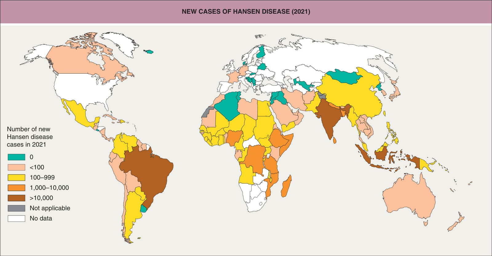
Fig. 75.1 New cases of Hansen disease, 2021. The World Health Organization (WHO) has achieved its goal of a prevalence rate of less than 1 case per 10 000 persons in all but a few countries. Over half of cases in the US are from seven states – Florida, Louisiana, Texas, California, New York, Hawaii, and Georgia. Reproduced from the World Health Organization, https://apps.who.int/neglected_diseases/ntddata/leprosy/leprosy.html
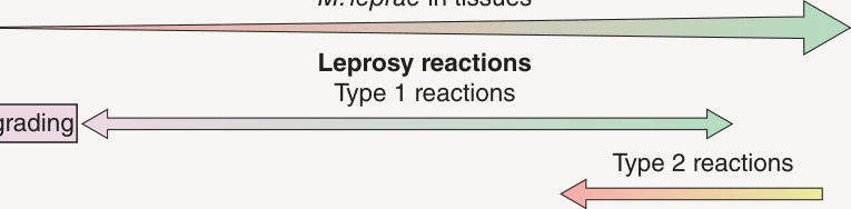
Fig. 75.2 The clinical–immunologic spectrum of Hansen disease. This reflects the underlying host immunity as measured by the T lymphocyte and antibody responses to Mycobacterium leprae. TT, tuberculoid leprosy; BT, borderline tuberculoid leprosy; BB, mid-borderline leprosy; BL, borderline lepromatous leprosy; LL, lepromatous leprosy; IFN, interferon; IL, interleukin. Adapted from Britton WJ. Leprosy. In Cohen J, Powderly WG (eds). Infectious Diseases. London: Mosby, 2003. With permission of Elsevier.
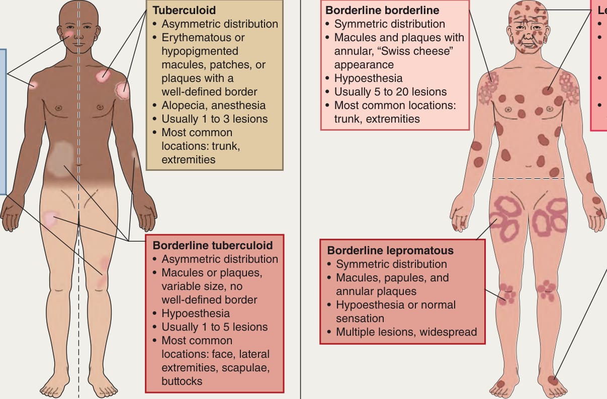
Fig. 75.3 Cutaneous findings in paucibacillary and multibacillary leprosy.A Paucibacillary disease – indeterminate, tuberculoid, and borderline tuberculoid. B Multibacillary disease – borderline, borderline lepromatous, and lepromatous.
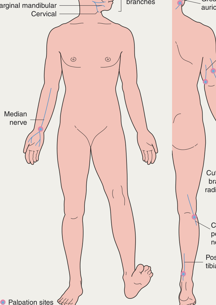
Fig. 75.4 Nerve examination sites.
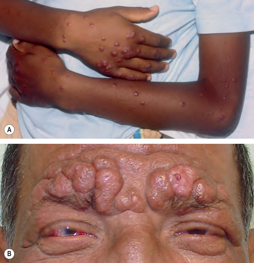
Fig. 75.5 Lepromatous form of Hansen disease.A Numerous erythematous papules and nodules on the forearms and hands. B Infiltrated nodules coalescing on the forehead with leonine facies and madarosis. Note the ocular involvement.
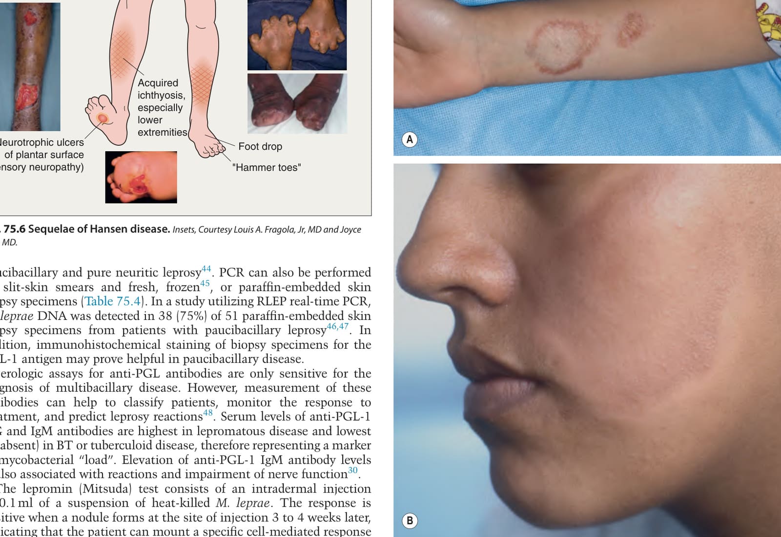
Fig. 75.6 Sequelae of Hansen disease.Insets, Courtesy Louis A. Fragola, Jr, MD and Joyce Rico, MD.
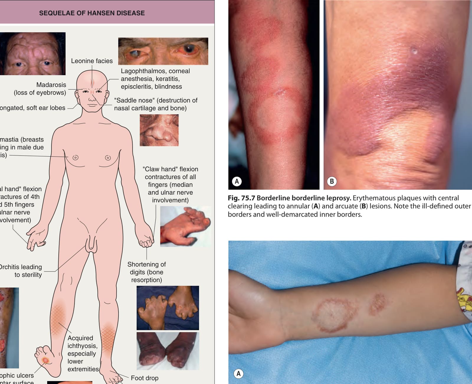
Fig. 75.7 Borderline borderline leprosy. Erythematous plaques with central clearing leading to annular (A) and arcuate (B) lesions. Note the ill-defined outer borders and well-demarcated inner borders.
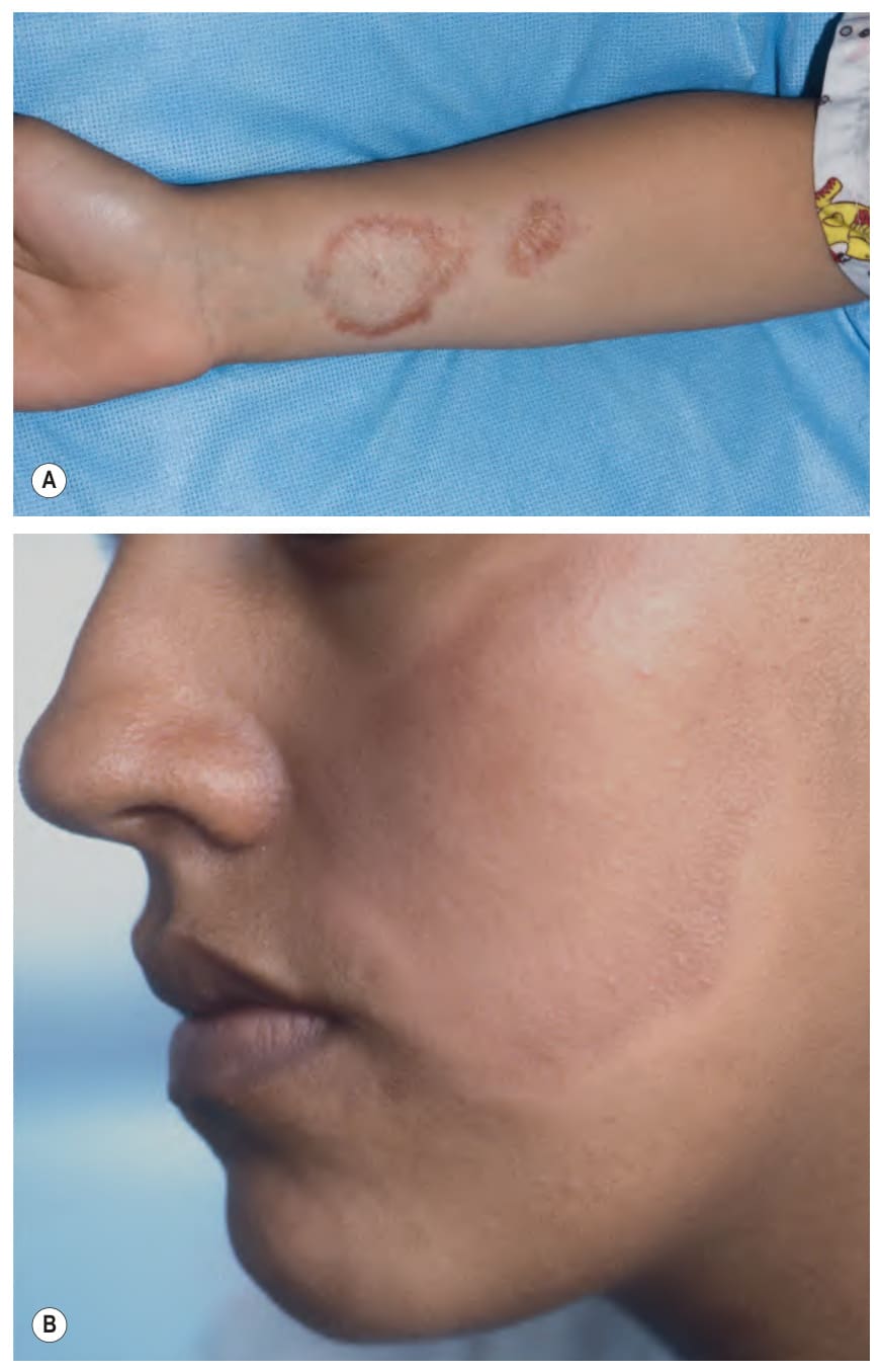
Fig. 75.8 Tuberculoid form of Hansen disease.A Two plaques on the forearm of a child that have dull pink–brown raised borders and central atrophy with hypopigmentation. B Large annular lesion on the cheek with a red–brown color and rim of hypopigmentation. A, Courtesy Edward Cowen, MD.
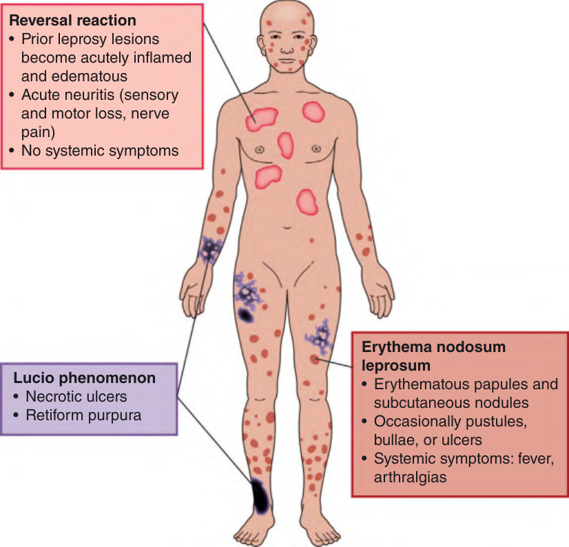
Fig. 75.9 Leprosy reactions – reversal reaction (type 1), erythema nodosum leprosum (type 2), and Lucio phenomenon.
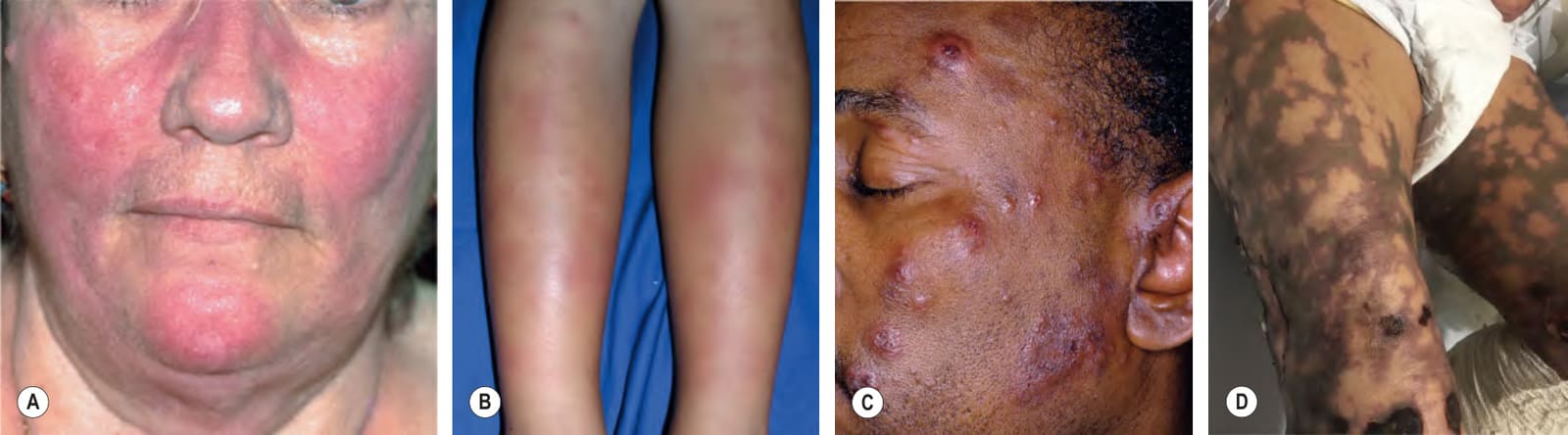
Fig. 75.10 Leprosy reactions.A Type 1 “upgrading” reaction with lupus erythematosus-like facial involvement in a “butterfly” distribution with marked inflammation. B Erythema nodosum leprosum, a type 2 reaction, presenting with erythematous nodules on the legs. C Type 2 reaction presenting with red facial papulonodules due to immune complex-mediated small vessel vasculitis in patients with the lepromatous form of Hansen disease. D Lucio phenomenon presenting with retiform purpura and necrotic ulcers. C, Courtesy Louis A. Fragola, Jr, MD; D, Courtesy Carlos G. Wambier, MD, PhD.
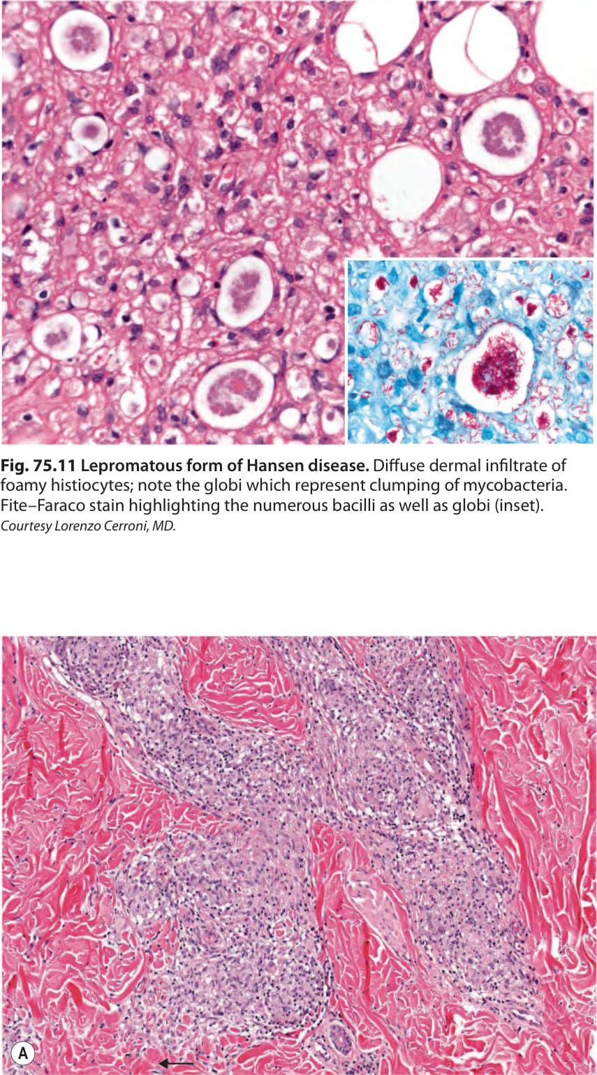
Fig. 75.11 Lepromatous form of Hansen disease. Diffuse dermal infiltrate of foamy histiocytes; note the globi which represent clumping of mycobacteria. Fite–Faraco stain highlighting the numerous bacilli as well as globi (inset). Courtesy Lorenzo Cerroni, MD.
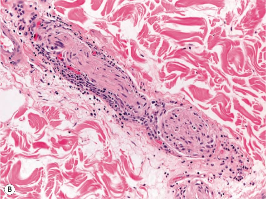
Fig. 75.12 Tuberculoid form of Hansen disease.A Granulomatous nodular infiltrate within the dermis. B Nerve involvement characterized by a sparse perineural infiltrate with lymphocytes and histiocytes. Courtesy Lorenzo Cerroni, MD.
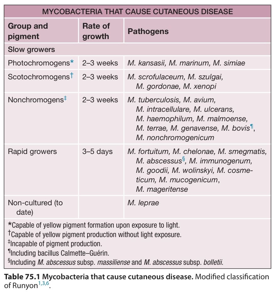
Table 75.1 Mycobacteria that cause cutaneous disease. Modified classification of Runyon.
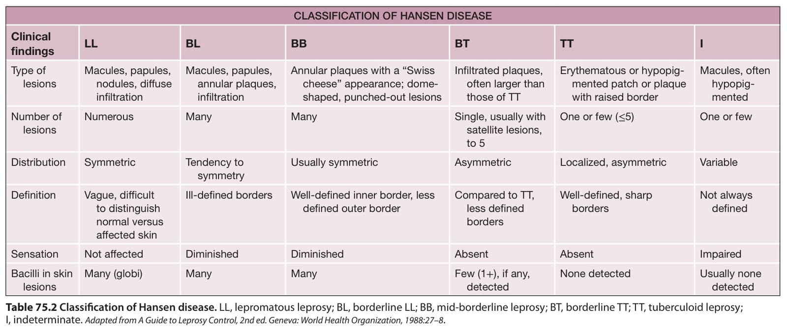
Table 75.2 Classification of Hansen disease. LL, lepromatous leprosy; BL, borderline LL; BB, mid-borderline leprosy; BT, borderline TT; TT, tuberculoid leprosy; I, indeterminate. Adapted from A Guide to Leprosy Control, 2nd ed. Geneva: World Health Organization, 1988:27–8.
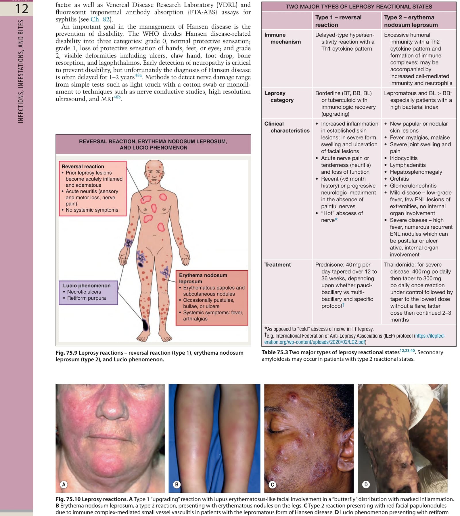
Table 75.3 Two major types of leprosy reactional states. Secondary amyloidosis may occur in patients with type 2 reactional states.
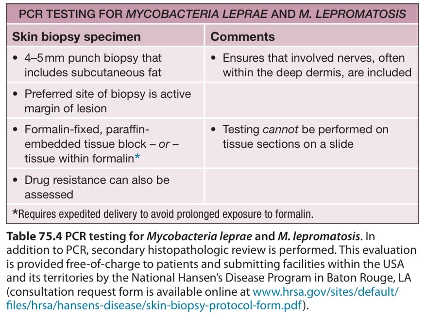
Table 75.4 PCR testing for Mycobacteria leprae and M. lepromatosis. In addition to PCR, secondary histopathologic review is performed. This evaluation is provided free-of-charge to patients and submitting facilities within the USA and its territories by the National Hansen’s Disease Program in Baton Rouge, LA (consultation request form is available online at www.hrsa.gov/sites/default/ files/hrsa/hansens-disease/skin-biopsy-protocol-form.pdf).
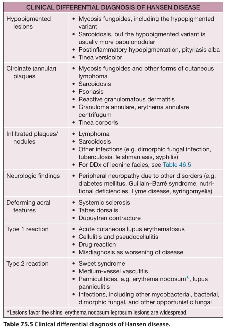
Table 75.5 Clinical differential diagnosis of Hansen disease.
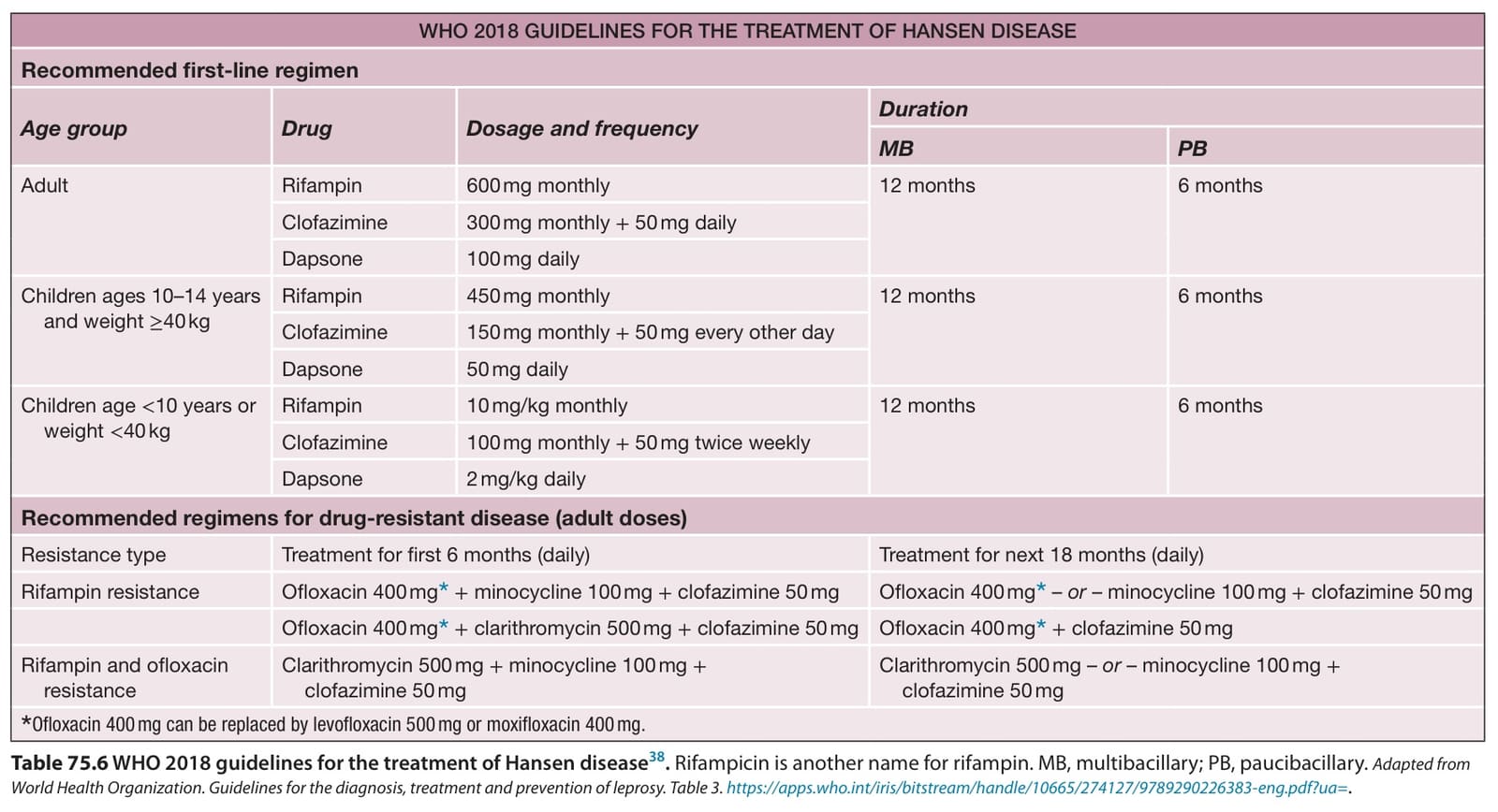
Table 75.6 WHO 2018 guidelines for the treatment of Hansen disease. Rifampicin is another name for rifampin. MB, multibacillary; PB, paucibacillary. Adapted from World Health Organization. Guidelines for the diagnosis, treatment and prevention of leprosy. Table 3. https://apps.who.int/iris/bitstream/handle/10665/274127/9789290226383-eng.pdf?ua=.