CUTANEOUS TUBERCULOSIS
Key features
Infection of the skin due to Mycobacterium tuberculosis, an acid- and alcohol-fast bacillus
A resurgence of tuberculosis (TB) in the late 1980s and early 1990s resulted from the HIV epidemic, emergence of resistant strains of M. tuberculosis, and a decline in TB control efforts
Exogenous exposure produces primarily the tuberculous chancre and TB verrucosa cutis
Endogenous infection can lead to scrofuloderma, miliary TB, and lupus vulgaris
Tuberculids include a papulonecrotic eruption, lichen scrofu- losorum, and erythema induratum
Introduction
Tuberculosis (TB) has affected humans since prehistoric times. There was, however, a worldwide explosion in its incidence in the late 1980s and early 1990s, and consequently a resurgence of cutaneous TB. This phenomenon has been attributed to the HIV epidemic, the emergence of resistant strains of Mycobacterium tuberculosis, increased use of immunosuppressive therapies, more migration of people around the world, and a decline in TB control efforts, superimposed on the pre-existing factors of poverty and malnutrition.
History
There are paleopathologic findings that suggest there were M. tuberculosis infections as early as 3700 BCE in Egypt, and 2500–1500 BCE in Europe. Utilizing PCR, M. tuberculosis DNA was detected in mummified remains from ancient times. In 1826, Laennec first reported cutaneous TB in the form of a “prosector’s wart”. Rokitansky and Virchow subsequently described the histologic features of cutaneous TB in detail. The discovery of M. tuberculosis by Koch in 1882 and the advances in descriptive pathology during the nineteenth century helped establish cutaneous TB as part of the spectrum of this infectious disease.
Epidemiology
M. tuberculosis is not particularly virulent, as only 5% to 10% of infections lead to clinical disease in immunocompetent hosts. This bacterium has a worldwide distribution (Fig. 75.13), with >6 million people newly diagnosed with TB in 2021. Cutaneous involvement is relatively uncommon, accounting for ~1%–2% of extracutaneous manifestations of TB. The incidence of cutaneous TB parallels that of pulmonary TB and is therefore higher in low-income countries and impoverished populations. HIV infection, immunosuppressive therapies (especially TNF inhibitors; see Ch. 128), and innate defects of the IL-12/IFN-γ axis (see Ch. 60) place individuals at greater risk for the development and reactivation of TB.
≥500
Pathogenesis
M. tuberculosis is the predominant etiologic agent of cutaneous TB. Occasionally, M. bovis and bacille Calmette–Guérin (BCG), an attenuated strain of M. bovis, may produce skin lesions.
M. tuberculosis is a non-motile, aerobic, non-spore-forming, filamentous rod. It is an acid- and alcohol-fast bacillus that has a waxy coating with a high lipid content, which makes the organism resistant to degradation after phagocytosis. M. tuberculosis produces multiple proteins involved in immune system invasion, allowing it to survive inside phagocytic cells.
M. tuberculosis disseminates primarily via inhalation of aerosolized droplets of saliva from individuals with active disease; transmission by ingestion or inoculation can also occur. Intact skin provides an effective protective barrier against invasion of the organism, but a break in the mucocutaneous barrier can facilitate entrance. Untreated infected individuals who do not have underlying medical problems have a 5% to 10% lifetime risk of progression to active TB; this risk increases substantially with immunosuppression, e.g. due to HIV infection or treatment with medications such as TNF inhibitors.
Studies using a model of zebrafish infected with M. marinum have demonstrated that granulomas, which are classically considered as host-protective structures formed to control infection, actually contribute to early bacterial growth and facilitate spread of infection (Fig. 75.14). Mycobacteria induce granuloma formation by releasing the early secreted antigenic target 6 (ESAT-6) protein, which stimulates neighboring epithelial cells to produce matrix metalloproteinase-9 (MMP-9). MMP-9 enhances recruitment of macrophages that contribute to bacterial proliferation and spread as well as maturation of the nascent granuloma. Increased production of other MMPs (e.g. MMP-1, MMP-3) has been shown to drive tissue destruction in TB. Eventually, development of adaptive immunity results in the curtailment, but not eradication, of bacterial growth by CD4+ and CD8+ effector T cells. The mature granuloma therefore represents an equilibrium between mycobacterial growth and the host immune response, and MMPs represent potential targets for antimycobacterial therapy.
The interaction of T cells and mycobacterial antigens, which are displayed on the surface of antigen-presenting cells, induces the liberation of interferons and other cytokines. These substances promote the activation and expression of MHC class II on antigen-presenting cells as well as the IL-2 receptor on T cells. During the initial sensitization, memory T cells are generated and remain for decades in lymphoid organs and the circulation. In mice, the intracellular pathogen resistance 1 gene (Ipr1) within the supersusceptibility to tuberculosis 1 (sst1) locus encodes a protein that mediates innate immunity to M. tuberculosis and other intracellular organisms. Ipr1 is expressed in macrophages and limits the multiplication of M. tuberculosis within these cells. IFN-γ- induced nitric oxide production also contributes to defense against M. tuberculosis by modulating macrophage responses (e.g. via activation of hypoxia inducible factor-1α) and repressing a neutrophil recruitment cascade that promotes bacterial replication.
The sensitization status of the host to mycobacterial antigens (e.g. previously infected versus never exposed), the degree of cell-mediated immunity of the host, the route of infection, and the pathogenicity of the infective strain of mycobacteria determine the resulting infection (Table 75.7). In immunocompromised hosts, cell-mediated immunity is impaired; as a consequence, there may be reactivation of quiescent disease.
Clinical Features and Pathology
Cutaneous TB has a wide variety of clinical presentations. Direct inoculation of M. tuberculosis from an exogenous source can lead to tuberculous chancre, TB verrucosa cutis, and occasionally, lupus vulgaris. Skin involvement due to endogenous infection may appear as scrofuloderma, acute miliary TB, a tuberculous gumma, orificial TB, and lupus vulgaris (see Table 75.7). In addition, there are cutaneous tuberculids, which represent immune reactions against M. tuberculosis or its antigens.
Cutaneous tuberculosis
A tuberculous chancre develops 2 to 4 weeks after the inoculation of M. tuberculosis into the skin of an individual without a history of TB.
It is a painless, firm, red–brown papulonodule that slowly enlarges, eventually eroding to form a sharply demarcated ulcer. Frequently there is spread to draining lymphatics and regional lymph nodes; the combination of the latter with a tuberculous chancre is analogous to the Ghon complex in the lung. The skin lesion usually heals spontaneously within 3 to 12 months, leaving an atrophic scar and calcified regional lymph nodes. The tuberculous chancre may occasionally evolve into verrucous plaques, scrofuloderma-like lesions, or lupus vulgaris.
Tuberculosis verrucosa cutis results from exogenous inoculation of M. tuberculosis at sites prone to trauma in previously infected individuals. The lesion begins as a small, asymptomatic, indurated wart-like papule with a subtle inflammatory rim (Fig. 75.15). It gradually enlarges, often in a serpiginous manner, to form a firm reddish–brown verrucous plaque. The center of the lesion may become fluctuant, with pus and keratinaceous debris expressed by slight pressure. After several years, the plaque can heal spontaneously.
Scrofuloderma begins as a firm, deep-seated, subcutaneous nodule that has accumulated inflammatory material and necrotic tissue, best characterized as a “cold abscess”. The suppurative nodule later becomes fluctuant and drains, with ulceration and sinus tract formation (Fig. 75.16). Because of the drainage, the bacilli secondarily infect the overlying dermis. The borders of the ulcers are often blue, the margins are undermined, and the base is covered with soft granulation tissue. Multiple ulcers can appear. After healing, keloids and retracted tethered scars develop at the sites of infection. The original focus of TB is usually underlying lymph nodes or bones, but it may be joints or the epididymis.
Orificial tuberculosis usually results from autoinoculation of M. tuberculosis, although occasionally it has an exogenous origin. Affected individuals typically have advanced systemic TB in the setting of impaired cell-mediated immunity (CMI). The mucosal or cutaneous lesions occur within or adjacent to a natural orifice that is draining an active tuberculous infection, primarily pulmonary, intestinal, or anogenital. The most common location is the mouth, especially the tongue. The initial lesion is an edematous red papule that ulcerates (Fig. 75.17) and develops undermined edges. The ulcers are painful, recalcitrant to treatment, and do not tend to heal spontaneously.
Lupus vulgaris is a form of cutaneous TB that occurs in previously sensitized individuals who have a strongly positive delayedtype hypersensitivity reaction to tuberculin due to intact CMI. It can develop secondarily from TB verrucosa cutis or scrofuloderma as well as from other endogenous and exogenous sources; lupus vulgaris also occasionally arises following BCG inoculation. The typical lesion is a red–brown plaque composed of papulonodules with an “apple-jelly” color on diascopy (Fig. 75.18). As the plaque expands, central scarring often develops. This can lead to substantial tissue destruction over a period of years. Clinical presentations of lupus vulgaris include: (1) plaque-type; (2) ulcerative or mutilating; (3) vegetating; (4) tumor-like; and (5) papulonodular. The head and neck region is the most commonly affected site, in particular the nose, cheeks, and ear lobes. Mucosa, especially oral, can also be involved.
Scrofuloderma. Plaques and nodules with central ulceration as well as resultant scarring with retraction.
The initial lesions of miliary tuberculosis are pinhead-sized, bluishred papules capped by minute vesicles. The vesicles develop a tiny central umbilication followed by crusting. When the papules heal, they leave a residual white scar with a brownish rim. The cutaneous lesions are the result of mycobacteremia, and the primary focus is often in the lungs.
A tuberculous gumma is also due to mycobacteremia with cutaneous seeding. It can present as either a firm subcutaneous nodule that slowly softens or an ill-defined fluctuant swelling. The overlying skin gradually breaks down to form an undermined ulcer, often with sinus tract formation. The extremities are more often affected than the trunk.
A skin biopsy of early lesions of cutaneous tuberculosis typically shows a mixed infiltrate that includes lymphocytes, neutrophils, and plasma cells. This is followed after a few weeks by granulomatous inflammation (tuberculoid granulomas; see Fig. 0.26 A), sometimes with central caseation. Acid-fast bacilli are usually abundant in early lesions, but are difficult to find when granulomas develop.
Tuberculids
The tuberculids represent a group of disorders that classically were associated with systemic TB. They are considered immune reactions within the skin due to hematogenous dissemination of M. tuberculosis or its antigens from a primary source, in an individual with strong antituberculous CMI. Often beginning as an immune complex-mediated reaction, they evolve into a granulomatous inflammatory response.
There is an ongoing debate as to the pathogenesis of tuberculids. Arguments in favor of an association with M. tuberculosis include a positive tuberculin skin reaction in affected patients; detection by PCR of M. tuberculosis DNA within lesions of erythema induratum, lichen scrofulosorum, and papulonecrotic tuberculid; and improvement with antituberculous therapy. In addition, many skin lesions demonstrate granulomatous
inflammation. However, there are multiple alternative causes of granulomatous inflammation and an active focus of TB is usually not found in patients with tuberculids; only a positive tuberculin test or IFN-γ release assay reflecting past exposure to TB is a consistent finding. The argument that corticosteroids can improve these eruptions is not a particularly strong one against an association, since other infectious diseases, e.g. viral hepatitis, can improve with such therapy and one would expect an antigendriven inflammatory process to respond to corticosteroids. Although a positive PCR is not absolute proof of pathogenesis, in the setting of appropriate controls, it provides strong evidence in favor of an association.
Papulonecrotic tuberculid is seen most frequently in children and young adults. It presents with dusky red papules or papulopustules that are widely distributed in a somewhat symmetric pattern and favor the extensor aspects of the extremities and buttocks (Fig. 75.19). Individual lesions may have central necrosis and are usually asymptomatic; occasionally there is associated pruritus. Usually there is spontaneous healing of individual lesions, often with scar formation. Multiple cyclic eruptions are characteristic, even after antimycobacterial therapy. PCR for mycobacterial DNA and the tuberculin test are positive. Histologically, there is evidence of leukocytoclastic or granulomatous vasculitis and wedge-shaped necrosis in the dermis. The history and clinical appearance aid in the diagnosis, which is bolstered by the histologic findings. The differential diagnosis often includes pityriasis lichenoides et varioliformis acuta (PLEVA) and small vessel vasculitis.
The early lesions of lichen scrofulosorum, a rare disorder, are firm, typically perifollicular, pink or yellow–brown, tiny flat-topped papules with variable scale. These asymptomatic papules are grouped in clusters, predominantly on the trunk. Lesions may persist for months and then disappear without scarring. Although it is seen in all age groups, children with nodal or skeletal TB are most frequently affected. This type of tuberculid has also been reported after BCG vaccination and can be triggered by tuberculin testing in highly reactive patients. Histologic examination demonstrates superficial granulomas around hair follicles and sweat ducts, with little or no caseation necrosis. No bacilli are found; however, PCR testing has demonstrated M. tuberculosis DNA within lesions. The clinical differential diagnosis includes lichen nitidus, lichen planus, and other lichenoid dermatoses (see Ch. 11) as well as “id” reactions, Blau syndrome, sarcoidosis, and secondary syphilis.
Erythema induratum (nodular vasculitis) is a form of lobular panniculitis associated with M. tuberculosis infection in a subset of affected individuals, especially in countries with a high prevalence of TB (see Ch. 100). The term erythema induratum of Bazin is reserved for cases related to M. tuberculosis. Both immune complex deposition, which may play a role in the vasculitic component, and a delayed-type hypersensitivity reaction to mycobacterial antigens are thought to be involved in the pathogenesis. Women are most commonly affected (80%–90% of patients), with peaks in incidence during early adolescence and around menopause.
Symptoms often begin after exposure to low temperatures. Typically, subcutaneous nodules appear on the calves of both legs; they may involute or break down, creating irregular deep ulcers with undermined
bluish borders. Healing results in atrophic hyperpigmented scars. Histologically, there is a lobular panniculitis with or without septal involvement. A neutrophilic vasculitis with mural thickening of subcutaneous arteries and veins and perivascular cuffing is often evident. Fat necrosis and granulomas can sometimes be observed. Bacilli are absent and caseation is rare.
Other entities such as erythema nodosum, lupus miliaris disseminatus faciei, granulomatous rosacea, and lichenoid tuberculid were formerly considered tuberculids, but are no longer included in this category.
BCG inoculation
BCG vaccination is performed with attenuated M. bovis. By increasing the degree of protection against M. tuberculosis infection, this vaccine has reduced the worldwide incidence of TB considerably. It can, however, cause complications, which include localized or generalized tuberculids, lupus vulgaris, scrofuloderma, and other nonspecific reactions such as fever, local inflammation, subcutaneous abscesses with or without ulceration, severe regional lymphadenitis, osteitis, and tuberculous foci in distant organs (Fig. 75.20). Rarely, it leads to a disseminated or even fatal infection, especially in individuals with a primary immunodeficiency affecting the IL-12/IFN-γ axis (e.g. Mendelian susceptibility to mycobacterial disease, hypohidrotic ectodermal dysplasia with immunodeficiency; see Ch. 60). Injections of methanol extraction residue (MER) of BCG have been used as adjuvant immunotherapy in patients with melanoma and can also result in nodules and subcutaneous abscesses (Fig. 75.21).
Diagnosis
In addition to the use of tuberculin skin tests, M. tuberculosis infection can be diagnosed with IFN-γ release assays (IGRAs; e.g. QuantiFERON® TB Gold Plus, T-SPOT®.TB). These assays determine whether exposure to recombinant peptides from M. tuberculosis (ESAT-6,
BCG. Nodules, some of which have ulcerated, arranged in a linear “lymphatic” pattern in a patient with a high-risk extremity melanoma who had received an injection of MER of BCG as adjuvant immunotherapy.
culture filtrate protein [CFP]-10, ± TB7.7) stimulates IFN-γ production by T cells within patient blood samples. In general, IGRAs have similar sensitivity (~80%–85%, less in immunocompromised individuals) and greater specificity compared to tuberculin skin tests. Current guidelines from the US Centers for Disease Control and Prevention (CDC) and National TB Controllers Association state that IGRAs may be used in place of a tuberculin skin test in all situations where the latter would be employed, with a general preference for IGRAs in patients ≥2–5 years of age with a low or intermediate risk of disease progression. Advantages and disadvantages of these methods of TB detection as well as situations where one type of test would be preferred are listed in Table 75.8. Of note, these tests are negative in a subset of individuals with active TB disease and should not be used to exclude TB in patients with clinical evidence of infection.
Methods to detect M. tuberculosis in samples of respiratory secretions and infected tissues such as the skin include staining for acid-fast bacteria, culture, and PCR-based assays. The latter have variable sensitivities but are rapid and specific for organisms in the M. tuberculosis complex (which includes BCG). The Xpert MTB/RIF® is an automated, fast, semi-quantitative PCR method that simultaneously detects organisms in the M. tuberculosis complex and resistance to rifampin (rifampicin) in liquid clinical samples (e.g. sputum, lymph node aspirate) over a period of two hours. Genotyping methods based on spacer oligonucleotides (spoligotyping), mycobacterial interspersed repetitive unit-variable number tandem repeats (MIRU-VNTRs), and restriction fragment-length polymorphisms (RFLPs) can separate
M. tuberculosis from non-tuberculous mycobacteria and detect mutants associated with drug resistance.
Treatment
Medications used to treat cutaneous forms of TB are the same as those for systemic TB (Table 75.9 & Fig. 75.22). Administration of appropriate therapy also assists in preventing bacterial resistance and reducing transmission of M. tuberculosis. In addition to the increase in multidrug-resistant TB that accompanied the resurgence of this disease in the late 1980s and early 1990s, extensively drug-resistant TB strains that are even more difficult to treat have also emerged in all regions of the world (Table 75.10). For latent TB infection, recommended regimens include isoniazid plus rifapentine weekly for 3 months, isoniazid plus rifampin daily for 3 months, and rifampin daily for 4 months.
In addition to “repurposing” existing antibiotics (e.g. quinolones, amoxicillin with clavulanate, linezolid, clarithromycin) for use in patients with extensively drug-resistant TB, novel drugs have been developed. Bedaquiline, a diarylquinoline that inhibits mycobacterial ATP synthase, was FDA-approved in 2012 as a part of combination therapy for multidrug-resistant TB. Although bedaquiline was found to increase the rate of sputum culture conversion, this drug can lead to QT prolongation and more deaths were observed when compared to the placebo group. Pre-screening electrocardiography is recommended, and an initial 8-week course of bedaquiline plus linezolid for rifampinsensitive TB was recently reported as noninferior to standard therapy, notably with limited toxic effects. Pretomanid, a nitroimidazole, was FDA-approved in 2019 to treat extensively drug-resistant TB in combination with bedaqualine and linezolid. Delamanid, a dihydro-nitroimidazooxazole derivative that inhibits synthesis of the mycolic acid component of mycobacterial cell walls, was approved in Europe in 2014 for treatment of multidrug-resistant TB. The antidiabetic drug metformin may have an adjunctive role in TB treatment by increasing reactive oxygen species production, enhancing specific immune responses, improving the efficacy of conventional anti-TB drugs, and decreasing detrimental inflammation.
The development of new TB vaccines with effective adjuvants represents a priority of the WHO. Sequential administration of BCG and a recombinant modified vaccinia virus Ankara (MVA) expressing antigen 85 A can induce robust antimycobacterial immunity. Several TB subunit and virus-vectored vaccines are currently being evaluated in clinical trials. Vaccines found to have efficacy in controlled studies include the adjuvanted protein subunit vaccine M72/AS01E (~50% efficacy after 3 years), a non-tuberculous Mycobacterium-derived inactivated whole-cell vaccine, an adenovirus-vectored vaccine containing three M. tuberculosis antigens (85 A, 85B, and TB10.4), and the recombinant “next-generation” BCG vaccine VPM1002.
Although the production of nitric oxide by macrophages is known to help control infection by M. tuberculosis, the organisms can persist within these cells. Proper function of the mycobacterial proteosome appears to play an important defensive role, and, in the future, inhibition of proteosome function could provide a novel therapeutic approach.
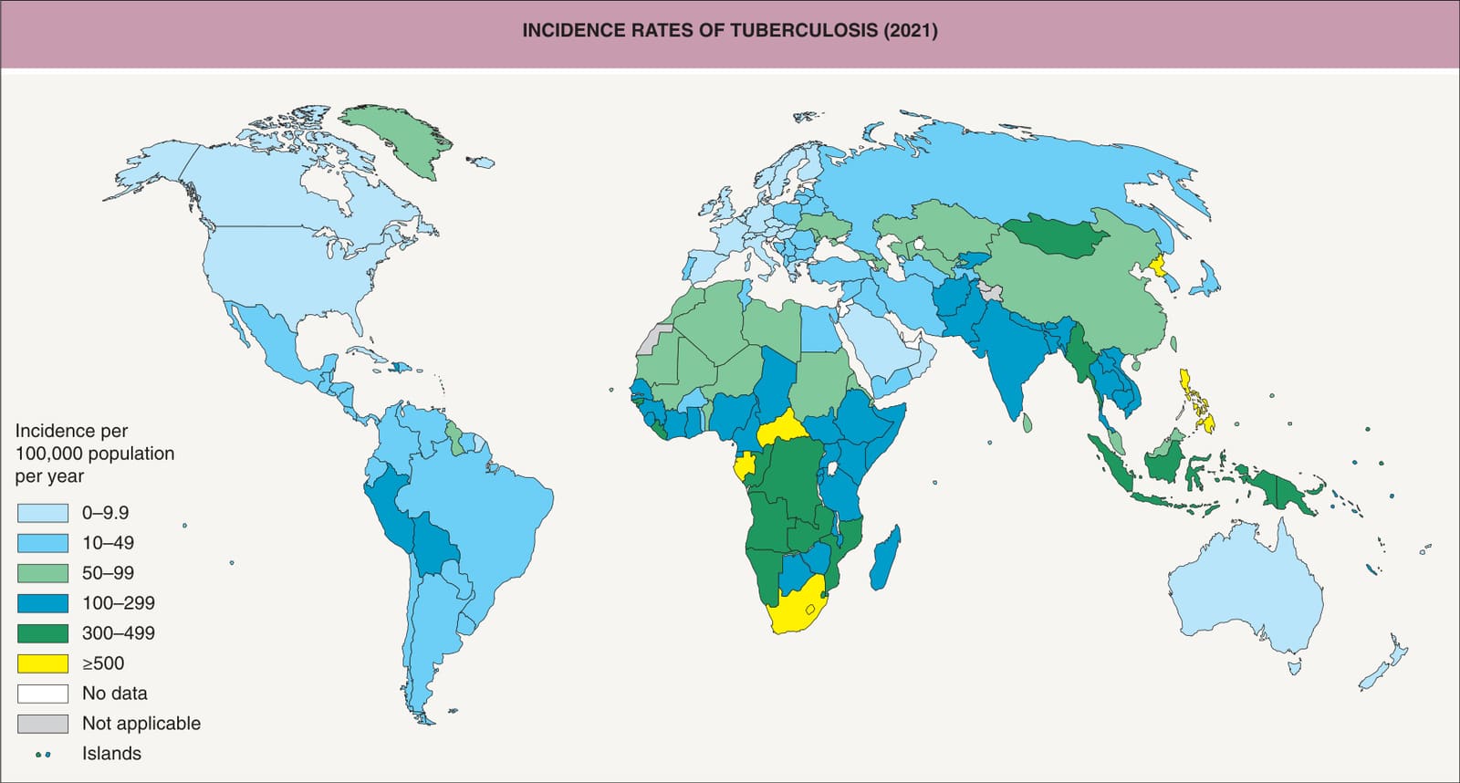
Fig. 75.13 Tuberculosis incidence rates, 2021.Reproduced from the Global Tuberculosis Report 2022. Geneva: World Health Organization, 2022. https://www.who.int/teams/ global-tuberculosis-programme/tb-reports/global-tuberculosis-report-2022/tb-disease-burden/2-1-tb-incidence
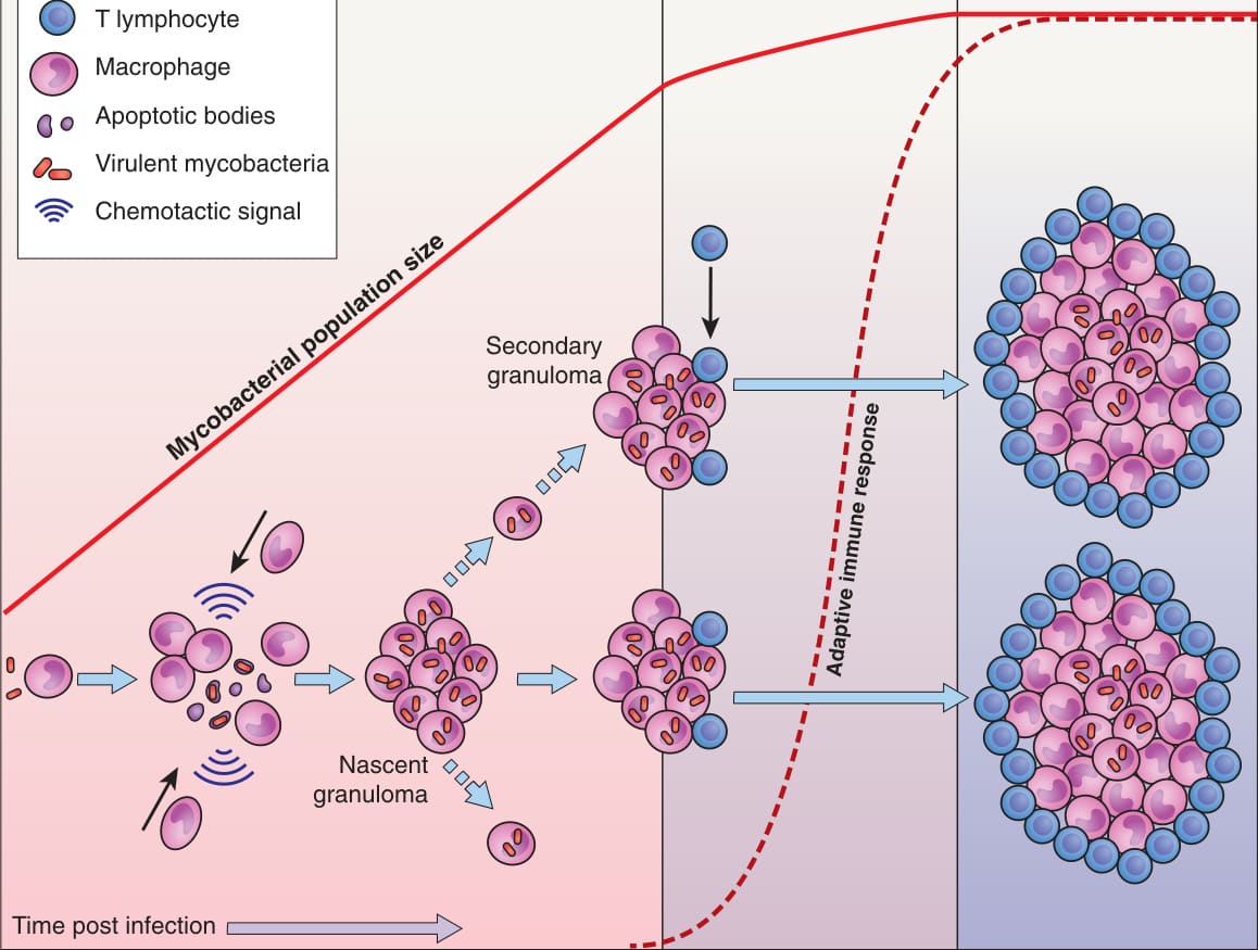
Fig. 75.14 Stages of granuloma formation in tuberculosis. The initial stage of tuberculosis is characterized by expansion of the mycobacterial population in the absence of adaptive immunity. Mycobacterial multiplication and spread are facilitated by the formation of the nascent granuloma. Infected macrophages undergo apoptosis and recruit additional macrophages, which phagocytose remnants of infected cells and their mycobacterial contents. Some of these newly infected macrophages then seed secondary granuloma formation. When adaptive immunity eventually occurs, CD4+ and CD8+ effector T cells curtail mycobacterial growth. Although this controls the infection, it is not eradicated. The mature granuloma therefore represents an equilibrium between mycobacterial proliferation and the host response. Adapted with permission from Bold TB, Ernst JD. Who benefits from granulomas, mycobacteria or host? Cell 2009;136:18–19.
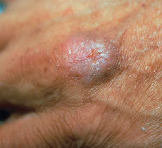
Fig. 75.15 Tuberculosis verrucosa cutis. A wart-like papule at the site of exogenous inoculation in a patient with immunity against M. tuberculosis.
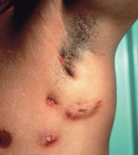
Fig. 75.16
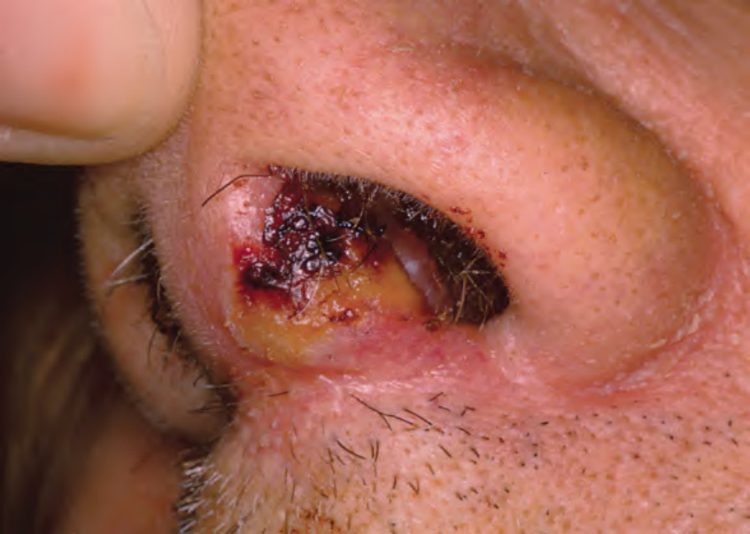
Fig. 75.17 Orificial tuberculosis. A non-healing ulcer of the nasal mucosa. Courtesy Louis A. Fragola, Jr, MD.
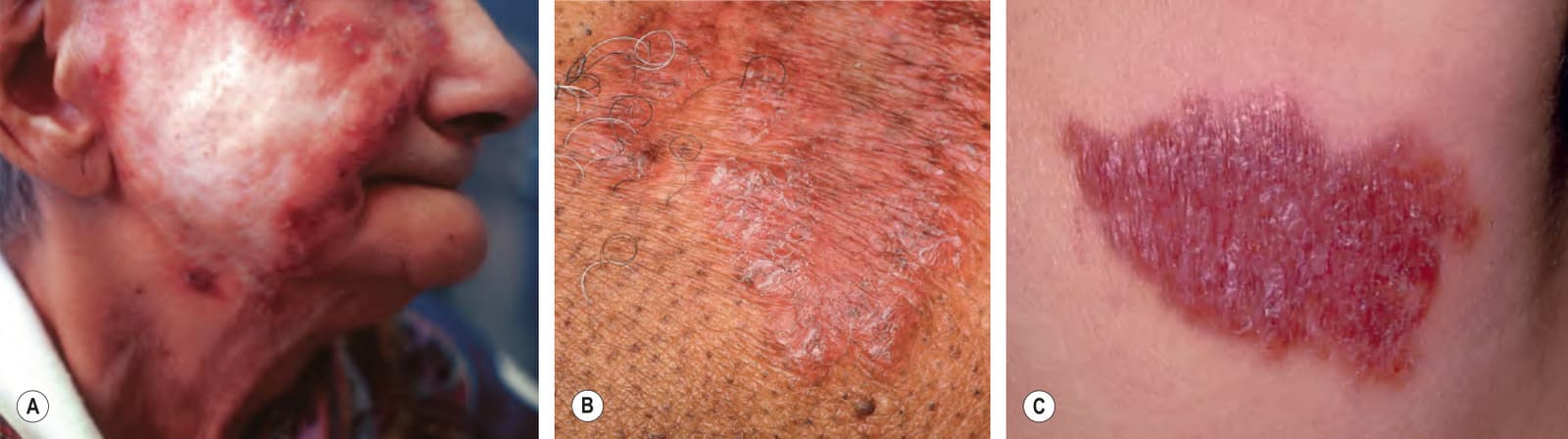
Fig. 75.18 Lupus vulgaris.A Annular granulomatous plaque with central scarring. B Coalescing pink–brown papules. C Red–brown plaque on the neck. C, Courtesy Eugene Mirrer, MD.
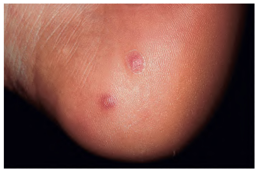
Fig. 75.19 Papulonecrotic tuberculid. Erythematous papules and papulopustules on the heel.
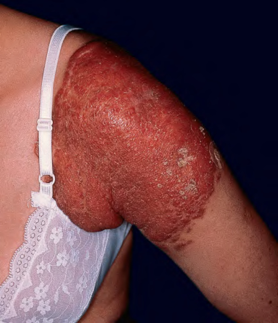
Fig. 75.20 Site of a BCG vaccination. Enlarging granulomatous plaque (BCGitis).
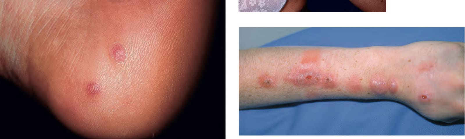
Fig. 75.21 Complication of injection of methanol extraction residue (MER) of
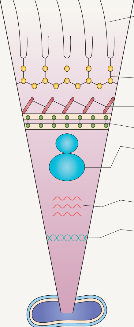
Fig. 75.22 Targets of the antituberculous agents.Adapted from Grange JM, Zumla A. Antituberculous agents. In: Cohen J, Powderly WG (eds). Infectious Diseases. London: Mosby, 2003. With permission from Elsevier.
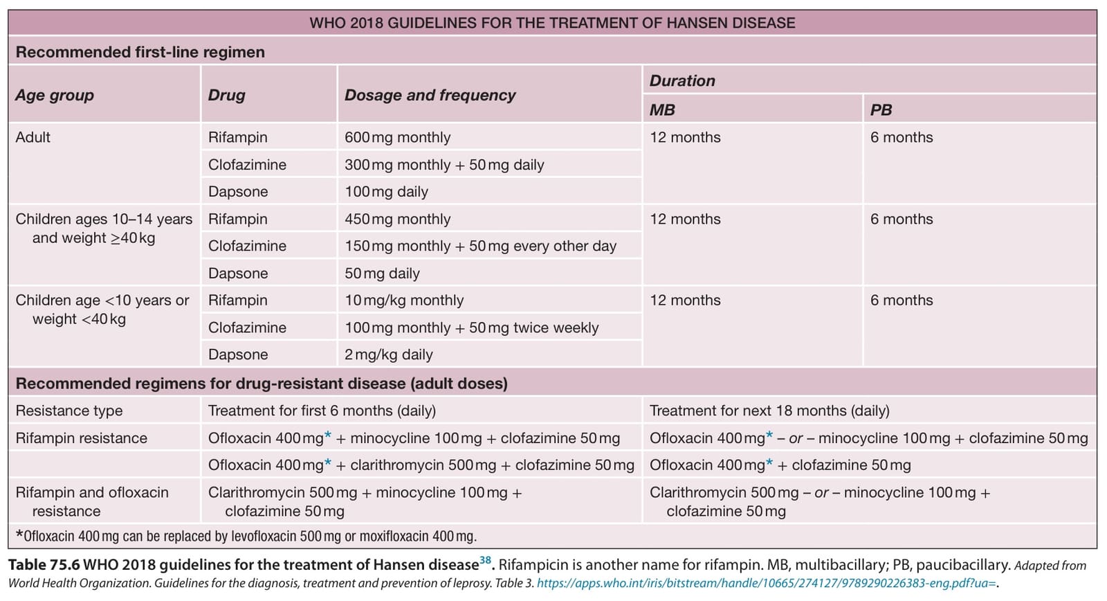
Table 75.6 WHO 2018 guidelines for the treatment of Hansen disease. Rifampicin is another name for rifampin. MB, multibacillary; PB, paucibacillary. Adapted from World Health Organization. Guidelines for the diagnosis, treatment and prevention of leprosy. Table 3. https://apps.who.int/iris/bitstream/handle/10665/274127/9789290226383-eng.pdf?ua=.
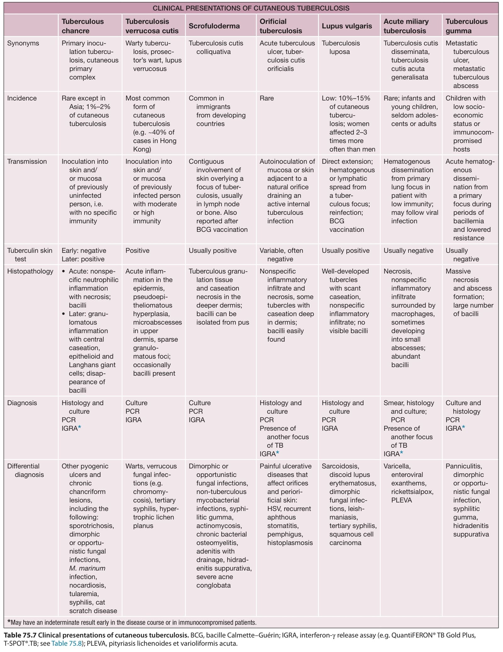
Table 75.7 Clinical presentations of cutaneous tuberculosis. BCG, bacille Calmette–Guérin; IGRA, interferon-γ release assay (e.g. QuantiFERON® TB Gold Plus, T-SPOT®.TB; see Table 75.8); PLEVA, pityriasis lichenoides et varioliformis acuta.
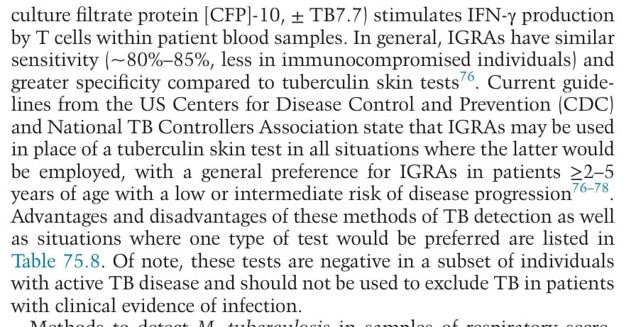
Table 75.8 Advantages and disadvantages of interferon-γ (IFN-γ) release assays and tuberculin skin testing. Both types of tests may be negative in patients with early active tuberculosis. Indeterminate IFN-γ release assay results due to failure of the internal positive control (i.e. a “low mitogen” response) are more common in immunocompromised individuals, young children, and older adults. Indeterminate results due to inappropriately high IFN-γ levels in the negative control (“high nil”) can also occur. Testing with a second method after an initial negative test may be useful when the risk of infection/progression is high or when there is clinical suspicion of active tuberculosis. The QuantiFERON® TB Gold Plus test directly measures IFN-γ levels, whereas the T-SPOT®.TB test determines the number of IFN-γ-producing T cells; both use a peripheral blood sample. BCG, bacille Calmette–Guérin.
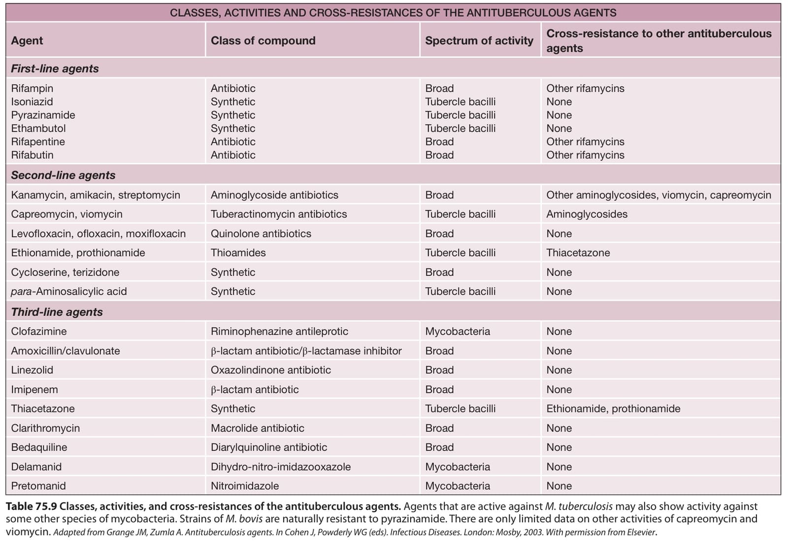
Table 75.9 Classes, activities, and cross-resistances of the antituberculous agents. Agents that are active against M. tuberculosis may also show activity against some other species of mycobacteria. Strains of M. bovis are naturally resistant to pyrazinamide. There are only limited data on other activities of capreomycin and viomycin. Adapted from Grange JM, Zumla A. Antituberculosis agents. In Cohen J, Powderly WG (eds). Infectious Diseases. London: Mosby, 2003. With permission from Elsevier.
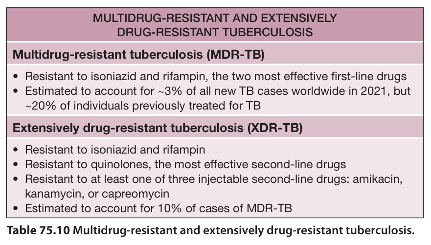
Table 75.10 Multidrug-resistant and extensively drug-resistant tuberculosis.