NON-TUBERCULOUS MYCOBACTERIOSES
Synonyms: Atypical mycobacterioses Mycobacteria other than tuberculosis (MOTT) Environmental mycobacterioses Anonymous mycobacterioses Opportunistic mycobacterioses
Key features
Over a dozen species of mycobacteria, found in the natural environment and transmitted via inoculation into the skin, can produce cutaneous disease
Clinical presentations include pustules, keratotic plaques and nodules (with or without suppuration), a sporotrichoid (lympho cutaneous) pattern, and ulcers with draining sinuses
Disseminated infections usually occur in immunocompromised hosts
Although suggested by a history of exposure, the diagnosis rests on isolation or identification of the responsible organism
Introduction
Non-tuberculous (“atypical”) mycobacteria (NTM) were first isolated in 1931 by Pinners, who also discovered that these organisms were different from M. tuberculosis – they were not virulent in the guinea pig model, and they responded poorly to antituberculous therapy. Their importance as human pathogens, however, was not appreciated until the 1950s92.
These environmental mycobacteria are found in water, wet soil, house dust, dairy products, cold-blooded animals, vegetation, and human feces. NTM are transmitted by inhalation, ingestion, or percutaneous penetration, which can result in pulmonary, lymph node, or skin disease. The type of disease depends on the species of mycobacteria, the route and degree of exposure, and the immune status of the host. Individuals with innate defects of the IL-12/IFN-γ axis (≥10 genes implicated; e.g. STAT1, ISG15 [IFN-stimulated gene 15]), GATA2 deficiency, and anti-IFN-γ antibodies are predisposed to severe and disseminated infections with NTM (see Ch. 60).
The incidence of infections by NTM appears to be increasing due to factors such as greater use of immunosuppressive medications and more frequent surgical procedures as well as improved detection via PCR-based techniques. Although these infections should be considered in patients with indolent ulcers, nodules, or plaques, especially in the setting of immunosuppression, they are often misdiagnosed because of their nonspecific or sometimes subtle clinical presentations, combined with inadequate biopsy and culture techniques. The ability of some NTM to survive sterilization procedures and to even contaminate antiseptic solutions can lead to infections in surgical patients. These infections have been described in association with cutaneous surgical procedures (e.g. laser resurfacing, botulinum toxin injections, hair transplantation, liposuction), other surgical procedures
(e.g. cardiothoracic, ocular, breast reconstruction), and acupuncture, body piercing, and tattooing.
Antibiotic susceptibility and therefore selection of therapy depends on the NTM species (Table 75.11). Empiric treatment with clarithromycin can be initiated while awaiting culture and sensitivity results in patients with clinical presentations suggestive of cutaneous NTM infection. However, multiplex PCR-based assays (e.g. targeting 16 S rRNA and hsp65) are available to more rapidly identify NTM species of clinical importance and help direct therapy.
In 1954, Runyon proposed the first classification system for NTM based upon rate of growth in vitro and pigment production following exposure to light (see Table 75.1). Due to advances in genomic analysis, at least 200 species of mycobacteria are now recognized. Selected NTM of dermatologic interest will be discussed in this chapter.
Mycobacterium ulcerans
Synonyms: Buruli ulcer Bairnsdale ulcer Searle’s ulcer
Introduction and history
M. ulcerans is a slow-growing, acid-fast bacillus that characteristically infects the skin and subcutaneous tissues, producing indolent ulcers. After Hansen disease and tuberculosis, the “Buruli ulcer” is the third most common mycobacteriosis worldwide in immunocompetent hosts, and it is classified as a neglected tropical disease by the WHO. In 1897, Cook first described patients with huge cutaneous ulcers from the Buruli county of Uganda. It was not until the late 1940s, in Australia, that the agent was isolated and characterized by MacCallum and colleagues.
Epidemiology
M. ulcerans infections have been reported in >30 countries worldwide, with primary endemic foci near freshwater wetlands in tropical and subtropical countries. Although the exact mode of transmission is not known, exposure to water or mud from rivers, ponds, or swamps is thought to play a role. The organism is harbored by snails, fish, and water insects, and it can be isolated from aquatic plants. In Australia, koalas and opossums are naturally infected.
Agricultural activities and consequent deforestation have contributed to increases in the incidence of M. ulcerans infections, especially in West Africa. Focal outbreaks have followed floods and human migrations, and HIV co-infection has been associated with more rapid progression into severe ulcerative stages. The countries most affected are in Central Africa (e.g. Uganda, Democratic Republic of the Congo) and West Africa (e.g. Ivory Coast, Ghana, Benin). Japan, Southeast Asian countries, and Australia have major foci of infection, and there have been a few reports from South America and Mexico.
M. ulcerans is typically introduced into the skin by minor trauma, and the disease is rarely transmitted from patient to patient. Children under 15 years of age are most commonly affected in West Africa, but infection can occur in all age groups.
Pathogenesis
After inoculation into the skin, M. ulcerans organisms proliferate and form abundant clumps of extracellular acid-fast bacilli. The organisms
are found at the base of the ulcer and within adjacent necrotic subcutaneous tissue as well as in surrounding normal-appearing skin.
M. ulcerans produces mycolactone, a macrolide exotoxin produced by polyketide synthases that are encoded by the virulence plasmid pMUM. Mycolactone is able to: (1) decrease inflammation via inhibition of protein translocation into the endoplasmic reticulum (ER), leading to suppression of cytokine/chemokine production, dendritic cell function, and L-selectin-mediated lymphocyte homing; (2) promote cell death via anoikis by disrupting actin dynamics through hyperactivation of the Wiskott–Aldrich syndrome protein (WASP); and (3) inhibit pain perception by binding to angiotensin II type 2 receptors on neurons, leading to potassium-dependent hyperpolarization. These effects explain the severe tissue destruction with minimal pain and inflammation observed in Buruli ulcers, and different geographical lineages of M. ulcerans produce forms of mycolactone with varying virulence. In addition, mycolactone itself represents a potential diagnostic/prognostic biomarker and therapeutic target.
Clinical features
The initial lesion is usually a single, asymptomatic, firm, non-tender, mobile subcutaneous nodule that measures 1 to 2 cm in diameter. After 1 or 2 months, the nodule becomes fluctuant and ulcerates. The resultant painless ulcer has a deep necrotic base with undermined edges, and often expands to 15 cm or more in diameter. Most lesions are on the extremities, especially the leg. There is little to no regional lymphadenopathy or systemic manifestations, although edema can develop in adjacent tissues. Without treatment, ulcers may remain small and heal as depressed scars, or they may spread rapidly over large areas of skin causing extensive scarring and deformity. Structures such as the eye, breast, or genitalia are sometimes severely damaged. Underlying bone can become involved, and progressive osteomyelitis may require amputation.
The WHO divides M. ulcerans disease into three categories of severity: (1) single lesion <5 cm in diameter; (2) single lesion (± ulceration) or area of edema 5–15 cm in diameter; (3) lesion >15 cm in diameter, multiple lesions, osteomyelitis, or joint involvement.
Pathology
Histologically, mature lesions show extensive necrosis with destruction of nerves, appendages, and blood vessels. There are few inflammatory cells in active lesions, but a granulomatous response may develop during healing. Many acid-fast bacilli can be seen in characteristic clumps, particularly in the center of the lesion.
Diagnosis and differential diagnosis
Clumps of acid-fast bacilli are typically seen with Ziehl–Neelsen staining of smears and biopsy specimens from the necrotic ulcer base or under-mined ulcer edge. M. ulcerans can be cultured, but visible growth often requires 6 to 8 weeks of incubation at 32°C. PCR-based detection of the M. ulcerans-specific insertion sequence 2404 (IS2404), which is highly sensitive and specific, has become the gold standard for Buruli ulcer diagnosis. Additional diagnostic tests that are under development include the loop-mediated isothermal amplification (LAMP) test, thin-layer chromatography for the detection of mycolactone, and antigen detection assays. In a series of 70 patients with Buruli ulcers, a modified PCR assay had a sensitivity of 98%, while the sensitivities of direct smear, culture, and histology were 42%, 49%, and 82%, respectively.
The differential diagnosis depends upon the stage of disease. During the initial phase, when a subcutaneous nodule is present, M. ulcerans infection must be differentiated clinically from other forms of panniculitis (infectious and non-infectious; see Ch. 100), cysts, nodular fasciitis, foreign body granulomas, and other granulomatous diseases. During the ulcerative phase, considerations may include fungal infections, pyoderma gangrenosum, suppurative panniculitis, and other causes of chronic ulcers (see Ch. 105).
Treatment
Antibiotic therapy can be effective when used alone or in combination with surgery (debridement or more extensive procedures [see below]). Treatment with oral rifampin (10 mg/kg daily) plus clarithromycin (7.5 mg/kg twice daily or 15 mg/kg extended release daily) is now recommended as first-line treatment by the WHO. A randomized controlled trial demonstrated that 8 weeks of this regimen was not inferior to and better tolerated than the same oral rifampin dose plus intramuscular streptomycin (15 mg/kg) daily in patients with a single lesion ≤10 cm in diameter. Other antibiotic protocols (e.g. in Australia) utilize rifampin plus moxifloxacin or ciprofloxacin for 6–8 weeks. Of note, ~10%–20% of patients with Buruli ulcers (especially those associated with a high bacterial load) experience a paradoxical reaction upon initiation of antibiotic therapy, with temporary worsening of ulceration (peak at 6–8 weeks) and subsequent healing.
While surgical excision represents the traditional treatment of choice for ulcers (see Table 75.11), this is obviously easier to accomplish when the ulcers are small, as large ulcers often require skin grafts or, rarely, amputation. Continuous local heating to 40°C by circulating water jackets promotes healing, even without excision, and hyperbaric oxygen may also be useful. Physical therapy is important when the patient is at risk for contractures. There is no effective prophylaxis, but vaccination with BCG may lead to limited, relatively short-lived protection.
Mycobacterium marinum
Synonyms: Fish tank granuloma Swimming pool granuloma Infection by M. balnei
Introduction and history
M. marinum is a free-living organism that causes disease in freshwater and saltwater fish and sporadically in humans. The latter results from exposure to contaminated water when there are breaks in the skin barrier. M. marinum was first isolated from saltwater fish in the Philadelphia Aquarium in 1926 by Aronso. The first report of human disease was in 1951, when the agent was found in granulomatous skin lesions of individuals who swam in a contaminated swimming pool. There is scientific interest in M. marinum because of its phylogenetic similarity to M. tuberculosis.
Epidemiology and pathogenesis
M. marinum is found in aquatic environments, including fresh, salt, and brackish water. When cleaning fish tanks at home or in the workplace (e.g. restaurants, fish markets), trauma to the hands may result in exposure to M. marinum. The infection can also affect swimming pool bathers, as this organism seems to be rather resistant to chlorine.
Cutaneous infection with M. marinum requires a portal of entry, e.g. abraded or traumatized skin. The incubation period is typically 2 to 3 weeks, but it can range from 1 week to 2 months. In several case series of skin infections due to NTM from higher-income countries, M. marinum was the most common species identified.
Clinical features
The initial lesion is a single bluish-red inflammatory nodule or pustule at the site of inoculation, which is often on an upper extremity (Fig. 75.23A). It then forms a crusted ulcer, suppurative abscess, or verrucous nodule. Multiple lesions can develop along the course of the draining lymphatics, i.e. sporotrichoid (lymphocutaneous) spread (Fig. 75.23B; see Table 77.17). Among the NTM, M. marinum is the most common cause of this pattern. Occasionally, deeper infections are complicated by tenosynovitis, septic arthritis, or rarely, osteomyelitis. As with other NTM, atypical or disseminated lesions can occur in immunocompromised hosts, including those treated with TNF inhibitors (Fig. 75.23C).
Pathology
The histologic features, as in other mycobacterial infections, range from acute and chronic inflammation to well-formed tuberculoid granulomas. Fibrinoid changes and caseation necrosis can occur. In immunocompetent individuals, organisms are often difficult to find.
Diagnosis and differential diagnosis
The clinical features combined with a history of trauma in a wet environment or exposure to fish tanks suggests the possibility of M. marinum infection. The diagnosis is confirmed by culture, which is positive in 70% to 80% of cases. When this infection is suspected, the laboratory should be notified in advance and care must be taken to provide proper culture conditions. M. marinum grows best at 31°C, in contrast to the optimal incubation temperature of 37°C for many other mycobacteria; growth becomes evident after a mean of ~3 weeks, and identification of the species and its antimicrobial susceptibility pattern often requires 2 months. PCR can also be performed to enable a rapid diagnosis.
Infection due to M. marinum should be differentiated from sporotrichosis and the entities outlined in Table 77.17 as well as tuberculosis verrucosa cutis. It is not associated with marked regional lymphadenitis.
Treatment
M. marinum is usually sensitive to the antimicrobial drugs listed in Table 75.11; however, sensitivity testing should be requested as part of the laboratory evaluation. Based on usual in vitro susceptibility patterns and clinical responsiveness, initial empiric treatment with clarithromycin (pending culture and sensitivity results) has been recommended when there is suspicion of an M. marinum infection. A general approach is to treat with two agents (e.g. clarithromycin plus ethambutol or rifampin) until 1–2 months after resolution of clinical findings, often for a total of 3–4 months; a single agent is an option for milder disease, and longer-term treatment may be necessary in immunosuppressed patients.
Mycobacterium kansasii
Introduction and history
M. kansasii most often causes lung disease, which can be clinically and radiologically indistinguishable from tuberculosis. There is also evidence that infection with this organism provides increased immunity against tuberculosis.
M. kansasii was first reported in 1953 as the “yellow bacillus”. This characteristic pigmentation is due to the deposition of β-carotene crystals after exposure to light.
Epidemiology and pathogenesis
This organism is found throughout the world, but it is more prevalent in temperate zones within the US, South America, China, and Europe. It has been isolated from cattle and swine, but the natural reservoir may be water. Middle-class, middle-aged White men living in urban areas are most commonly affected. Cutaneous infection is usually acquired via minor trauma such as puncture wounds.
Clinical features
Verrucous plaques, ulcers, and nodules are observed, and the lesions may be arranged in a lymphocutaneous (sporotrichoid) pattern. The majority of patients with cutaneous involvement have some alteration in immune status. Papulonecrotic lesions and cellulitis are less common presentations. Iijima and Sasaki divided cutaneous mycobacteriosis due to M. kansasii into three types: (1) primary cutaneous chronic granulomatous; (2) primary cutaneous pyogenic; and (3) disseminated acute pyogenic, which occurs primarily in immunocompromised individuals.
Pathology
Histologic features are as variable as the clinical presentations. Typical tuberculoid granulomas may be seen, or a dense neutrophilic infiltrate may be evident, with or without abscess formation and epidermal
necrosis. In some patients, the findings are indistinguishable from those of M. tuberculosis infection.
Diagnosis and differential diagnosis
M. kansasii can be isolated from lesional skin biopsy specimens, and the laboratory should be advised to provide appropriate culture conditions to detect slow-growing mycobacteria. The rarity of diagnosis of primary cutaneous M. kansasii infections may reflect failure to grow the organisms in culture as well as difficulties in phenotyping. PCR analysis may circumvent these potential problems because of its higher sensitivity and specificity.
Table 75.7 and the M. ulcerans section above outline the differential diagnosis of verrucous plaques and ulcers, respectively. Sporotrichosis and other NTM infections need to be considered when there is a sporotrichoid pattern (see Table 77.17). A combination of histologic examination, special stains, and culture for bacteria and fungi usually distinguishes these entities, but PCR may be required.
Treatment
A combination of antituberculous medications should be used (see Table 75.11), especially for disseminated infections. Because resistance to rifampin and other drugs may occur, sensitivity testing should be performed to optimize therapy.
Mycobacterium fortuitum, Mycobacterium chelonae, and Mycobacterium abscessus
Synonyms:M. chelonei=M. chelonae Turtle tubercle bacillus
Introduction and history
These rapidly growing mycobacteria are members of either the M. fortuitum group or the M. chelonae/M. abscessus complex. Although the individual organisms can be distinguished on the basis of their DNA and serologic analyses, they result in infections with similar clinical features.
In 1903, Friedmann isolated M. chelonae. Two subspecies (chelonae and abscessus) were initially identified, but M. abscessus was reclassified as a separate species in 1992. Costa-Cruz used the name M. fortuitum in 1938 for what he believed to be a new species isolated from a skin abscess that occurred after an injection. The M. fortuitum group currently includes the following additional species reported to cause human infections – M. alvei, M. boenickei, M. conceptionense, M. farcinogenes, M. houstonense, M. neworleansense, M. peregrinum, M. porcinum, M. senegalense, M. septicum, M. setense. The M. chelonae/M. abscessus complex now consists of M. chelonae, M. abscessus subsp. abscessus, M. abscessus subsp. bolletii, M. abscessus subsp. massiliense, M. immunogenum, and M. salmoniphilum.
Epidemiology and pathogenesis
The organisms exist as saprophytes, and they are found in water, soil, dust, and animals. Cutaneous infections are uncommon, and immunocompromised patients are more susceptible to severe disease caused by these species of mycobacteria.
Infections typically occur following trauma, surgery, or other procedures, including contact with contaminated medical instruments (e.g. liposuction, mesotherapy), placement of implants (e.g. prosthetic
breast implants), and tattoo ink or acupuncture needles. A post-injection abscess is another frequent presentation, e.g. after injection of botulinum toxin, dermal filler, phosphatidylcholine/deoxycholate, or vaccines. Furunculosis of the lower extremities has also been observed following footbaths in nail salon customers. Less often, cutaneous infection is the result of dissemination from an endogenous source.
Clinical features
This group of mycobacteria can produce a variety of clinical diseases, including non-cavitary pneumonia, keratitis, endocarditis, lymphadenitis, osteomyelitis, and skin infections. The cutaneous manifestations range from cellulitis, abscesses, and papulopustules to sinuses and ulcers with extensive subcutaneous necrosis and serosanguineous or purulent discharge (Fig. 75.24 & 75.25A). However, the most common presentation is multiple erythematous subcutaneous nodules, frequently occurring on the distal limbs or in a sporotrichoid pattern (see Fig. 75.24).
Pathology
The presence of both neutrophilic microabscesses and granuloma formation with foreign body-type giant cells is characteristic (Fig. 75.25B). Necrosis may occur.
Diagnosis and differential diagnosis
Culture of biopsy material must be performed. When an abscess is present, a biopsy specimen that includes the wall is preferred, rather than aspiration of pus. The organisms grow on routine bacterial culture media, such as 5% sheep blood agar or chocolate agar, within 7 days. Molecular methods utilizing PCR, gene sequencing, and matrixassisted laser desorption ionization–time of flight mass spectrometry are available to identify these NTM.
Infection with these organisms should be considered in patients with therapy-resistant or “cold” abscesses. It can also be confused with foreign body reactions, subcutaneous mycoses, or osteomyelitis.
Treatment
Treatment is dependent on the type and extent of the lesions. These organisms are resistant to antituberculous drugs, but nearly all M. chelonae and ~80% of M. fortuitum isolates are sensitive to clarithromycin. Excision and debridement may be required for abscesses and ulcers. Recommended antibiotics, either alone or in combination, are listed in Table 75.11; prolonged courses may be required. Successful treatment of refractory pulmonary and disseminated M. abscessus infections by intravenous administration of engineered bacteriophages has been reported in patients with advanced cystic fibrosis lung disease.
Mycobacterium avium Complex (MAC)
Synonyms:M. aviumM. avium-intracellulare (MAI) M. intracellulare
Introduction
In the 1980s, two closely related pathogens, M. avium and M. intracellulare, were designated as M. avium-intracellulare (MAI). Currently, the term M. avium complex (MAC) is utilized for these organisms as well as a growing number of additional species, including the emerging pathogen M. chimaera and others that have rarely been identified in human infections (M. arosiense, M. bouchedurhonense, M. colombiense, M. marseillense, M. timonense, M. vulneris, M. yongonense).
MAC organisms are facultative pathogens that only occasionally produced human disease prior to the HIV epidemic, when these mycobacteria produced disseminated disease in 15%–40% of patients with AIDS.
Epidemiology and pathogenesis
MAC organisms are found in the environment, including fresh and salt water, soil, dairy products, and domestic animals. They may be transmitted via inhalation into the lungs, or via water and food into the gastrointestinal tract, without person-to-person transmission. As many as 30% of samples of normal human feces yield isolates of MAC.
Chronic pulmonary infection is the most common clinical manifestation of MAC infection in those who are not co-infected with HIV, followed in frequency by cervical and inguinal lymphadenitis and osteomyelitis. Lymphadenitis more frequently occurs in children. Cutaneous lesions may be primary after a traumatic inoculation, which is rare, or secondary to a disseminated infection in an immunocompromised host.
Clinical features
In patients with AIDS, there is often disseminated disease characterized by nonspecific symptoms such as fever, night sweats, weight loss, bone pain, hepatosplenomegaly, and lymphadenopathy. Serum alkaline phosphatase levels may be elevated. Skin involvement is unusual and most often presents with papulopustules and multiple purulent leg ulcers. Nodules with abscess formation (Fig. 75.26), panniculitis, sinus tracts, folliculitis, and granulomatous plaques may also be seen. A papulonecrotic tuberculid-like eruption due to disseminated MAC infection has been reported in patients with AIDS.
Pathology
Macrophages may contain large numbers of bacilli without necrosis, and spindle cell transformation of macrophages with formation of a histoid-like lesion (as in Hansen disease) can occur. The histologic features can resemble those of the lepromatous form of Hansen disease (see above) or a spindle cell neoplasm (mycobacterial spindle cell pseudotumor).
Diagnosis and differential diagnosis
In disseminated disease, the diagnosis is often established by culture of the blood or from bone marrow or liver biopsies. Depending on the site(s) of involvement, cultures can also be performed on sputum or biopsies of skin and lymph nodes; in addition, highly sensitive and specific PCR analysis is available.
The differential diagnosis includes pyoderma, folliculitis, other causes of panniculitis and ulcers, dimorphic fungal infections, the lepromatous form of Hansen disease, cutaneous tuberculosis, and a spindle cell neoplasm.
Treatment
Table 75.11 provides a list of the medications that have been used alone or in combination to treat MAC infections. Immunocompetent patients should continue treatment until culture-negative for at least one year, whereas antimicrobial therapy should be continued for life in immunocompromised individuals with disseminated infection.
Mycobacterium haemophilum
Introduction
M. haemophilum is a short, slightly curved rod that occurs singly or in a cord-like arrangement. It is a fastidious organism that requires ferric ions to grow, provided as hemin, hematin, hemoglobin, or ferric ammonium citrate. In initial studies, growth was only observed with the addition of hemolyzed sheep erythrocytes; as a consequence, the organism was given the name M. haemophilum.
Epidemiology and pathogenesis
M. haemophilum infection occurs primarily in immunocompromised hosts, including people living with HIV, organ transplant recipients, and individuals receiving immunosuppressive medications. However, this organism can also cause cervical lymphadenitis in healthy children, and cutaneous infections following tattooing (including permanent make-up) and acupuncture have been reported. The majority of affected individuals are from cities located near the ocean, the Mediterranean Sea, or the Great Lakes of North America.The natural habitat of M. haemophilum has not yet been identified, although water reservoirs are suspected. M. leprae and M. haemophilum are phylogenetically related, which may explain why some individuals infected with M. haemophilum experience immune reconstitution events analogous to leprosy reactions (see above).
Clinical features
This bacillus can produce several different types of cutaneous lesions. Immunosuppressed adults typically present with one or more bluishred tender pustules, papulonodules, or plaques, which evolve into abscesses and/or ulcers. Lesions are usually located on the extremities, often overlying joints. M. haemophilum can also cause contiguous septic arthritis, osteomyelitis, and pneumonia. In immunocompetent individuals, cutaneous inoculation can lead to papulonodules and pustules. Immunocompetent children infected by this organism frequently have isolated cervical, submandibular, or perihilar lymphadenitis.
Pathology
A mixed reaction with both suppurative and granulomatous inflammation is commonly seen, but other cases have a nonspecific pattern. The bacilli are sometimes found aggregated in globi, resembling those seen in the lepromatous form of Hansen disease.
Diagnosis and differential diagnosis
The diagnosis is established by culture of the bacillus from tissue or synovial fluid; in some patients, blood cultures are positive. However, this organism requires a special environment to grow, with a source of iron and a temperature between 30°C and 32°C. Due to these stringent requirements, it may not be isolated using routine techniques. Highly sensitive and specific PCR analysis is also available.
In immunocompetent children with cervical or submandibular lymphadenitis, the possibility of MAC infection should be considered, along with the more common causes of lymphadenitis (e.g. EBV, CMV). The differential diagnosis of the cutaneous lesions is similar to that for infections with other NTM (see above, M. fortuitum and MAC).
Treatment
M. haemophilum is often resistant to antituberculous drugs, except for the rifamycins, such as rifampin and rifabutin. The combination of rifampin and clarithromycin (± amikacin) has been used successfully (see Table 75.11). The duration of therapy should be at least 6–24 months, with longer courses in immunosuppressed patients. Surgical excision is also an option when the disease is localized, e.g. adenitis in an immunocompetent child.
Mycobacterium scrofulaceum
Introduction
M. scrofulaceum was formerly grouped with MAC in a complex called MAIS (M. avium-intracellulare-scrofulaceum) but was separated due to its distinct clinical manifestations. M. scrofulaceum produces self-limited disease, particularly lymphadenitis, while MAC typically causes chronic lung disease and, less often, osteomyelitis. M. scrofulaceum may also cause cervical lymphadenitis with sinus formation clinically indistinguishable from scrofuloderma.
Epidemiology and pathogenesis
This organism is most prevalent in the southeastern US. It is widely distributed and has been isolated from raw milk, other dairy products, oysters, soil, and water. Bacilli can be found in environments with: a warm temperature; low pH and dissolved oxygen level; and high soluble zinc, humic acid, and fulvic acid levels. It has also been found on the skin of healthy people and in cutaneous lesions of Hansen disease.
In general, infection by M. scrofulaceum affects children and occurs via inhalation or ingestion. Focal infection of lymph nodes due to contamination during surgical procedures may also occur. Cutaneous disease in a campground worker with corticosteroid-treated systemic lupus erythematosus and no history of preceding trauma was thought to result from subclinical bacteremia.
Clinical features
M. scrofulaceum may cause pulmonary disease and local lymphadenitis, primarily in the submandibular and submaxillary regions. It is usually a benign, self-limited condition with no systemic symptoms except for mild neck pain. The involved lymph nodes slowly enlarge over several weeks, and eventually ulceration and drainage occur with fistula formation. This organism can also produce skin lesions in a
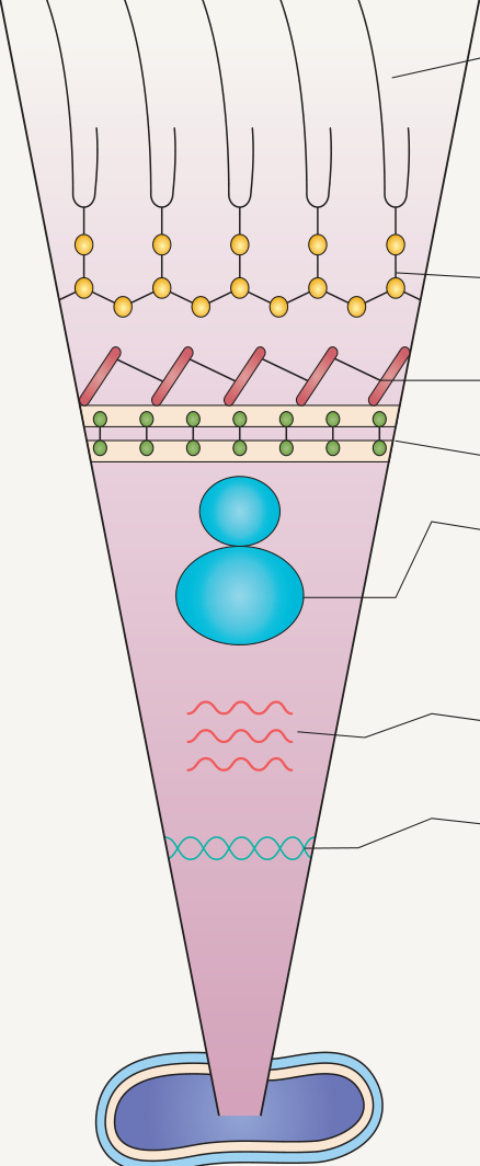
Fig. 75.22 Targets of the antituberculous agents.Adapted from Grange JM, Zumla A. Antituberculous agents. In: Cohen J, Powderly WG (eds). Infectious Diseases. London: Mosby, 2003. With permission from Elsevier.
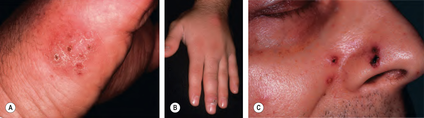
Fig. 75.23 Mycobacterium marinum infections. A range of presentations, including an erythematous plaque with scale-crust at the inoculation site on the lateral hand (A), a sporotrichoid pattern with the inoculation site on the distal third finger (B), and disseminated necrotic lesions on the face of an immunocompromised patient (C).
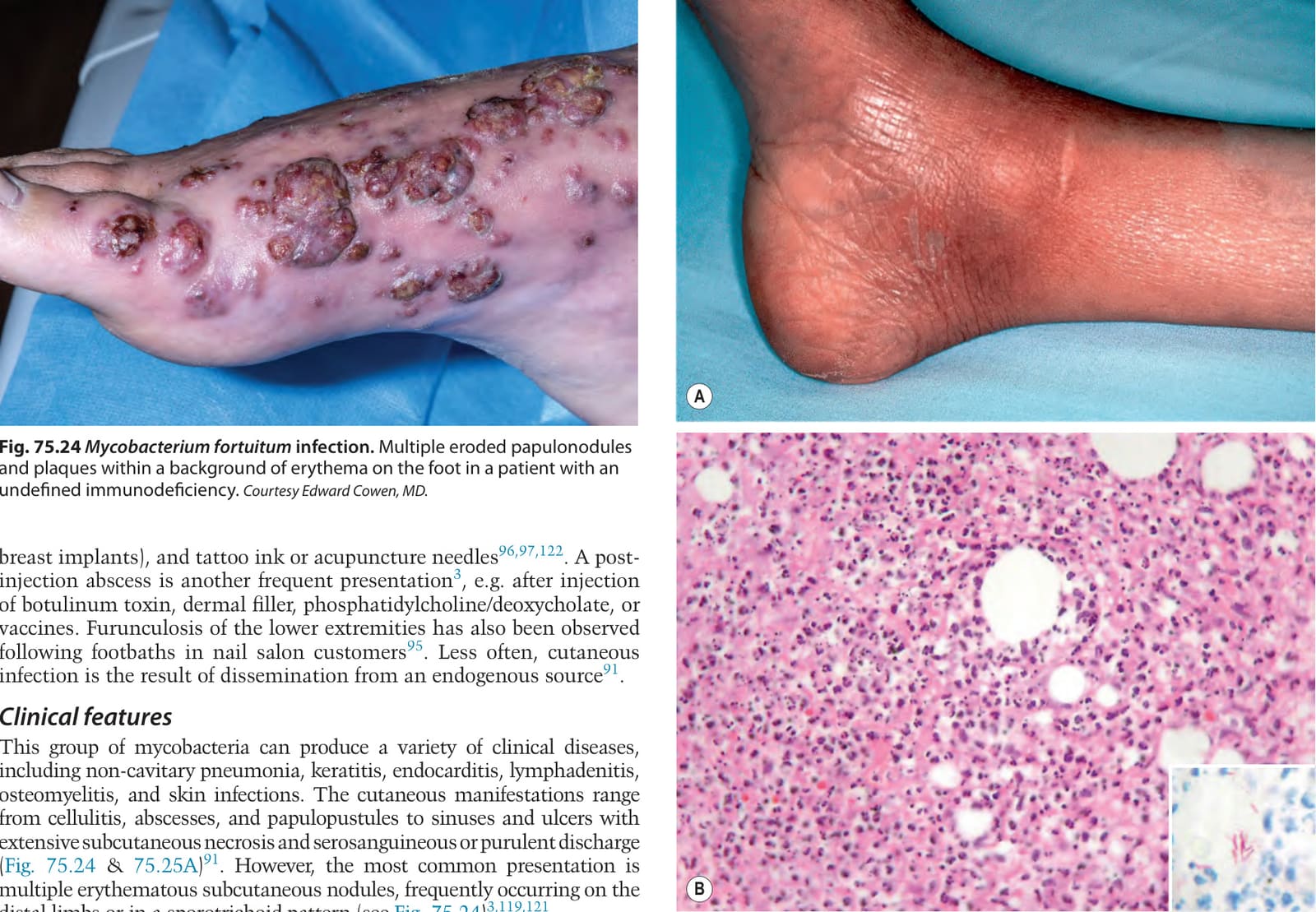
Fig. 75.24 Mycobacterium fortuitum infection. Multiple eroded papulonodules and plaques within a background of erythema on the foot in a patient with an undefined immunodeficiency. Courtesy Edward Cowen, MD.
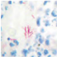
Fig. 75.25 Mycobacterium chelonae infection.A This immunosuppressed patient presented with an area of warmth and mild erythema on the ankle. B Mixed diffuse inflammatory infiltrate with granuloma formation. Curved acid-fast bacilli are seen with Fite staining (inset).
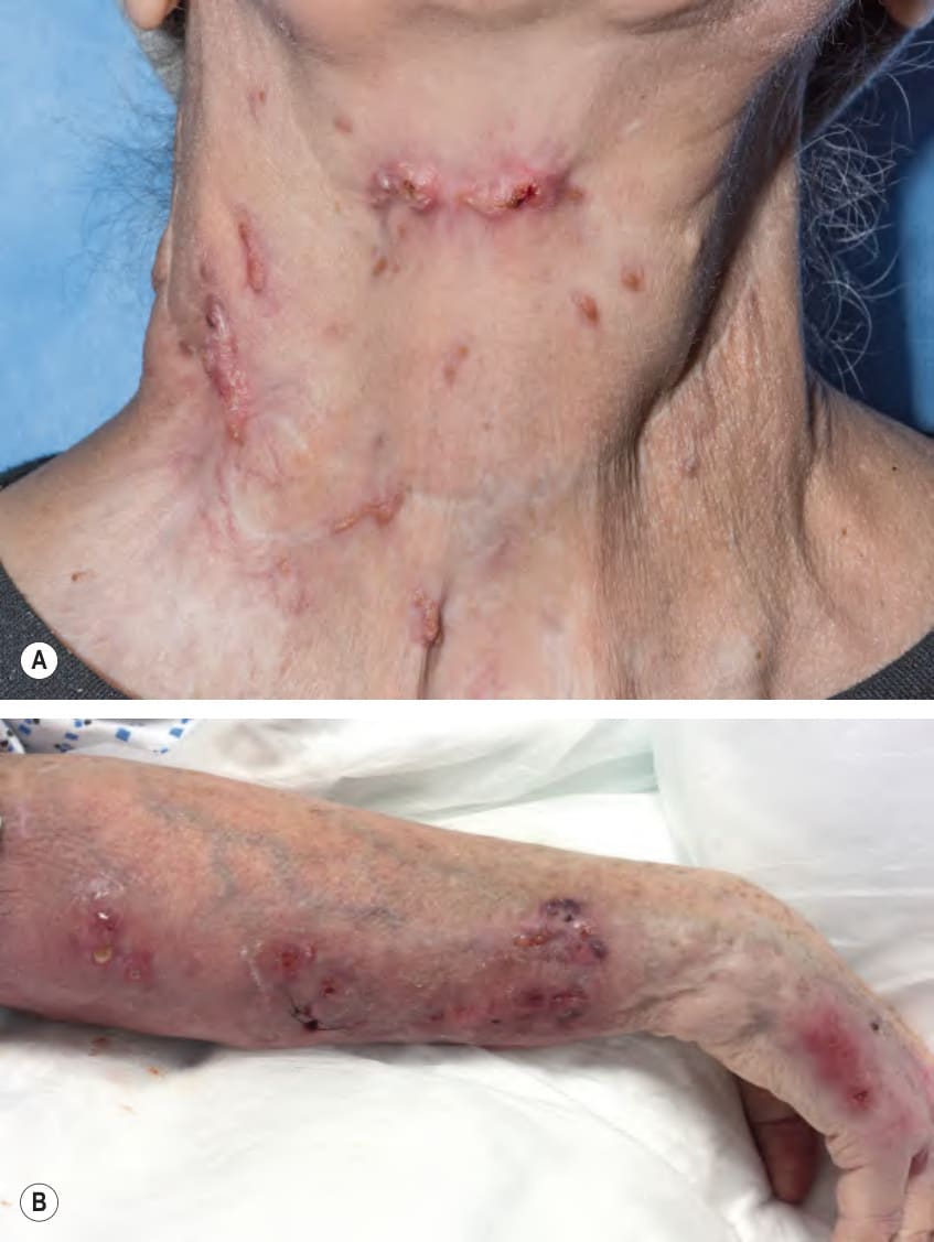
Fig. 75.26 Mycobacterium avium complex infections.A Papules and nodules on the neck in a patient with interferon gamma receptor deficiency. B Sporotrichoid (lymphocutaneous) pattern of the coalescing nodules with abscess formation in an immunocompromised patient. A, Courtesy Edward Cowen, MD.
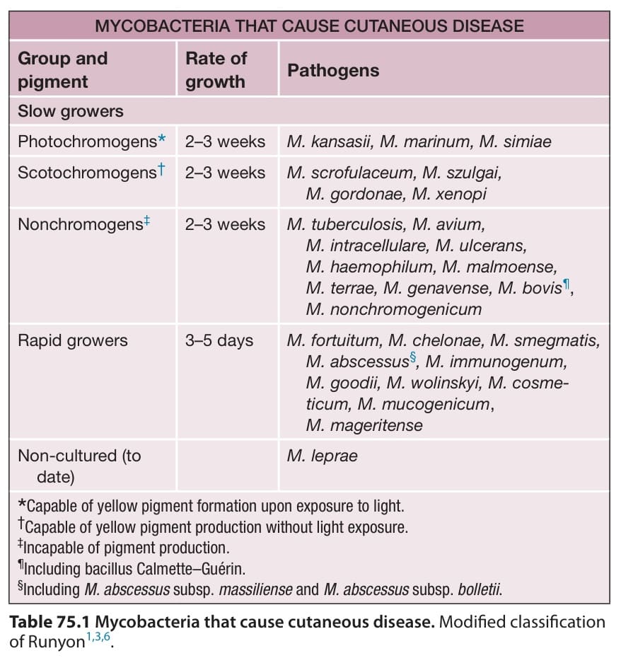
Table 75.1 Mycobacteria that cause cutaneous disease. Modified classification of Runyon.
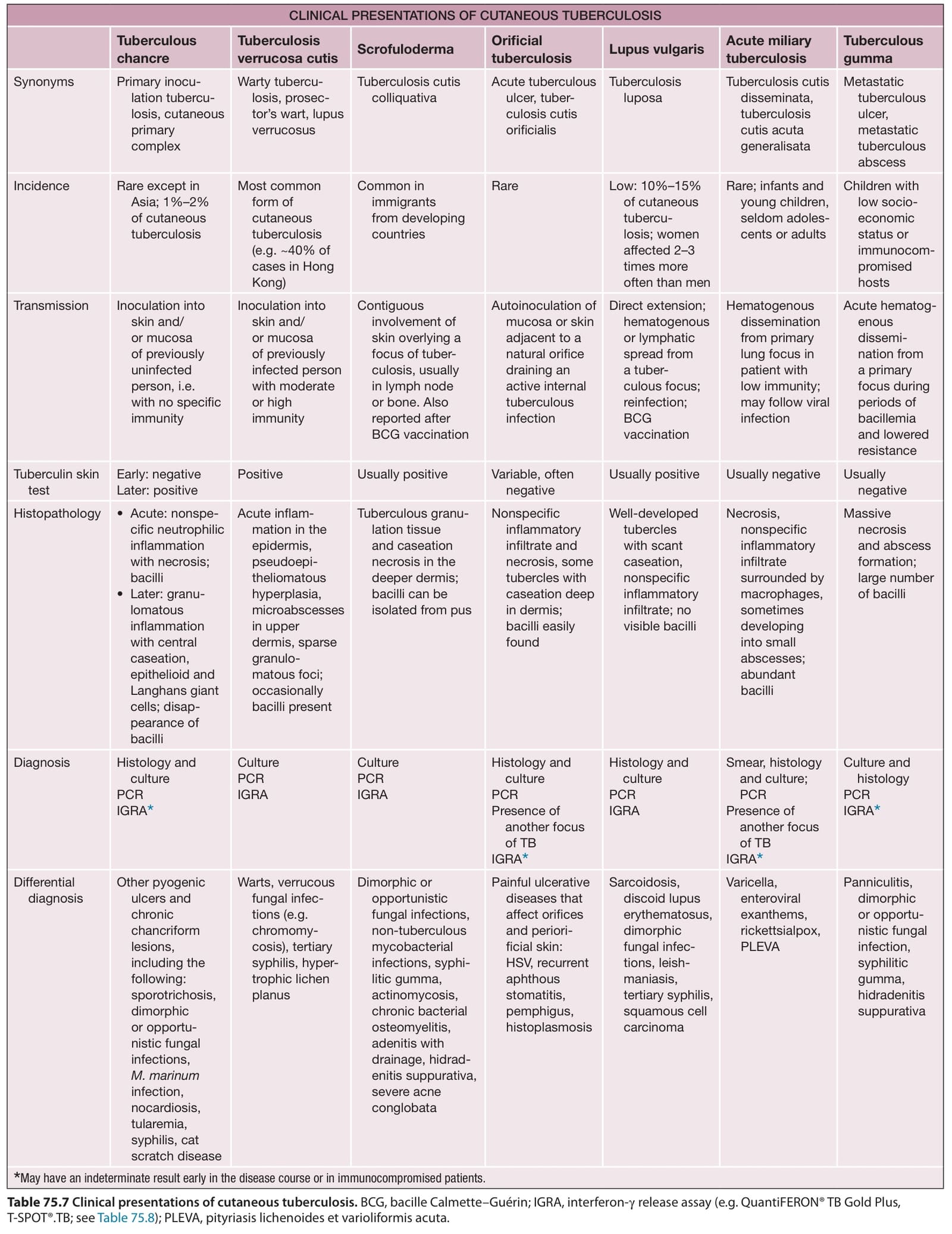
Table 75.7 Clinical presentations of cutaneous tuberculosis. BCG, bacille Calmette–Guérin; IGRA, interferon-γ release assay (e.g. QuantiFERON® TB Gold Plus, T-SPOT®.TB; see Table 75.8); PLEVA, pityriasis lichenoides et varioliformis acuta.
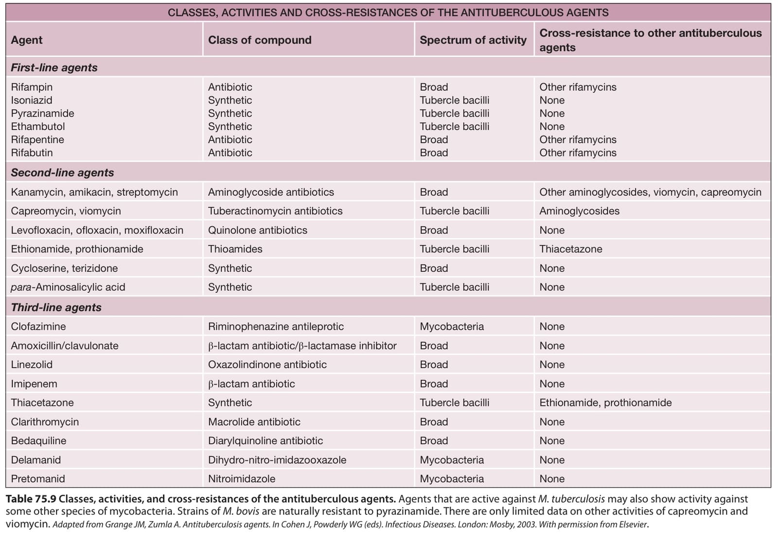
Table 75.9 Classes, activities, and cross-resistances of the antituberculous agents. Agents that are active against M. tuberculosis may also show activity against some other species of mycobacteria. Strains of M. bovis are naturally resistant to pyrazinamide. There are only limited data on other activities of capreomycin and viomycin. Adapted from Grange JM, Zumla A. Antituberculosis agents. In Cohen J, Powderly WG (eds). Infectious Diseases. London: Mosby, 2003. With permission from Elsevier.
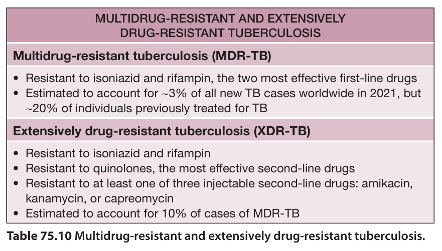
Table 75.10 Multidrug-resistant and extensively drug-resistant tuberculosis.
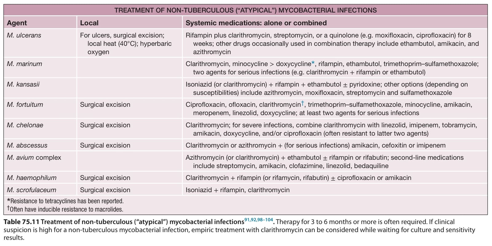
Table 75.11 Treatment of non-tuberculous (“atypical”) mycobacterial infections. Therapy for 3 to 6 months or more is often required. If clinical suspicion is high for a non-tuberculous mycobacterial infection, empiric treatment with clarithromycin can be considered while waiting for culture and sensitivity results.
sporotrichoid pattern, but it rarely causes disseminated infection. Primary cutaneous disease due to inoculation of M. scrofulaceum is unusual.
In the lymph nodes, widespread abscess formation is the typical finding; histiocytes and granulomas may be inconspicuous. It may be difficult to differentiate M. scrofulaceum lymphadenitis from that due to M. tuberculosis when there are tuberculoid granulomas with central necrosis and abscess formation. The bacilli are found within involved lymph nodes. In the skin, abscess formation is typically seen.
In addition to histologic examination, culture of sputum, skin, or involved lymph nodes helps to confirm the diagnosis. The infection should be distinguished from other causes of cervical lymphadenopathy (see M. haemophilum), particularly infection with M. tuberculosis. However, M. tuberculosis usually affects tonsillar and anterior cervical nodes, while M. scrofulaceum typically affects submandibular and submaxillary nodes. When lesions have a sporotrichoid pattern, M. marinum infections and other non-tuberculous mycobacterioses should be considered (see Table 77.17).
The treatment of choice is total excision of the involved lymph nodes because, in general, antibiotic therapy is unsatisfactory. However, promising results have been reported with isoniazid plus rifampin. Successful treatment with clarithromycin has also been described (see Table 75.11).
Additional figures and a table on Drug regimens for pulmonary tuberculosis, available in our eBook (see inside front cover for access code).
O, Harman R, eds. Clinical Tropical Dermatology. 2nd ed. Boston: Blackwell Scientific; 1992:165–200.20. Bhatia A, Katoch K, Narayanan R, et al. Clinical and histo-
Neves RG, Talhari S, eds. Dermatologia Tropical. Rio de Janeiro: Medsi; 1995:283–290.7. Han XY, Seo YH, Sizer KC, et al. A new Mycobacterium treatment and prevention of leprosy. <https://apps.who. int/iris/bitstream/handle/10665/274127/9789290 226383-eng.pdf?ua>.
Table 75.11 Treatment of non-tuberculous (“atypical”) mycobacterial infections. Therapy for 3 to 6 months or more is often required. If clinical suspicion is high for a non-tuberculous mycobacterial infection, empiric treatment with clarithromycin can be considered while waiting for culture and sensitivity results.