Q FEVER
Coxiella burnetii are obligately intracellular bacteria categorized in the order Legionellales. Although they reside within several tick species, these organisms are transmitted to humans primarily by inhalation of aerosols generated from the placenta and birth fluids of infected animals (see Table 76.1). The bacteria target macrophages, where they thrive in acidic phagolysosomes, and their resistance to heat, desiccation, and disinfectants allows them to survive in the environment.
Acute disease typically presents as a nonspecific febrile illness, with atypical pneumonia or granulomatous hepatitis occurring occasionally. Chronic Q fever most often manifests as “culture-negative” endocarditis affecting a previously damaged heart valve. Cutaneous manifestations of Q fever are rare (see Table 76.2). A truncal maculopapular rash, an urticarial eruption, erythema nodosum, palpable purpura, palatal petechiae, and vasculitis (reported in association with mixed cryoglobulinemia) have been described.
-
Blanton LS. The rickettsioses: a practical update. Infect
-
Yu XJ, Walker DH, Family I. Rickettsiaceae. In: Brenner DJ,
Kreig NR, Staley JT, editors. Bergey’s Manual of Systematic Bacteriology: Proteobacteria. v. 2. 2nd ed. New York: Springer-Verlag; 2005: p. 96–116.2. Walker DH. Rickettsiae and rickettsial infections: the
- Helmick CG, Bernard KW, D’Angelo LJ. Rocky Mountain
Dis Clin North Am. 2019;33:213–229.4. Sahni A, Fang R, Sahni SK, Walker DH. Pathogenesis of spotted fever: clinical, laboratory, and epidemiological features of 262 cases. J Infect Dis. 1984;150:480–488.6. Raoult D, Ndihokubwayo JB, Tissot-Dupont H, et al.
rickettsial diseases: pathogenic and immune mechanisms of an endotheliotropic infection. Annu Rev Pathol. 2019;14:127–152.
current state of knowledge. Clin Infect Dis. 2007;45S:39–44.
Outbreak of epidemic typhus associated with trench fever in Burundi. Lancet. 1998;353:353–358.
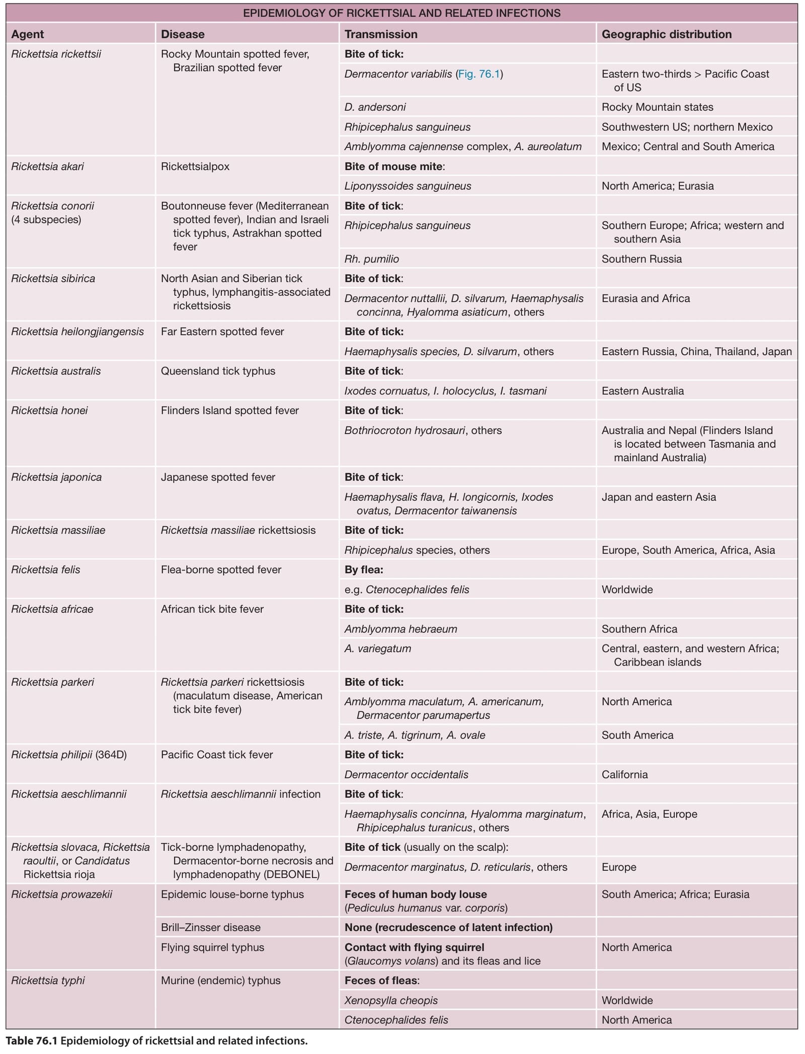
Table 76.1 Epidemiology of rickettsial and related infections.
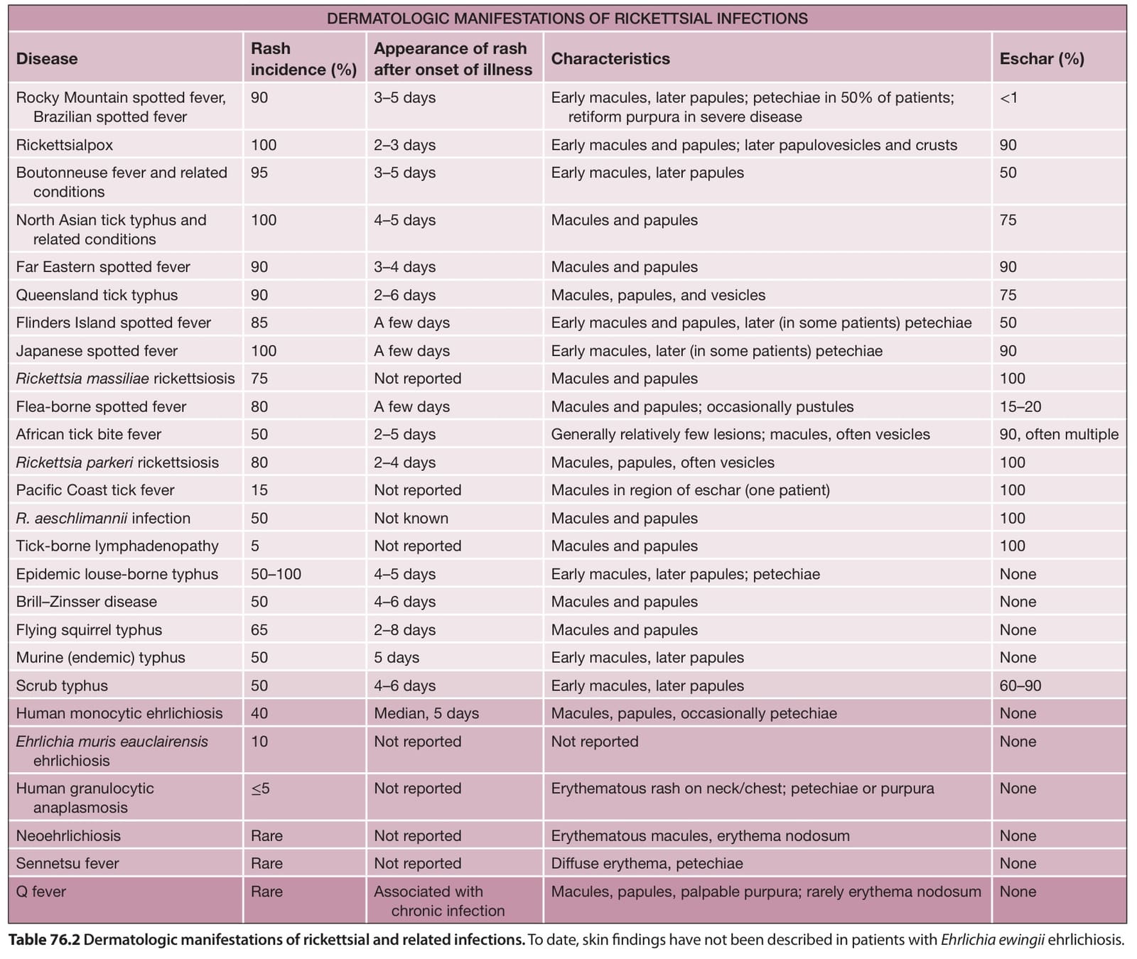
Table 76.2 Dermatologic manifestations of rickettsial and related infections. To date, skin findings have not been described in patients with Ehrlichia ewingii ehrlichiosis.
Several types of lesions may be present simultaneously or sequentially in an individual patient.
Approximately 30%–40% of patients with HME have cutaneous manifestations, which develop a median of 5 days after the onset of the illness and may be more common in children. Macular, maculopapular, and petechial eruptions involving the trunk and extremities are most often observed, but the morphology and distribution of skin lesions can vary considerably (Table 76.4). Ehrlichia ewingii ehrlichiosis has not been reported to have cutaneous manifestations. The differential diagnosis of ehrlichioses is similar to that of rickettsioses (see above).
Since Ehrlichia spp. and Anaplasma phagocytophilum are found within circulating monocytes or neutrophils, PCR-based assays of peripheral blood samples have greater sensitivity than for rickettsial infections. However, empiric treatment should be initiated based on clinical suspicion rather than awaiting laboratory confirmation. Doxycycline is the drug of choice. In vitro evidence suggests that chloramphenicol is not an effective therapy for these infections.
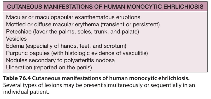
Table 76.4 Cutaneous manifestations of human monocytic ehrlichiosis.