SUPERFICIAL MYCOSES
Introduction
Superficial mycoses are due to fungi that only invade fully keratinized tissues, i.e. stratum corneum, hair, and nails. They can be further subdivided into those that induce minimal, if any, inflammatory response, e.g. tinea (pityriasis) versicolor, and those that lead to more substantial cutaneous inflammation, e.g. dermatophytoses (Table 77.2).
Non-inflammatory Superficial Mycoses
Synonyms: Tinea nigra: tinea nigra palmaris et plantaris, super-ficial phaeohyphomycosis Piedra: molestia de Beigel, trichomycosis nodularis Tinea versicolor: pityriasis versicolor, dermatomycosis furfuracea, tinea flava
History
In 1846, Eichstedt first noted the disease known today as tinea (pityriasis) versicolor. Over the ensuing 150 years, Malassezia furfur (previously known as Pityrosporum ovale and P. orbiculare) came to be recognized as the causative microbe. More recently, however, studies have pointed to M. globosa as a major causative organism. In 1865, Beigel first described piedra after isolating a fungus, which was likely actually a contaminant, from a wig. Tinea nigra was first described in the 1890s by Cerqueira, who named it “keratomycosis nigricans palmaris”.
Epidemiology
Tinea nigra and piedra most often occur in tropical climates of Central and South America, Africa and Asia, but also occasionally develop in the southeastern US. While any age, ethnicity, or sex may be affected, the typical patient is a young adult. Trichosporon spp., which were previously collectively known as T. beigelii, cause white piedra and can also lead to disseminated trichosporonosis in immunocompromised individuals (see Table 77.2).
Malassezia spp. are found worldwide (Table 77.3). In fact, they are part of the normal flora (microbiota) of human skin (predominantly M. sympodialis, M. globosa, and M. restricta). Although tinea versicolor occurs most frequently in tropical climates with high ambient temperatures and high humidity, it is also common in temperate climates. No racial or sex differences have been observed. Tinea versicolor favors adolescents and young adults but can occur at any age. Malassezia requires oil to grow, accounting for its predilection for sebumrich areas of the skin. Malassezia has been implicated in several other skin diseases, including seborrheic dermatitis and a variant of atopic dermatitis that primarily involves the head and neck. Neonatal cephalic pustulosis (neonatal acne) may also be associated with Malassezia spp., particularly M. sympodialis (see Ch. 34).
Pathogenesis
Hortaea werneckii (formerly Exophiala werneckii) and Piedraia hortae are both environmental pathogens. H. werneckii (tinea nigra) can be found in soil, sewage, and even shower stalls under humid conditions. The source of exposure to P. hortae (black piedra) is thought to be the soil. There is no known transmission of these organisms from human to human. Trichosporon spp. (white piedra) are also acquired from the environment; in addition, they are occasionally part of the normal flora of the mucous membranes and skin, particularly inguinal and axillary areas.
Malassezia spp. normally live on human skin in amounts too minute to be detectable on potassium hydroxide (KOH) examination of stratum corneum. Tinea versicolor occurs when the round yeast form transforms to the mycelial form. Factors that can promote this conversion include high ambient temperatures and humidity, oily skin, excessive sweating, immunodeficiency, poor nutrition, pregnancy, and corticosteroid use. Because this yeast is lipophilic, application of oils (especially organic products such as olive oil) to the skin may also encourage its growth. Risk factors for Malassezia (Pityrosporum) folliculitis include chronic antibiotic use, immunosuppression, and local occlusion.
Clinical features
“Piedra” means “stone”, reflecting the fungal elements’ adherence to one another to form nodules along the hair shaft. Piedra is a super-ficial infection that begins under the cuticle of the hair and extends outward, which can lead to weakening and breakage of the hair. As the nodules enlarge, they can even envelope the hair shaft (Fig. 77.1). The two major forms – black piedra and white piedra – are distinguished by their clinical appearance and findings on microscopic examination (Table 77.4).
Patients with black piedra develop asymptomatic brown to black nodules along the hair shaft. White piedra manifests with soft, less adherent nodules that are typically white but may also be red, green, or light brown in color. In addition to white piedra, Trichosporon spp., especially T. asahii, can cause trichosporonosis in immunocompromised patients. This serious systemic infection presents with fungemia, fever, pulmonary infiltrates, and skin lesions. The latter are typically papulovesicular and purpuric, often with central necrosis; another potential finding is creamy white plaques resembling the in vitro fungal colonies. Trichosporon endocarditis, usually involving prosthetic valves, can also occur.
After a 10- to 15-day incubation period, tinea nigra most commonly presents as a single, sharply marginated, brown to gray–green macule or patch that can be velvety or have mild scale (Fig. 77.2). There are usually no associated symptoms (e.g. pruritus), and no predisposing factors have been identified. Although most frequently seen on the palms, tinea nigra can also appear on the soles, neck, and trunk. Palmoplantar lesions are said to resemble acquired acral melanocytic nevi, but the former are usually larger, lighter in color, and lack the linear striations of acral nevi. The advancing border of a tinea nigra lesion may also be more darkly pigmented than its center. While the disease tends to be chronic, recurrence after effective treatment is infrequent except in the case of re-exposure.
Patients usually present with multiple oval-to-round macules, patches, or thin plaques with mild, fine scale. Demonstration of this associated scale may require scratching or stretching the skin surface. The lesions are often confluent centrally within areas of involvement, which may be extensive. Seborrheic regions, in particular the upper trunk and shoulders, are the favored sites. Less frequently, lesions are seen on the face (especially in children), scalp, antecubital fossae, submammary region, and groin. Involvement of flexural areas is sometimes referred to as “inverse” tinea versicolor.
The most common colors are brown (hyperpigmented; Fig. 77.3A) and whitish-tan (hypopigmented; Fig. 77.3B); occasionally there is mild inflammation leading to a pink color (Fig. 77.3C). Hypopigmentation may be secondary to either reduced tanning or inhibition of melanin production by dicarboxylic acids that result from metabolism of skin surface lipids by the yeast. In general, tinea versicolor is asymptomatic and the major concern is its appearance.
This condition is most commonly seen in young women and is characterized by pruritic, monomorphic follicular papules and pustules on the upper trunk, arms, neck, and face (especially on the forehead and along the hairline). It is due to excessive growth of M. furfur and M. globosa within the hair follicle, with resulting inflammation due to yeast products as well as free fatty acids generated by fungal lipases. Only yeast forms are observed, i.e. no hyphal forms as in tinea versicolor.
Pathology and fungal culture
For black and white piedra, cut hair shafts are placed in KOH and a “crush preparation” is examined microscopically. In a black piedra nodule, dematiaceous hyphae are seen around an organized cluster of asci, each of which contains eight ascospores. The ascospores represent the sexual phase of P. hortae. P. hortae grows very slowly when cultured and yields a green to black colony with a velvety texture (asexual phase).
KOH preparation of a crushed white piedra nodule reveals non-dematiaceous hyphae, blastoconidia and arthroconidia, representing the asexual state (Fig. 77.4). When cultured, Trichosporon spp. grow rapidly, forming moist, cream-colored, yeast-like colonies that
resemble “butter cream frosting”. On Mycosel® agar, these organisms are inhibited by the presence of cycloheximide. When Trichosporon spp. are isolated from skin and nail specimens, correlation with clinical findings is necessary to determine the medical significance.
Biopsies of tinea versicolor and tinea nigra are usually not performed, as KOH examination of associated scale is typically diagnostic. In tinea versicolor, both hyphal and yeast forms are seen; although likened to “spaghetti and meatballs”, the findings more closely resemble “ziti and meatballs” (Fig. 77.5A). In tinea nigra, KOH examination reveals septate pigmented hyphae. Dermoscopy of tinea nigra also shows curvilinear brown strands representing pigmented hyphae. When biopsy specimens
Single, sharply demarcated brown macule on the finger. Courtesy Frank Samarin, MD.
are obtained, similar findings are observed within the stratum corneum (Fig. 77.3D). In KOH examination of expressed follicular contents or biopsy specimens of Malassezia (Pityrosporum) folliculitis, only yeast forms are seen. Cultures of H. werneckii first appear as pasty,
green–black colonies with a yeast-like appearance, and after approximately 2 weeks become a fuzzy, dematiaceous (dark in color) mold. Culture of Malassezia is generally not required; if necessary, the plate must be overlaid with sterile oil because of the organism’s lipophilic nature.
Differential diagnosis
Piedra is generally diagnosed by clinical and microscopic inspection of a hair shaft and must be distinguished from pediculosis (nits), hair casts, trichorrhexis nodosa, trichomycosis axillaris (see Fig. 77.1), and the scales of psoriasis and eczema. Unlike eczema and psoriasis, the scalp typically appears normal in piedra.
In most patients, the diagnosis of tinea nigra is made clinically and confirmed via KOH examination and/or fungal culture. Occasionally, tinea nigra could be confused with melanocytic nevi (see above), a fixed drug eruption, postinflammatory hyperpigmentation, or staining from chemicals, pigments, and dyes. Cutaneous melanoma has even been clinically misdiagnosed as tinea nigra.
Clinical examination is often sufficient to recognize tinea versicolor. However, pityriasis alba or other forms of postinflammatory hypopigmentation, vitiligo, seborrheic dermatitis, pityriasis rosea, tinea corporis, and secondary syphilis may be diagnostic considerations. Progressive macular hypomelanosis, which presents as non-scaly hypopigmented macules on the trunk of young adults and may be caused by Cutibacterium (formerly Propionibacterium) acnes, is often misdiagnosed as residual hypopigmentation related to tinea versicolor. Wood light examination revealing bright yellow fluorescence and then direct microscopy of scales can confirm the diagnosis of tinea versicolor. Malassezia (Pityrosporum) folliculitis must be differentiated from other causes of folliculitis (see Table 38.1), in particular itching folliculitis, as well as acne vulgaris.
Treatment
Clipping hairs with adherent nodules as well as shampooing the affected hairs with 2% ketoconazole shampoo is usually an effective treatment for piedra (see Table 77.4). Oral terbinafine may also be of therapeutic benefit. For treatment of tinea nigra, topical keratolytic agents such as Whitfield ointment (typically 6% benzoic acid plus 3% salicylic acid) are effective, as are topical azole or allylamine antifungal medications. Several weeks of therapy may be required to prevent recurrence. Systemic therapy is generally not indicated, and griseofulvin is not effective.
Tinea versicolor usually responds to topical antifungal therapy, but oral treatment with fluconazole or itraconazole can be considered when there is more extensive involvement (Table 77.5; see Ch. 127). Residual pigmentary changes often require weeks to months to resolve. The rate of recurrence of tinea versicolor is very high, especially in hot humid climates, and maintenance therapy may be helpful (see Table 77.5). The treatment of Malassezia (Pityrosporum) folliculitis is reviewed in Chapter 38.
Dermatophytoses
Synonyms: Tinea corporis: ringworm, tinea circinata, tinea glabrosa Tinea cruris: ringworm of the groin, eczema marginatum, jock itch, gym itch Tinea barbae: tinea sycosis, barber’s itch, ringworm of the beard Tinea capitis: ringworm of the scalp/hair, tinea tonsurans, herpes tonsurans Tinea pedis: ringworm of the foot, athlete’s foot Tinea unguium: ringworm of the nail, dermatophytic onychomycosis
Introduction
Dermatophytoses are fungal infections caused by three genera of fungi that have the unique ability to invade and multiply within keratinized tissue (hair, skin, and nails). These fungi, collectively called “dermatophytes”, are alike in their physiology, morphology, and pathogenicity. The three genera are Microsporum, Trichophyton, and Epidermophyton; species within these genera that do not invade keratinized tissue in animals or humans are not considered as dermatophytes. In naming clinical infections due to dermatophytes, “tinea” precedes the Latin name for the involved body site, e.g. “tinea pedis” refers to a dermatophyte infection of the foot. Overall, ten species of dermatophytes are commonly isolated and an additional 20 are occasionally identified (Table 77.6).
History
Dermatophyte infections have been described since ancient times. In 1910, Sabouraud published a categorization scheme that is currently employed in which dermatophytes are classified by genera. A revised and simplified nomenclature system was proposed in 2017.
Epidemiology
Some dermatophytes are restricted geographically, while others are found worldwide. Trichophyton rubrum, for example, is the most common dermatophyte and has a global distribution, while T. concentricum is endemic to certain parts of the South Pacific and South America. Human travel and migration, coupled with antifungal therapy and the development of drug resistance, have brought about significant changes in the geographic distribution of dermatophytes. Terbinafine-resistant T. indotineae (formerly T. mentagrophytes var. VIII), which was recognized in 2020, emerged in India with subsequent global spread12a,12b. One theory for its emergence is the misuse of potent topical corticosteroids in settings where they are available without a prescription. In general, dermatophyte infections are more common in tropical environments. Other important epidemiologic factors include socioeconomic status, occupation, air conditioning, and the use of footwear.
In general, dermatophytoses occur most frequently in postpubertal hosts. One major exception is tinea capitis (dermatophytosis of the scalp), which occurs primarily in children. Risk factors for the development of a dermatophyte infection during childhood include household exposure to tinea capitis or tinea pedis; environmental factors such as contaminated hats, brushes, and barber instruments; and (especially for tinea unguium) Down syndrome. Individuals with chronic mucocutaneous candidiasis, common variable immunodeficiency, or HIV infection have a propensity to develop severe, chronic, or recurrent dermatophytoses (see Chs. 60 & 78). Men more frequently have tinea pedis, tinea cruris, and tinea unguium than women.
Pathogenesis
Transmission of dermatophytes to humans occurs via three major sources, each resulting in infections with characteristic features (Table 77.7). While dermatophytes are not especially virulent and typically invade only the outer, cornified layers of the skin, they can be responsible for considerable morbidity. The first stage of infection involves contact with and adherence of the infectious elements of the fungus (arthroconidia) to the skin. The ability of certain fungi to adhere to a particular host arises from a variety of microbial mechanisms and host factors.
Dermatophytes, unlike most other fungi, produce keratinases (enzymes that break down keratin), which allow invasion of the fungi into the stratum corneum. Mannans in the cell walls of dermatophytes have immuno-inhibitory effects. In T. rubrum, the mannans may also decrease epidermal proliferation, thereby reducing the likelihood of the fungus being sloughed off prior to invasion. This mechanism is thought to contribute to the chronicity of infections caused by T. rubrum. However, host factors such as protease inhibitors may limit the extent of invasion. If invasion is successful, disease occurs.
The severity of clinical disease is also affected by several host factors. Sebum has an inhibitory effect on dermatophytes, and the degree of disease activity may be related to the number and activity of sebaceous glands in a particular body region. Breaks in the skin barrier or macerated skin encourage dermatophyte invasion, and increased susceptibility related to immunologic factors may be inherited. Once dermatophytes invade and begin to proliferate in the skin, several mechanisms aid in limiting the infection to keratinized tissue. These include the preference of dermatophytes for the cooler temperature at the skin’s surface, serum factors that inhibit dermatophyte growth (e.g. β-globulins, ferritin, other metal chelators), and the host immune response. Further invasion or dissemination is extremely unusual (see below). Other conditions influencing dermatophyte infections include skin disorders that affect cutaneous barrier function, such as ichthyoses.
Clinical features
Tinea corporis is a dermatophyte infection of the skin of the trunk and extremities, excluding the hair, nails, palms, soles, and groin. The infection is generally restricted to the stratum corneum and most commonly affects exposed skin. Any dermatophyte can potentially
cause tinea corporis, but T. rubrum is the most common pathogen worldwide, followed by T. mentagrophytes (Table 77.8).
Tinea corporis can result from human-to-human (including autoinoculation, e.g. from tinea capitis or pedis), animal-to-human (often transmitted by domestic animals), or soil-to-human spread (see Table 77.7). Sources include occupational or recreational exposure (e.g. military housing, gymnasiums, locker rooms, outdoor activities, wrestling) and contact with contaminated clothing or furniture.
There are multiple clinical presentations of tinea corporis, and they can mimic other dermatologic conditions (Table 77.9). As with most dermatophyte infections, the extent of inflammation depends on the causative pathogen and the immune response of the host. Because hair follicles serve as reservoirs for infection, hairier areas of the body may be more resistant to treatment.
The typical incubation period is 1 to 3 weeks. Infection spreads centrifugally from the point of skin invasion with central clearing of the fungus, typically resulting in annular lesions of varying sizes (Fig. 77.6A–D). Arcuate, circinate, concentric, and oval lesions can also develop (Fig. 77.6D–F). Although scale is usually present, it may be lessened or absent if topical corticosteroids have been used (tinea “incognito”) (Fig. 77.7). Pustules within the active border are suggestive of tinea (see Fig. 77.6E). Lesions are occasionally vesicular, granulomatous, or verrucous (Fig. 77.6G). Associated symptoms may include pruritus and burning.
Tinea profunda, nodular perifolliculitis (including Majocchi granuloma), and tinea imbricata represent clinical variants of tinea corporis. Tinea profunda results from an excessive inflammatory response to a dermatophyte, analogous to a kerion on the scalp. It may have a granulomatous or verrucous appearance and be mistaken for cutaneous tuberculosis or a dimorphic fungal infection. Nodular
perifolliculitis, usually caused by T. rubrum, is characterized by follicular papulopustules or granulomatous nodules that result from a deep dermatophyte folliculitis with disruption of the follicular wall (Fig. 77.8). This variant is most commonly seen in women who have tinea pedis or onychomycosis and shave their legs, and it can also occur in the setting of immunosuppression. The lesions may be extensive or even vegetating, and a prolonged granulomatous reaction (as described by Majocchi) can develop.
Tinea imbricata is a dermatophytosis caused by the anthropophilic dermatophyte T. concentricum. It causes chronic infections in an equatorial band encompassing the South Pacific, Asia, and Central and South America. The clinical presentation consists of concentric annular rings resembling erythema gyratum repens.
Tinea cruris is a dermatophyte infection of the inguinal region, in particular the inner aspects of the upper thighs (Fig. 77.9) and crural folds, with occasional extension onto the abdomen and buttocks. The three most common causative agents are Epidermophyton floccosum, T. rubrum, and T. mentagrophytes (Table 77.10).
This condition is more often seen in men than in women, since the scrotum provides a warm and moist environment that encourages fungal growth and men are more likely to have tinea pedis and onychomycosis as a source of dermatophytes. Other predisposing factors include obesity and excessive perspiration. Tinea cruris is frequently associated with tinea pedis because clothing that is brought over the feet is contaminated and then comes in contact with skin in the groin region.
The initial sign of infection is usually an area of erythema and pruritus in the fold between the scrotum and the inner thigh. Characteristic lesions are sharply demarcated with a raised, erythematous, scaly advancing border that may contain pustules or even vesicles. Lesions can initially be circinate and then become serpiginous. The disease may remain unilateral or become bilateral.
The duration of infection depends on the causative pathogen. Infections with T. rubrum and many other anthropophilic species tend to be chronic, sometimes with leathery and lichenified lesions. In contrast, the zoophilic form of T. mentagrophytes (previously T. mentagrophytes var. mentagrophytes) and other animal-acquired strains often cause acute infections with a prominent inflammatory component that may include pustules and vesicles (see Table 77.10). The scrotum itself is generally spared in tinea cruris. If the scrotum is involved or there are erosions or satellite pustules, cutaneous candidiasis should be considered. Measures to prevent recurrences of tinea cruris include wearing loose clothing, drying thoroughly after bathing, using topical powders, weight reduction (if obese), laundering contaminated clothing and linens, and treating concomitant tinea pedis.
Dermatophyte infections on the dorsal aspect of the hand have a clinical presentation similar to tinea corporis. However, dermatophyte infection of the palm and interdigital spaces has distinct characteristics and is referred to as tinea manuum. The reason for the two different clinical
pictures is thought to be related to the lack of sebaceous glands on the palms. The typical causative organisms are the same as those for tinea pedis and tinea cruris: T. rubrum, T. mentagrophytes, and E. floccosum. Infections with Neoscytalidium dimidiatum and N. hyalinum, two non-dermatophyte fungi, can have a tinea manuum-like appearance.
Moccasin-type tinea pedis is often present in patients with tinea manuum, and the two share clinical features such as chronicity and hyperkeratosis. Tinea manuum is usually non-inflammatory and often unilateral (“two feet and one hand syndrome”) (Fig. 77.10; see Fig. 77.16A); there is hyperkeratosis of the palms and digits, including within creases, that fails to respond to emollients. Tinea unguium of the involved hand can serve as a clue to the diagnosis, but infection of all the fingernails is uncommon. Other presentations include exfoliative, vesicular, and papular variants. The differential diagnosis may include psoriasis, irritant or allergic contact dermatitis, dyshidrosis, and a “dermatophytid” reaction.
Tinea barbae is a dermatophytosis that involves the bearded areas of the face and neck in men. Because the disease is often acquired from animals, the causative organisms are typically zoophilic dermatophytes, namely
A thin, “broken-up” erythematous plaque with an arciform papular border on the upper inner thigh.
T. mentagrophytes and T. verrucosum. Infection with Microsporum canis or T. rubrum is uncommon. In some geographic regions, other anthropophilic dermatophytes (T. schoenleinii, T. violaceum, and T. megninii) are endemic and cause tinea barbae. With the increased use of disposable razors and disinfectants, the incidence of tinea due to contaminated razors in barbershops has been dramatically reduced.
Since zoophilic organisms are the most common culprit and affected areas often have a large number of terminal hair follicles, the clinical presentation tends to be severe, with intense inflammation and multiple follicular pustules. Abscesses, sinus tracts, bacterial superinfection, and kerion-like boggy plaques can develop. Patients may have constitutional symptoms such as malaise as well as lymphadenopathy and even scarring alopecia. A second type of tinea barbae is superficial, less inflammatory, and similar to tinea corporis; T. rubrum is usually the causative agent in this variant (Fig. 77.11). Alopecia may be present in the center of the lesion, but it is reversible. Other conditions that can mimic tinea barbae include bacterial folliculitis, viral infections (herpes simplex or zoster), acne vulgaris, cervicofacial actinomycosis, and a dental sinus tract. Spontaneous resolution may occur if all of the infected hairs fall out.
While some dermatophyte infections of the face have classic features (e.g. scale, annular configuration, pustules in the border), others can be more difficult to diagnose clinically and require a high index of suspicion (Fig. 77.12, see Table 77.9).
Tinea capitis is a common dermatophyte infection of the scalp in children (Fig. 77.13), whereas adult infection occurs infrequently. The causative pathogens are members of only two genera: Trichophyton and Microsporum. T. tonsurans is currently the most common cause
of tinea capitis in the US (accounting for >90% of cases), with a predilection for individuals with Afro-textured hair, and M. canis is the second most frequent etiology. Globally, there is significant variation in the epidemiology of tinea capitis. While M. canis is a common cause in many countries, T. tonsurans has emerged as a major agent in Europe and elsewhere. The incidence of tinea capitis due to T. violaceum, which is endemic in Africa, has also recently increased in some areas of the US and Europe (especially western and Mediterranean regions), likely reflecting immigration patterns. The anthropophilic dermatophyte M. audouinii, which was once common and then subsequently diminished secondary to social and therapeutic advances, has more recently reappeared in Europe and may also potentially re-emerge in the US.
Different clinical presentations arise from the various causative organisms. For example, T. tonsurans causes an endothrix infection (see below), which classically results in “black dot” tinea capitis due to hair breakage near the scalp. In contrast, M. audouinii is an ectothrix form of tinea capitis that typically presents with dry, scaly patches of alopecia (“gray patch” tinea capitis). Both the pathogenicity of the culprit organism and the host immune response are factors that determine the severity of disease. Tinea capitis can range from non-inflammatory scaling that resembles seborrheic dermatitis (especially with T. tonsurans; Fig. 77.13A) to a severe pustular reaction with alopecia, known as a kerion. Alopecia with or without obvious scale is the most common presentation of tinea capitis (Fig. 77.13B–D). The alopecia may occur in discrete patches or involve the entire scalp. Use of dermoscopy can highlight “comma”, “corkscrew”, and dystrophic broken hairs that represent clues to the diagnosis of tinea capitis (see Ch. 69). Many patients have posterior cervical and posterior auricular lymphadenopathy, which is helpful in differentiating tinea capitis from less inflammatory causes of alopecia, such as alopecia areata.
A kerion results from advanced disease coupled with an exaggerated host response that leads to boggy, purulent plaques with abscess formation and associated alopecia (Fig. 77.13E,F). Some patients may even become systemically ill with extensive lymphadenopathy. The hair in the affected area usually returns, but the longer the infection persists, the more likely the alopecia will be permanent. If a kerion is misdiagnosed as a bacterial abscess and treated with antibiotics following incision and drainage, the infection will likely worsen, thereby increasing the likelihood of scarring alopecia.
The “carrier state” of T. tonsurans refers to a clinical situation in which there are no obvious signs or symptoms of a scalp infection, yet a positive fungal culture is obtained. Although also seen in children, this typically occurs in adults who have been exposed to infected children. The carriers shed the fungus and are considered contagious. Consequently, some experts advocate treating all carriers with oral or topical antifungals.
For those dermatophytes that invade hair, three patterns of invasion exist: endothrix, ectothrix, and favus (Fig. 77.14):
●The endothrix pattern results from infection with anthropophilic fungi in the genus Trichophyton and is characterized by non-fluorescent arthroconidia within the hair shaft (Fig. 77.13G). The clinical presentation varies from scaling to “black dots” with patchy alopecia to kerion formation. T. tonsurans and T. violaceum are important causes of endothrix infection.
●The ectothrix pattern occurs when arthroconidia are formed from fragmented hyphae outside the hair shaft (see Fig. 77.14). Cuticle destruction ensues. Ectothrix infection can be fluorescent (Microsporum) or non-fluorescent (Microsporum and Trichophyton), as determined by Wood’s lamp examination. Clinical features vary from patchy, scaly alopecia with little inflammation that may mimic alopecia areata to kerion formation.
●Favus is the most severe form of dermatophyte hair infection and is most frequently caused by T. schoenleinii. Hyphae and air spaces are observed within the hair shaft, and a bluish-white fluorescence by Wood’s light examination is typically seen. Favus presents as thick, yellow crusts composed of hyphae and skin debris (“scutula”). Scarring alopecia may develop in chronic infections (Fig. 77.15).
There are many scalp and hair disorders that lead to scaling or alopecia (scarring and non-scarring) that are not due to fungal infections (see Table 77.9). However, one should always consider and exclude a fungal infection (especially in children), because the treatment is typically quite simple and effective, and chronic untreated tinea capitis can lead to scarring alopecia. Oral therapy is required, as the drug must penetrate the hair follicle to be effective. Preventative measures are also important in the management of tinea capitis. Because the disease is contagious, all individuals residing with the infected patient should be examined for signs of tinea capitis and appropriately treated. Chronicity may develop if a child is continually re-exposed from untreated family members. Appropriate adjunctive treatment for household contacts as well as the patient includes regular (e.g. every other day) use of an antifungal shampoo, such as 2% ketoconazole or 2.5% selenium sulfide, until the patient is free of disease. Although routine screening in schools is difficult, screening children who have two or more infected classmates may be beneficial. Combs, brushes, and headwear used by the patient should be disinfected or preferably discarded.
Tinea pedis is a dermatophyte infection of the soles and interdigital web spaces of the feet. Infection of the dorsal aspect of the foot is considered tinea corporis. The feet are the most common location for dermatophyte infections, and the majority of the US adult population has experienced tinea pedis. This condition is more common in adults than children and is found around the world, affecting both sexes. The lack of sebaceous glands and the moist environment created by occlusive shoes are important factors in the development of tinea pedis. In fact, tinea pedis is uncommon in populations that do not wear shoes. However, the fungus may be acquired from going barefoot (locker rooms, gyms, public facilities).
The dermatophytes that are typically responsible for tinea pedis are T. rubrum, T. interdigitale (previously T. mentagrophytes var. interdigitale), T. mentagrophytes, E. floccosum, and T. tonsurans (in children). Non-dermatophyte pathogens that produce clinical findings identical with tinea pedis include Neoscytalidium dimidiatum and N. hyalinum (moccasin and interdigital types) and, occasionally, Candida spp. (interdigital type). There are four major clinical types of tinea pedis, as outlined in Table 77.11: moccasin (Fig. 77.16A), interdigital (Fig. 77.16B), inflammatory (Fig. 77.16C), and ulcerative. Each type has different associated morbidities and complications that can affect diagnostic considerations and therapeutic options. These include bacterial superinfection (the “dermatophytosis complex”), dermatophytid reactions, cellulitis (especially in patients who have venous hypertension, harvested saphenous veins, and chronic edema), and
even osteomyelitis leading to amputation in diabetics. Other conditions that can mimic tinea pedis are listed in Table 77.9. KOH examination and culture easily differentiate the moccasin and inflammatory forms of tinea pedis from these entities. Erythrasma can be diagnosed with Wood’s light examination because of its “coral red” fluorescence. Oral antifungal therapy should be considered in diabetics, immunocompromised patients, and those with moccasin-type tinea pedis. Finally, other dermatophyte infections often occur together with tinea pedis – in particular, tinea cruris, onychomycosis, and tinea manuum – and these sites should be examined (Fig. 77.17).
Onychomycosis is a term used to encompass all fungal infections of the nail and includes those due to dermatophytes as well as non-dermatophytes. It is divided into three patterns based upon the point of fungal entry into the nail unit:
●distal/lateral subungual, with invasion via the hyponychium (most common; Fig. 77.18A–C)
●superficial white, with direct penetration into (and usually confinement to) the dorsal surface of the nail plate (often due to T. interdigitale; Fig. 77.18D)
●proximal subungual, with invasion under the proximal nail fold (frequently in immunocompromised hosts)
●mixed pattern, with ≥2 of the above patterns in the same nail. Onychomycosis can be challenging to manage due to difficulties in diagnosis, the requirement for long treatment periods, potential side effects of systemic medications, and frequent recurrences. Although many consider onychomycosis solely as an annoyance, patients with the condition often complain of discomfort and pain associated with trimming the nails, running, and other activities such as dancing. In addition, serious complications such as cellulitis can result from onychomycosis, especially in patients who are diabetic or immunocompromised. Infections with non-dermatophytes such as Fusarium spp. (see below) are of particular concern in the latter group.
Tinea unguium refers specifically to dermatophyte infection of the nail unit. It occurs worldwide, affects men more often than women, and is frequently associated with chronic tinea pedis. Trauma and other nail disorders represent predisposing factors. Although all dermatophytes can cause tinea unguium, Microsporum spp. do so very rarely. The most common causative pathogens are T. rubrum, T. interdigitale, T. tonsurans (in children), and E. floccosum.
Toenail infections are considerably more common than fingernail infections, and only rarely does fingernail onychomycosis occur without concurrent toenail infection. A single nail may be involved, but more frequently, multiple nails on one or both hands or feet are affected. In the distal/lateral subungual type, invasion of the hyponychial region leads to hyperkeratosis of the nail bed. With further progression of infection, there
is yellowing and thickening of the distal nail plate as well as onycholysis (see Fig. 77.18A–C), which provides an ideal environment for further proximal invasion and growth of the dermatophyte. Eventually, the entire nail bed and plate may become involved (total dystrophic pattern).
The clinical appearance of the superficial white type of onychomycosis can vary based on host factors (e.g. immunosuppression) and the causative organism. Discrete white patches are seen in the classic form due to T. interdigitale (see Fig. 77.18D). However, transverse striate bands, origination from the proximal nail fold (which may overlap with the proximal subungual type if there is also invasion under the nail fold), and deeper invasion of the nail plate can also occur. These variants are more likely to be caused by T. rubrum, with more invasive forms observed in healthy children as well as people living with HIV; deep invasion of the nail plate can also result from non-dermatophyte molds such as Fusarium spp..
Although nail dystrophy can be the result of trauma, psoriasis, inherited disorders, and other conditions (see Ch. 71), an estimated 50% or more of cases are due to onychomycosis. Dermatophytes account for ~90% of cases of onychomycosis, and the remainder are due to yeasts or non-dermatophyte molds (Table 77.12). Candida spp. are often found in association with chronic paronychia (see Ch. 71), and infection of the nail may occur in this setting. The fingernails are usually affected, with ridging, yellow discoloration, and onycholysis. Candida spp. are a relatively common cause of onychomycosis in children less than 3 years of age, and nail involvement also represents a manifestation of chronic mucocutaneous candidiasis (see Ch. 60).
Pathology
Tinea pedis, manuum, faciei, cruris, and corporis are usually diagnosed via KOH examination (see Fig. 77.5B) and occasionally with fungal cultures. The chlorazol black E stain can help to highlight fungal elements in KOH preparations. Use of calcofluor, a fluorescent stain specific for the chitin in the fungal cell wall, and examination with a fluorescent microscope can also demonstrate fungi (apple-green fluorescence). PCR-based assays for dermatophytes are also commercially available.
If biopsies are performed of cutaneous dermatophyte infections, hyphae are seen within the stratum corneum; the fungi are made more apparent by PAS or silver stains. In dermatophytoses affecting glabrous skin, spongiosis, parakeratosis, and pustules are often seen. In onychomycosis, hyphae and arthroconidia may be visible in the nail plate and nail bed with PAS staining, and little to no inflammation is generally present. Histologic examination of formalin-fixed, PAS-stained nail plates is a quick and reliable method for diagnosing onychomycosis (Fig. 77.18E), with a sensitivity of 80% to >95%, compared with 35%–60% for culture alone. Biopsy specimens of tinea capitis, Majocchi granuloma, and tinea barbae can demonstrate hyphae or arthroconidia within hair shafts and are usually done if the diagnosis is suspected but KOH examination and culture are negative. However, false-negative results are a potential problem and tissue should also be sent for culture.
Differential diagnosis
Careful clinical examination is the first and most important step in diagnosing dermatophyte infections. Because many other conditions can mimic dermatophytoses (see Table 77.9), KOH examination and/ or culture are often necessary to confirm the diagnosis. Guidelines for proper specimen collection are outlined in Fig. 77.19. Because all dermatophytes have hyaline hyphae, they essentially look the same in KOH preparations. When identification of the particular species will provide helpful information or culture is needed to confirm the diagnosis, the specimen is cultured at 25–30°C (and in some instances also at 37°C) for 2 to 4 weeks.
Colonial morphology, microscopic examination of conidia (the asexual propagules formed de novo by the fungus), noting the growth rate/ requirements, and biochemical tests allow precise identification of the dermatophyte. The presence or absence of microconidia (usually small and unicellular) and macroconidia (usually larger and multicellular) and the typical features of each (e.g. shape, cell wall texture) remain fairly consistent among genera (Fig. 77.20). Other distinguishing microscopic features include the identification of arthroconidia (infective fungal elements) and chlamydoconidia. Sometimes, conidia may not be present in culture (“sterile” organism), and the hyphal patterns (spiral,
pectinate, antler, racquet, and nodular bodies) may be important in identification of the organism. The color, texture, and topography of the colony are features that are typical of, or even unique to, a particular species (see Table 77.6).
Dermatophytes are able to grow on media containing cycloheximide (e.g. Mycosel®, DTM [dermatophyte test medium]). However, some clinically relevant non-dermatophyte fungi do not, including Scopulariopsis brevicaulis, Aspergillus spp., Cryptococcus neoformans, Candida tropicalis, Pseudallescheria boydii/Scedosporium apiospermum, and Trichosporon spp.. These can potentially be missed if specimens, in particular from nails, are grown only on cycloheximide-containing media.
Treatment
Topical antifungals are the first-line treatment for many patients with uncomplicated, localized cutaneous dermatophyte infections such as tinea corporis, tinea cruris, and tinea pedis (see Ch. 127). The major potential adverse reaction is irritant (or, occasionally, allergic) contact dermatitis, usually from alcohols or other components in the vehicle. Systemic antifungal therapy, although associated with both a higher incidence of side effects including potentially severe adverse reactions and the potential for drug–drug interactions, is typically required to cure tinea manuum, capitis, and unguium. Suggested regimens are presented in Table 77.13. In general, oral treatment is also needed for infections involving extensive areas of skin, in hairy sites other than the scalp (e.g. tinea barbae), or associated with excessive inflammatory reactions. The adjunctive use of topical products containing glycolic acid, lactic acid, or urea may help to reduce the amount of hyperkeratosis in infections such as tinea manuum and pedis.
Treatment of tinea unguium deserves special consideration. Oral antifungal therapy is usually required to achieve a complete cure (see Table 77.13), with the occasional exception of classic superficial white onychomycosis. Topical therapies that have been FDA-approved for mild to moderate onychomycosis have combined clinical and mycologic cure rates after daily application for 48 weeks ranging from 6%–10% for tavaborole 5% and ciclopirox olamine 8% solutions to 15%–20% for efinaconazole 10% solution. Although mycologic cure rates of up to 80% (or higher with prolonged therapy) have been reported with oral antifungal drugs (terbinafine, itraconazole, and fluconazole), the combined clinical and mycologic cure rates are lower, i.e. ~20%–40% after 12 weeks for toenails. Recurrent disease is common, especially in the toenails and when the degree of involvement is severe (Table 77.14). Preventive measures include breathable footwear and socks, antifungal or absorbent powders, frequent nail clipping, and avoiding re-exposure (e.g. not going barefoot in locker rooms). Old shoes often harbor large numbers of infectious organisms and should be discarded or treated with disinfectants or antifungal powders.
Combination therapy consisting of a topical corticosteroid plus topical antifungal has been advocated by some clinicians for the treatment of dermatophyte infections. While a reduction in inflammation can be seen, the negative effects of high-potency corticosteroids on immune defenses coupled with restrictions on treatment duration to avoid side effects such as striae lead to an unacceptable failure rate. Dermatophyte resistance to antifungal therapy has emerged as a public health concern, especially with regard to squalene epoxidase mutations that lead to terbinafine resistance (see Fig. 127.9). Species affected include T. rubrum in the US as well as the recently recognized T. indotineae (T. mentagrophytes internal transcribed spacer [ITS] genotype VIII) that has resulted in an epidemic of resistant dermatophytosis in India, with subsequent global spread. This highlights the importance of confirmatory testing and antifungal stewardship, including limiting the use of combination topical corticosteroid-antifungal products and reserving agents such as posaconazole and voriconazole for more severe infections.
Invasive dermatophytosis
Rarely, dermatophytes proliferate within the dermis. Invasion or dissemination of dermatophytes typically occurs in the setting of a chronic dermatophyte infection (most commonly T. rubrum) in an individual with a primary immunodeficiency such as CARD9 deficiency (see Ch. 60) or iatrogenic immunosuppression. Hematogenous spread can lead to an acute onset of ulcerating or draining dermal and subcutaneous nodules. A more indolent process can also occur and most often presents as tender nodules on the extremities (Fig. 77.21). Currently, the recommended treatment is surgical excision (if localized) and systemic terbinafine, itraconazole, or posaconazole.
Mucocutaneous Candida Infections
Mucocutaneous candidiasis has a wide spectrum of clinical presentations. Early descriptions of oral candidiasis (thrush) were made by Hippocrates. In the 1940s, the association between oral antibiotic use and candidal infections was recognized. Oral candidiasis often presents as thrush, with the pseudomembranous form characterized by a white exudate resembling cottage cheese (Fig. 77.22A,B) and the chronic atrophic form by a patch of erythema; it can also lead to a chronic hyperplastic form with adherent white plaques or glossitis with painful atrophy of the dorsal tongue. Other presentations include denture stomatitis, angular cheilitis (perlèche) (Fig. 77.22C), vulvovaginitis, and balanitis (see Ch. 73). Predisposing factors for mucocutaneous candidiasis include diabetes mellitus, xerostomia, occlusion, hyperhidrosis, the use of corticosteroids or broad-spectrum antibiotics, and immunosuppression, including HIV infection. In the case of angular cheilitis, deep grooves due to overlap of the skin at the angles of the mouth, which is common in edentulous and older patients, is a key contributing factor, along with the use of orthodontic appliances, drooling, atopic dermatitis, and (occasionally) iron or vitamin deficiencies (e.g. B).
Cutaneous Candida infections present with markedly erythematous, sometimes erosive, patches that are often accompanied by satellite papules and pustules (Fig. 77.22D–F). The most common sites of involvement are intertriginous zones (e.g. submammary, beneath pannus, inguinal creases, intergluteal fold) and the scrotum, as well as the diaper area in infants. Candidiasis is sometimes superimposed on intertriginous seborrheic dermatitis or psoriasis. Candida can also infect periungual areas (e.g. in the setting of chronic paronychia), nails (see Tinea unguium above), and the web space between the third and fourth fingers (erosio interdigitalis blastomycetica; Fig. 77.22G), especially in individuals whose hands are frequently exposed to water.
Mucocutaneous candidiasis is most commonly due to C. albicans, followed by C. tropicalis. The diagnosis is established by the presence of budding yeast and pseudohyphae on KOH examination (see Fig. 77.5D) and by a positive fungal culture. C. albicans and C. stellatoidea can be distinguished by their ability to form chlamydoconidia on cornmeal- Tween 80 agar. Identifying and removing predisposing factors is very important in the management of mucocutaneous candidiasis. Treatment with topical nystatin or azole antifungals is often effective. When oral therapy is required for more extensive involvement, options include
fluconazole and itraconazole (see Ch. 127). Treatment regimens for various forms of mucocutaneous candidiasis are outlined in Table 77.15.
Chronic mucocutaneous candidiasis
This is discussed in detail in Chapter 60. Chronic mucocutaneous candidiasis is not a single entity but rather a clinical manifestation of a number of disorders, including primary immunodeficiencies. Patients have defects in the T helper 17 (Th17) response that prevent effective handling of Candida organisms, leading to chronic, recalcitrant infections of the skin (including granulomatous lesions), nails, and mucosae. Some patients have associated autoimmune endocrinopathies as well as alopecia areata and vitiligo. Therapy involves oral fluconazole and itraconazole (see Table 77.15).
Erosive Papulonodular Dermatosis: Granuloma Gluteale Infantum/Adultorum and Perianal Pseudoverrucous Papules/Nodules (see Ch. 15)
Granuloma gluteale infantum/adultorum and perianal pseudoverrucous papules/nodules are reactive conditions that occur in the setting of chronic, severe irritant contact dermatitis in the anogenital region of either infants with chronic diarrhea or children and adults with encopresis or urinary incontinence. They exist within the spectrum of erosive papulonodular dermatosis, which also includes Jacquet erosive dermatitis, a variant with prominent punched-out erosions. Erythematous to violaceous nodules and plaques, often ovoid in shape and sometimes eroded, develop on the vulva, lower buttocks, perianal region, and occasionally scrotum (Fig. 77.23). In addition to chronic irritation (e.g. from frequent loose stools), contributing factors may include candidal infection (in the past viewed as the key factor), occlusion, and topical corticosteroid use. Histologically, epidermal hyperplasia, a variably dense mixed inflammatory infiltrate in the dermis, and vascular proliferation are typically observed. Although barrier creams and topical antifungal agents may have some benefit, the condition tends to persist until the irritant trigger is eliminated (see Fig 15.6).
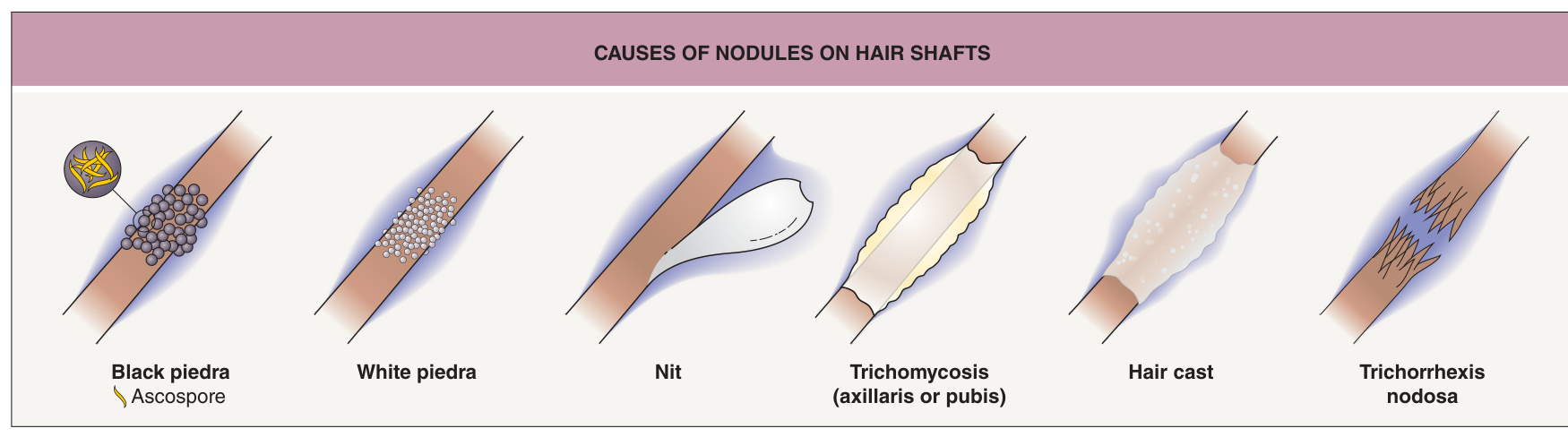
Fig. 77.1 Causes of nodules on hair shafts.
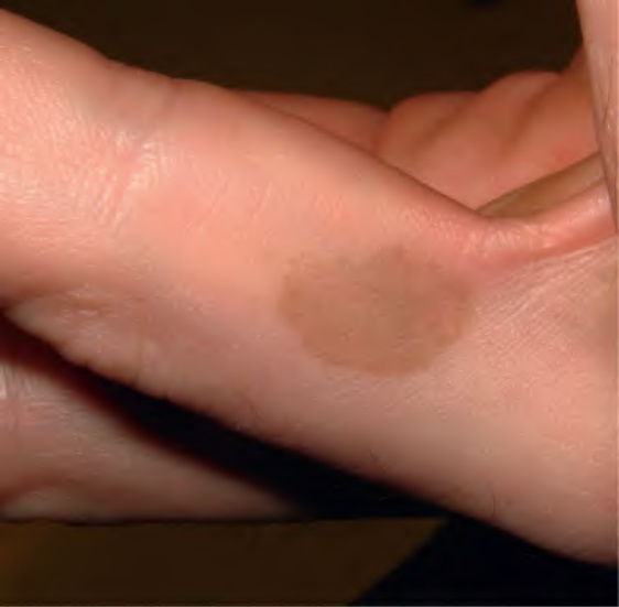
Fig. 77.2 Tinea nigra.
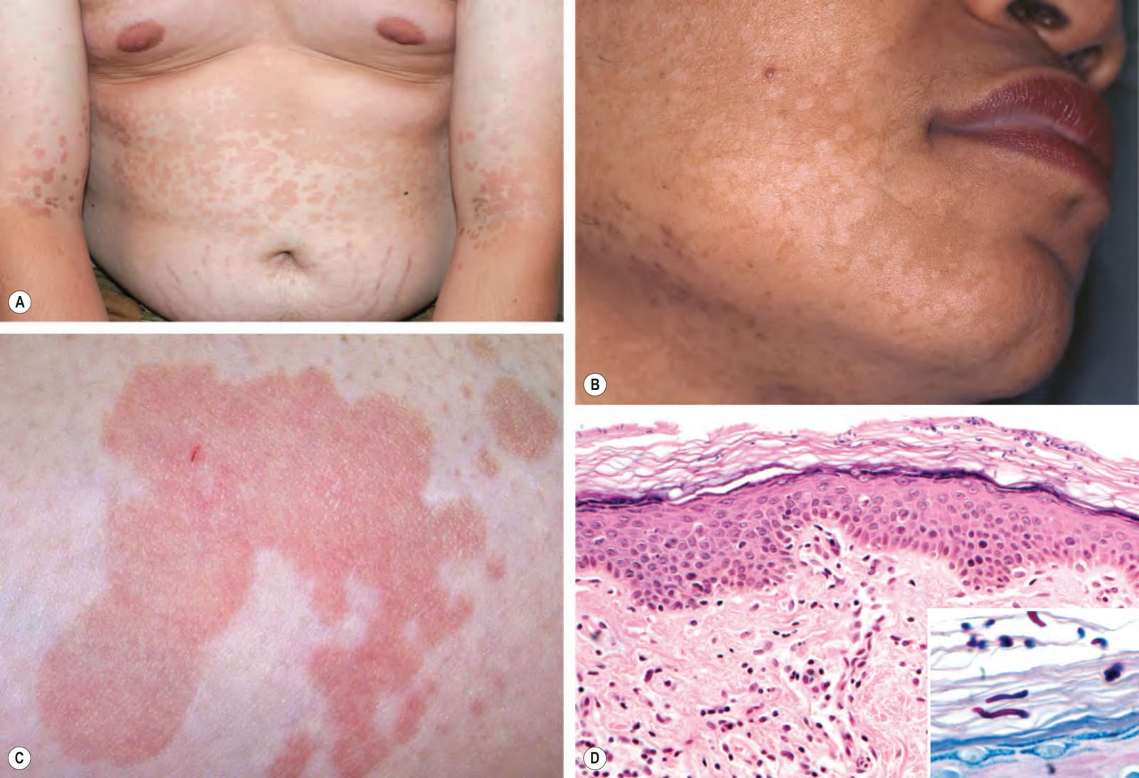
Fig. 77.3 Tinea (pityriasis) versicolor.A Hyperpigmented variant with confluence on the lower chest. B Hypopigmented variant on the face. C Coalescing pink lesions with fine scale. D Yeast and short hyphal forms in the stratum corneum highlighted by a PAS stain (inset). A, B, Courtesy Kalman Watsky, MD; D, Courtesy Lorenzo Cerroni, MD.
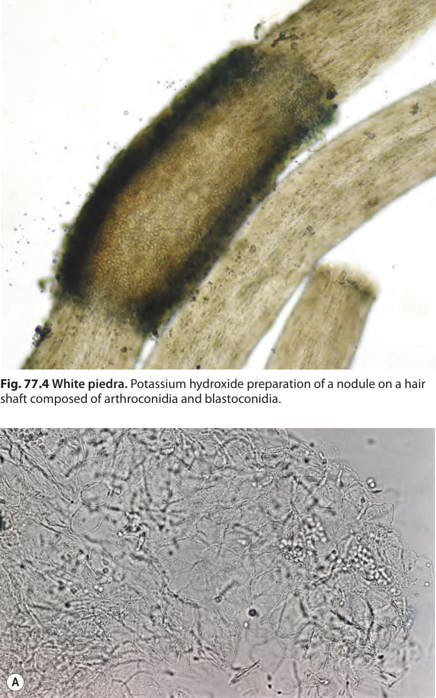
Fig. 77.4 White piedra. Potassium hydroxide preparation of a nodule on a hair shaft composed of arthroconidia and blastoconidia.
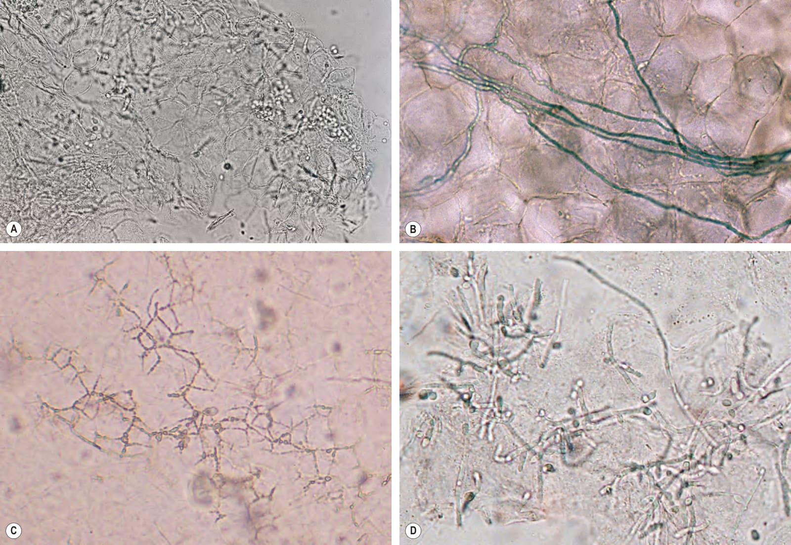
Fig. 77.5 Potassium hydroxide preparations.A Superficial skin scrapings from tinea (pityriasis) versicolor demonstrating clusters of yeast and short mycelial forms. B A dermatophyte, in this case Trichophyton tonsurans, demonstrating branching hyphae (chlorazol black stain). Note that the hyphae cross multiple squamous cells. C “Mosaic” pattern of cell walls that should not be misinterpreted as hyphae. D Yeast and pseudohyphae of candidiasis. A, Courtesy Ronald P. Rapini, MD; C, Courtesy Louis A. Fragola, Jr, MD; D, Courtesy Frank Samarin, MD.
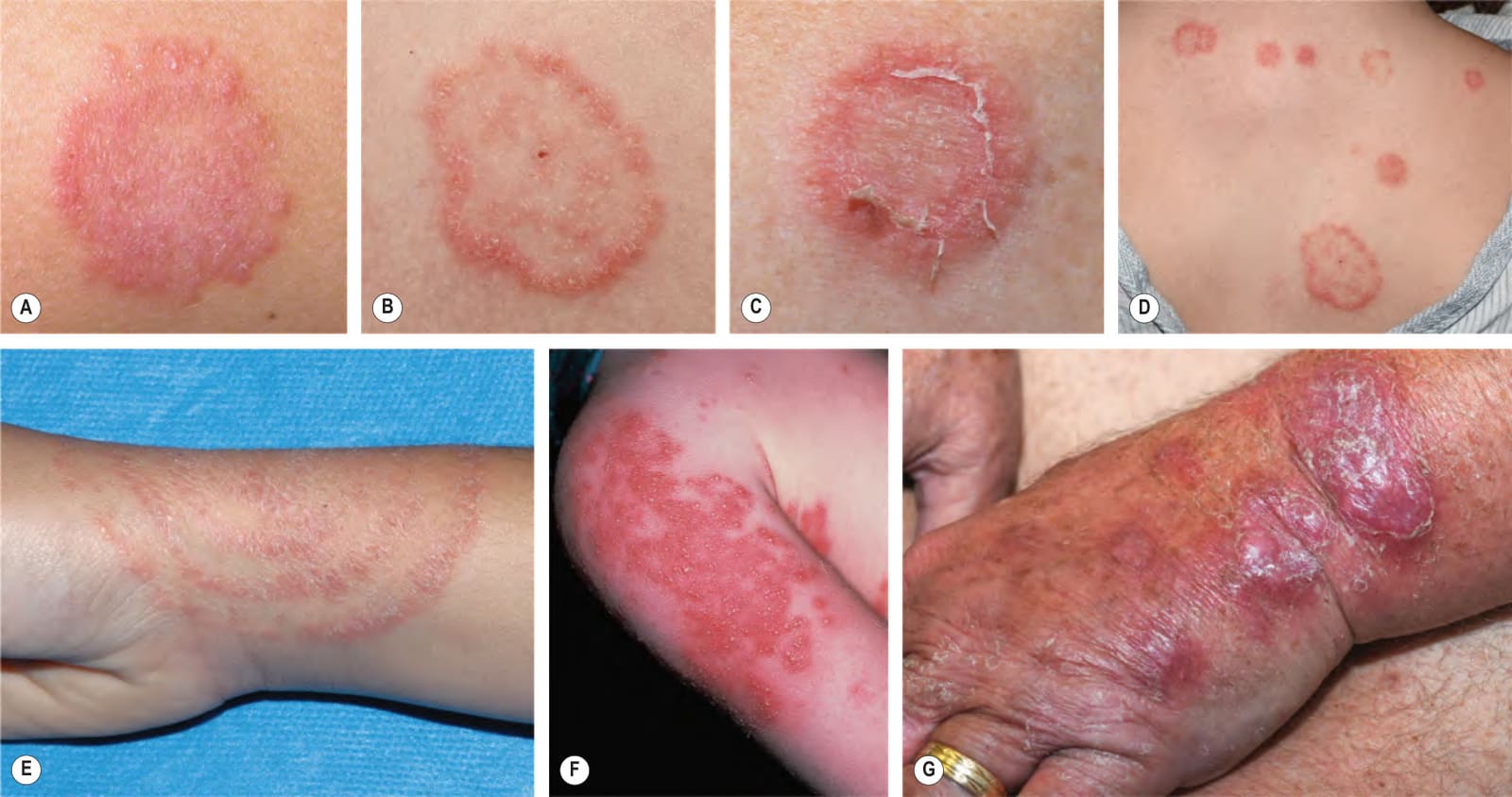
Fig. 77.6 Tinea corporis.A There is a subtle annular configuration with a border composed of individual, slightly scaly papules. B Classic annular lesion with a scaly raised border and central clearing. C Annular lesion with trailing scale reminiscent of erythema annulare centrifugum. D Multiple annular and circinate lesions of various sizes on the upper back. E Scaly concentric rings on the arm. F Pustules within multiple figurate lesions on the upper arm. G Inflammatory nodules on the dorsal hand and a thick granulomatous plaque on the distal forearm (tinea profunda). There is associated scale, including on the digits, and a few of the papulonodules are follicular (Majocchi granuloma). B, D, Courtesy Julie V. Schaffer, MD; C, G Courtesy Kalman Watsky, MD.
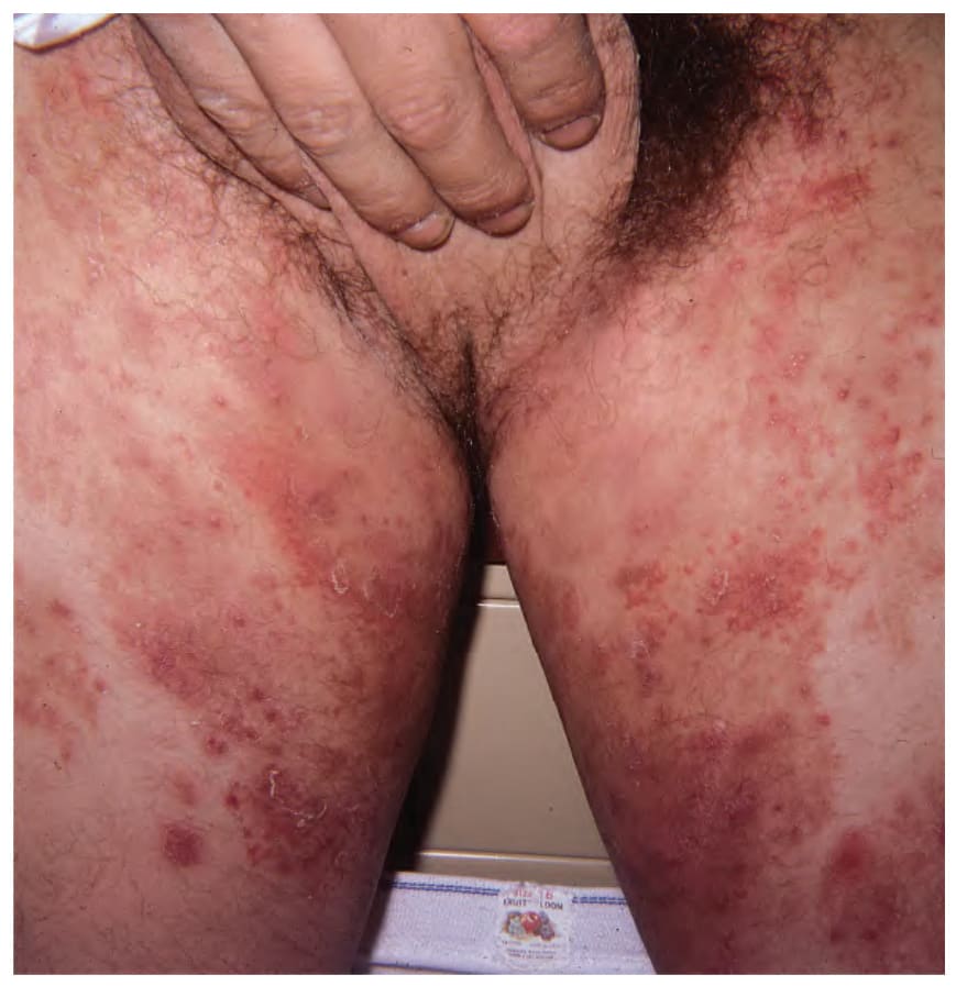
Fig. 77.7 Tinea incognito. Use of potent topical corticosteroids for the presumed diagnosis of dermatitis is a common clinical scenario. Numerous hyphae are usually seen on KOH examination. Courtesy Louis A. Fragola, Jr, MD.
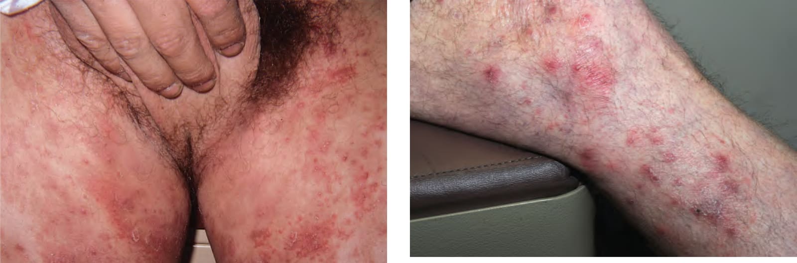
Fig. 77.8 Nodular perifolliculitis (Majocchi granuloma). Perifollicular inflammation and follicular pustules on the leg due to Trichophyton rubrum. A few of the lowermost papules have a granulomatous appearance. Courtesy Kalman Watsky, MD.
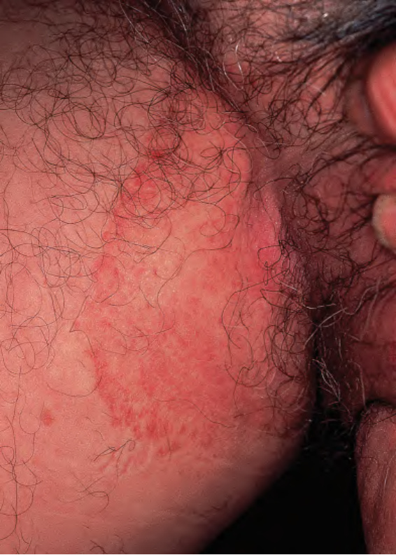
Fig. 77.9 Tinea cruris.
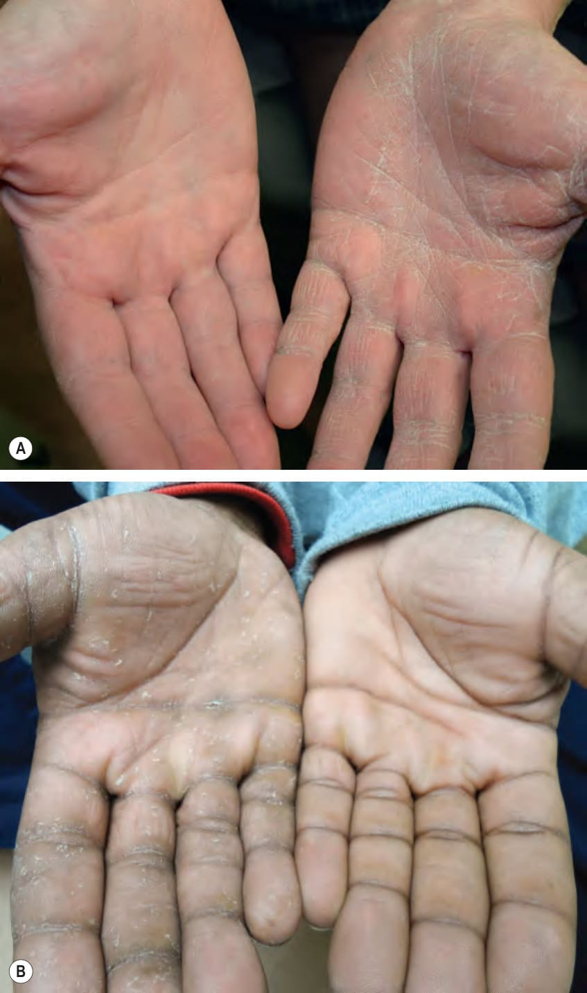
Fig. 77.10 Tinea manuum.A Diffuse scaling of the palm of one hand, with accentuation in the creases. B Multiple collarettes of scale reminiscent of keratolysis exfoliativa on one palm.
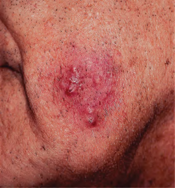
Fig. 77.11 Superficial form of tinea barbae due to Trichophyton rubrum. Several follicular pustules are evident. Courtesy Jean L. Bolognia, MD.
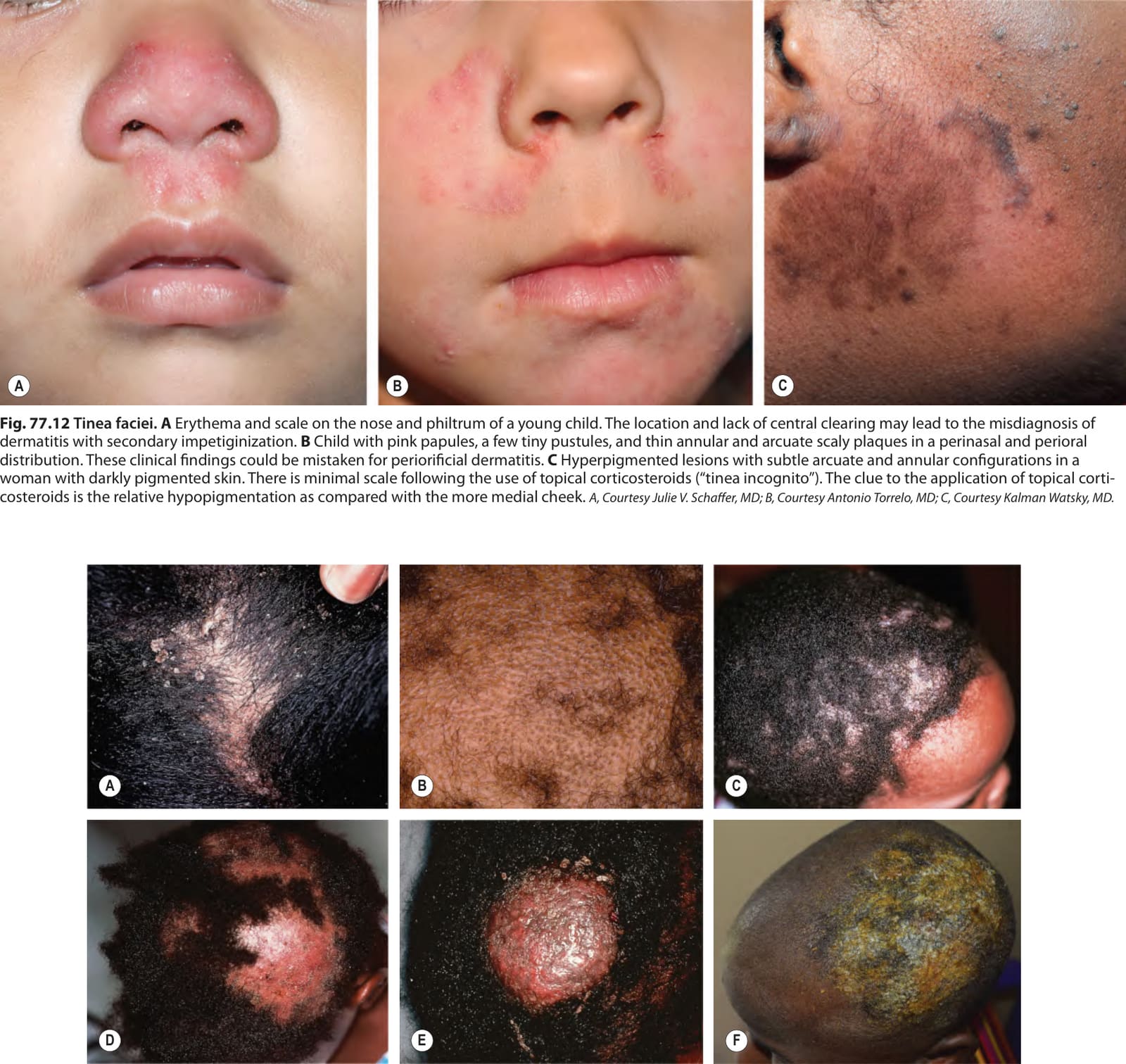
Fig. 77.12 Tinea faciei.A Erythema and scale on the nose and philtrum of a young child. The location and lack of central clearing may lead to the misdiagnosis of dermatitis with secondary impetiginization. B Child with pink papules, a few tiny pustules, and thin annular and arcuate scaly plaques in a perinasal and perioral distribution. These clinical findings could be mistaken for periorificial dermatitis. C Hyperpigmented lesions with subtle arcuate and annular configurations in a woman with darkly pigmented skin. There is minimal scale following the use of topical corticosteroids (“tinea incognito”). The clue to the application of topical corticosteroids is the relative hypopigmentation as compared with the more medial cheek. A, Courtesy Julie V. Schaffer, MD; B, Courtesy Antonio Torrelo, MD; C, Courtesy Kalman Watsky, MD.
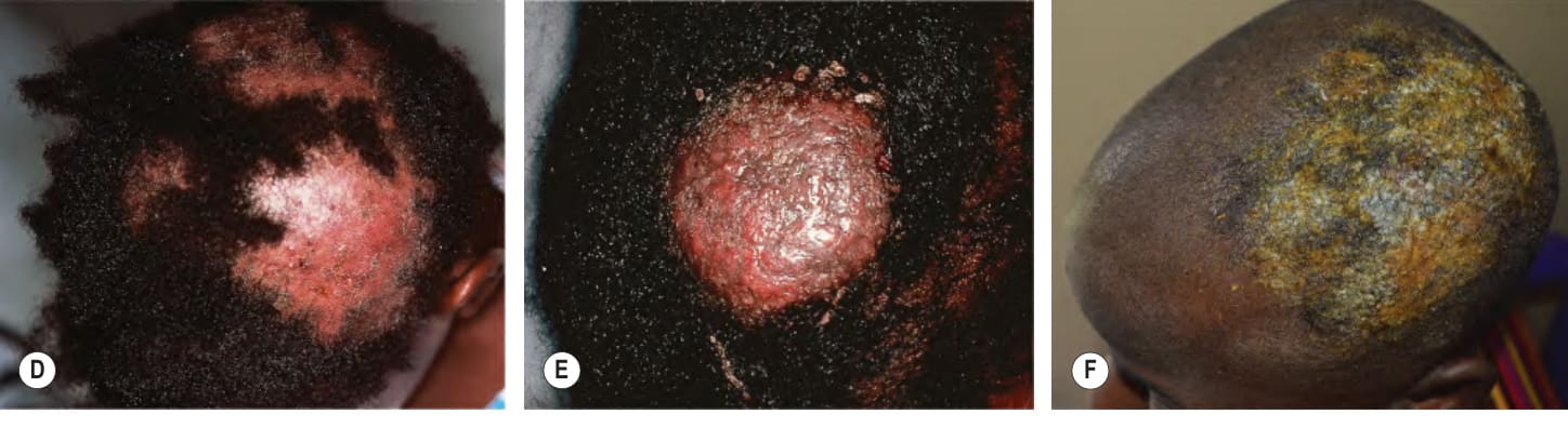
Fig. 77.13 Tinea capitis. The range of clinical presentations of tinea capitis due to Trichophyton tonsurans, from mild scalp scaling (A) to patchy alopecia with black dots (B) or scale (C) to large areas of alopecia with pustules and scale-crust (D). E, F Kerion formation due to T. tonsurans. G Microscopic examination of involved hairs demonstrates an endothrix pattern (KOH–chlorazol black stain). H Histologic examination shows arthroconidia and hyphae within hair shafts to the level of Adamson’s fringe (limit of the zone of keratinization; inset). B, Courtesy Louis A. Fragola, Jr, MD; F, Courtesy Robert Browdell, MD; H, Courtesy Lorenzo Cerroni, MD.
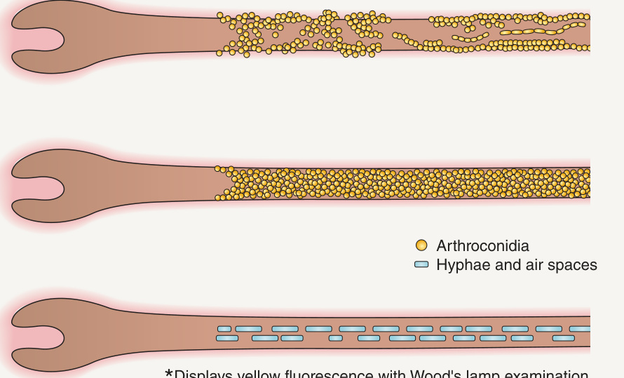
Fig. 77.14 The three patterns of hair invasion and the causative dermatophytes.
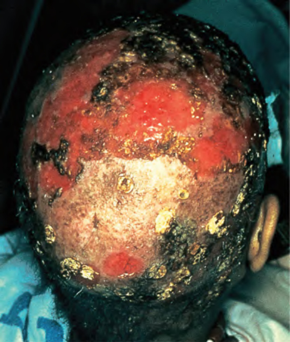
Fig. 77.15 Favus due to Trichophyton schoenleinii. Scarring alopecia with erosions and several scutula on the occipital scalp. The latter represent masses of keratin plus fungi. Courtesy Israel Dvoretzky, MD.
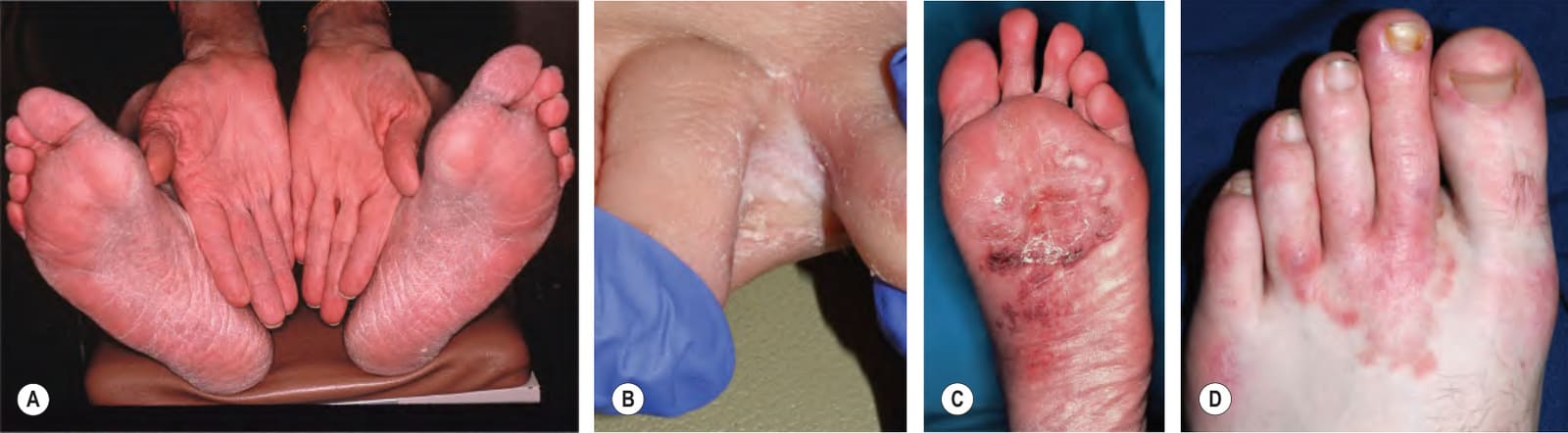
Fig. 77.16 Tinea pedis A Diffuse scaling on both feet (moccasin type) as well as on the right hand, representing “one hand–two feet” tinea. B Maceration and scaling between the fourth and fifth toes in the interdigital form. C Erythema, scale-crust, and bullae in the inflammatory form. D Extension of tinea pedis onto the dorsal foot in a serpiginous configuration of erythema and papules. Scale is minimal due to the patient’s use of a potent topical corticosteroid for presumed dermatitis. B, D, Courtesy Julie V. Schaffer, MD.
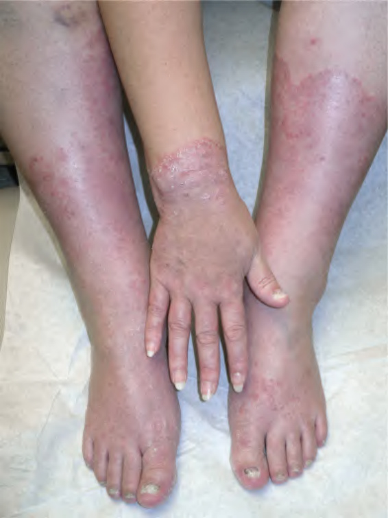
Fig. 77.17 Extensive tinea corporis emanating from tinea manuum and tinea pedis. Note the confluent involvement, serpiginous scaly borders, and associated tinea unguium of the toenails and one fingernail.
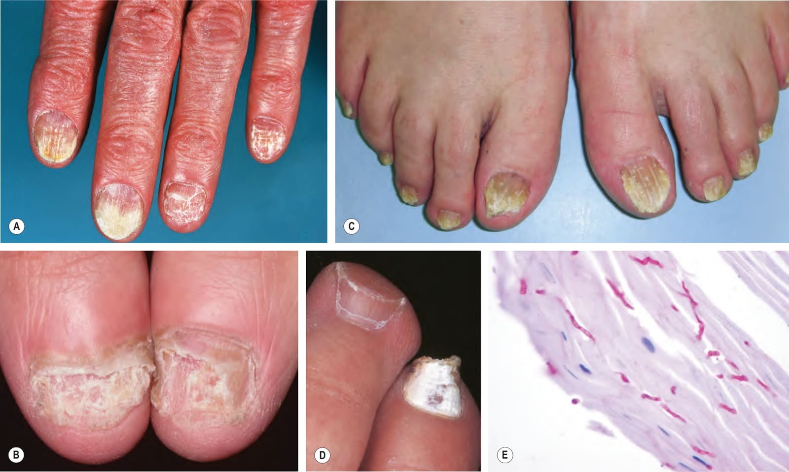
Fig. 77.18 Tinea unguium. Onycholysis, yellowing, crumbling and thickening of the fingernails (A), thumb nails (B), and toenails (C) in the distal/lateral subungual variant. D White discoloration of the toenail in the superficial white variant. E Hyphae within a formalin-fixed, PAS-stained nail plate. A, D Courtesy Jean L. Bolognia, MD; B, Courtesy Louis A. Fragola, Jr, MD; E, Courtesy Mary Stone, MD.
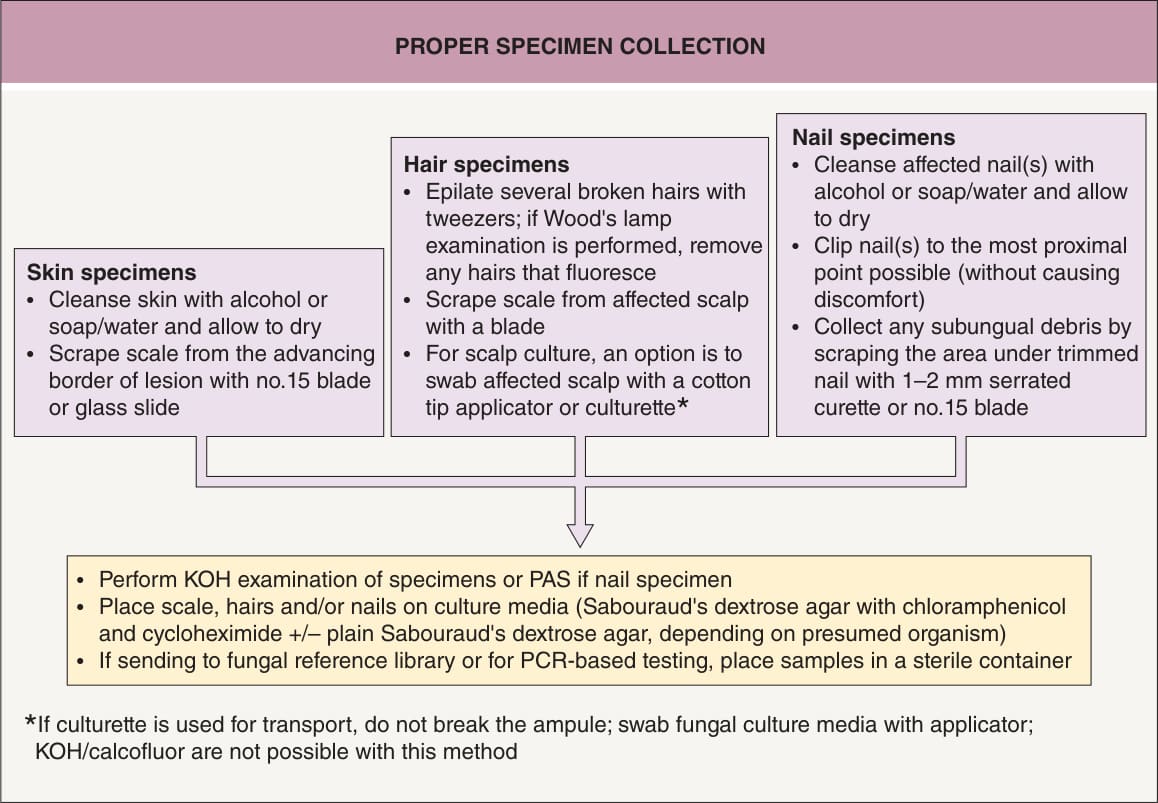
Fig. 77.19 Proper specimen collection. Proper collection of skin, hair, and nails is important. Following these simple guidelines will help the clinician to achieve the most accurate diagnosis. Courtesy Judy Warner.
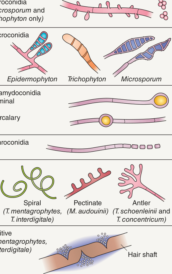
Fig. 77.20 Microscopic appearance of various forms of conidia and hyphae and the in vitro hair perforation test.
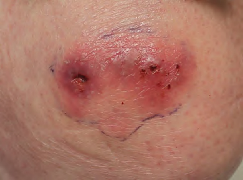
Fig. 77.21 Invasive dermatophytosis due to Trichophyton rubrum in an immunocompromised host.Courtesy Evelyn Lilly, MD.
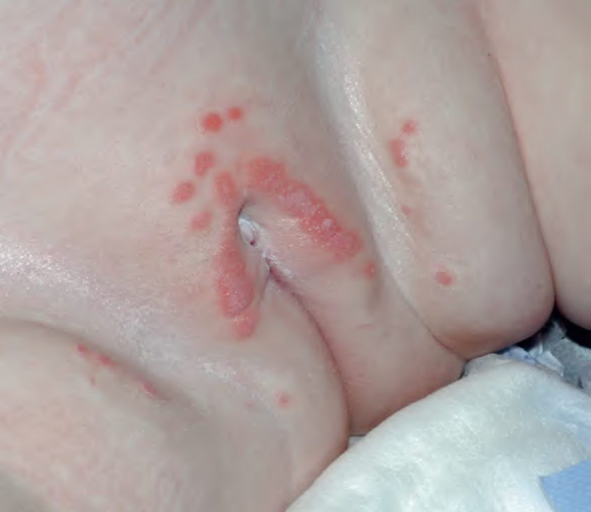
Fig. 77.23 Granuloma gluteale infantum. Coalescing moist, pink papules on the vulva and suprapubic area of an infant. Courtesy Julie V. Schaffer, MD.
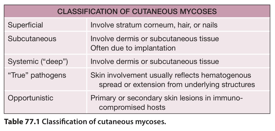
Table 77.1 Classification of cutaneous mycoses.
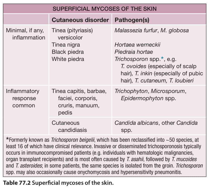
Table 77.2 Superficial mycoses of the skin.
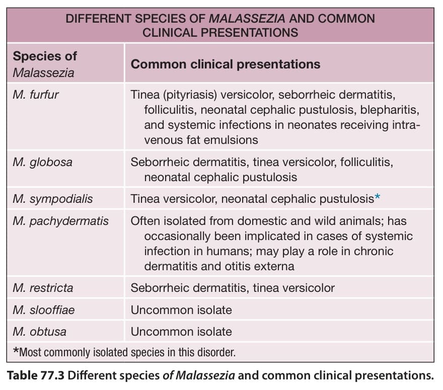
Table 77.3 Different species of Malassezia and common clinical presentations.
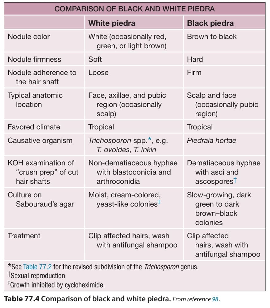
Table 77.4 Comparison of black and white piedra.From reference .
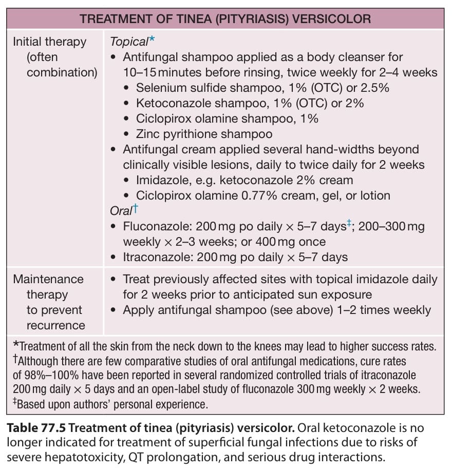
Table 77.5 Treatment of tinea (pityriasis) versicolor. Oral ketoconazole is no longer indicated for treatment of superficial fungal infections due to risks of severe hepatotoxicity, QT prolongation, and serious drug interactions.
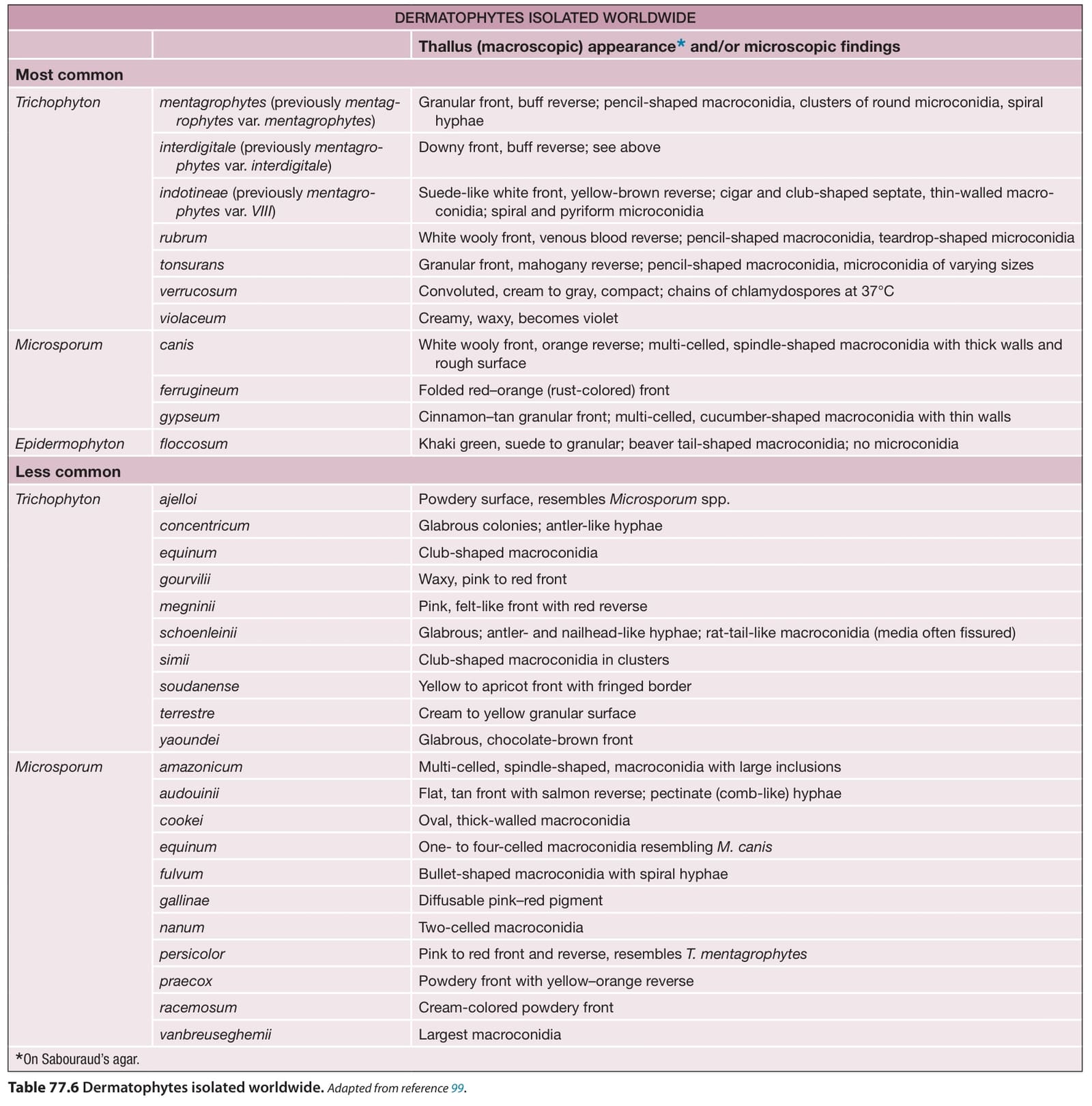
Table 77.6 Dermatophytes isolated worldwide.Adapted from reference .
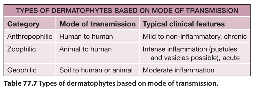
Table 77.7 Types of dermatophytes based on mode of transmission.
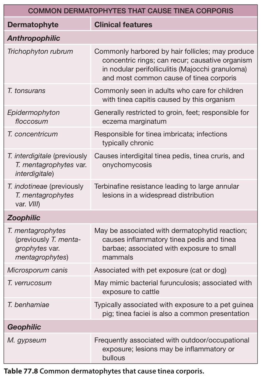
Table 77.8 Common dermatophytes that cause tinea corporis.
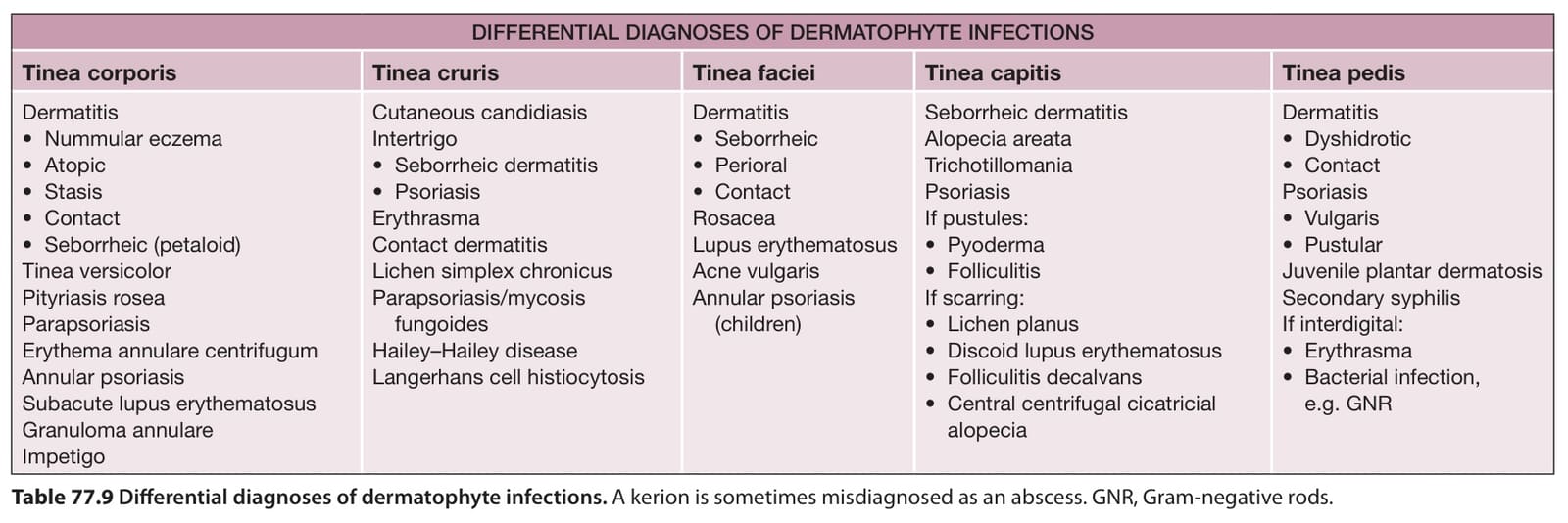
Table 77.9 Differential diagnoses of dermatophyte infections. A kerion is sometimes misdiagnosed as an abscess. GNR, Gram-negative rods.
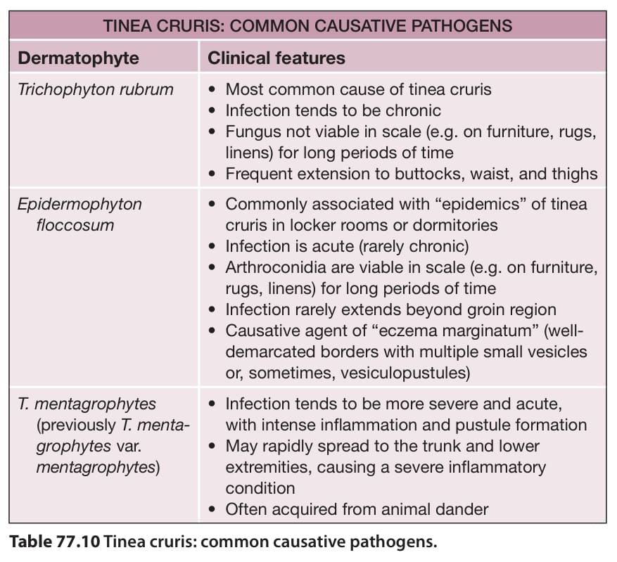
Table 77.10 Tinea cruris: common causative pathogens.
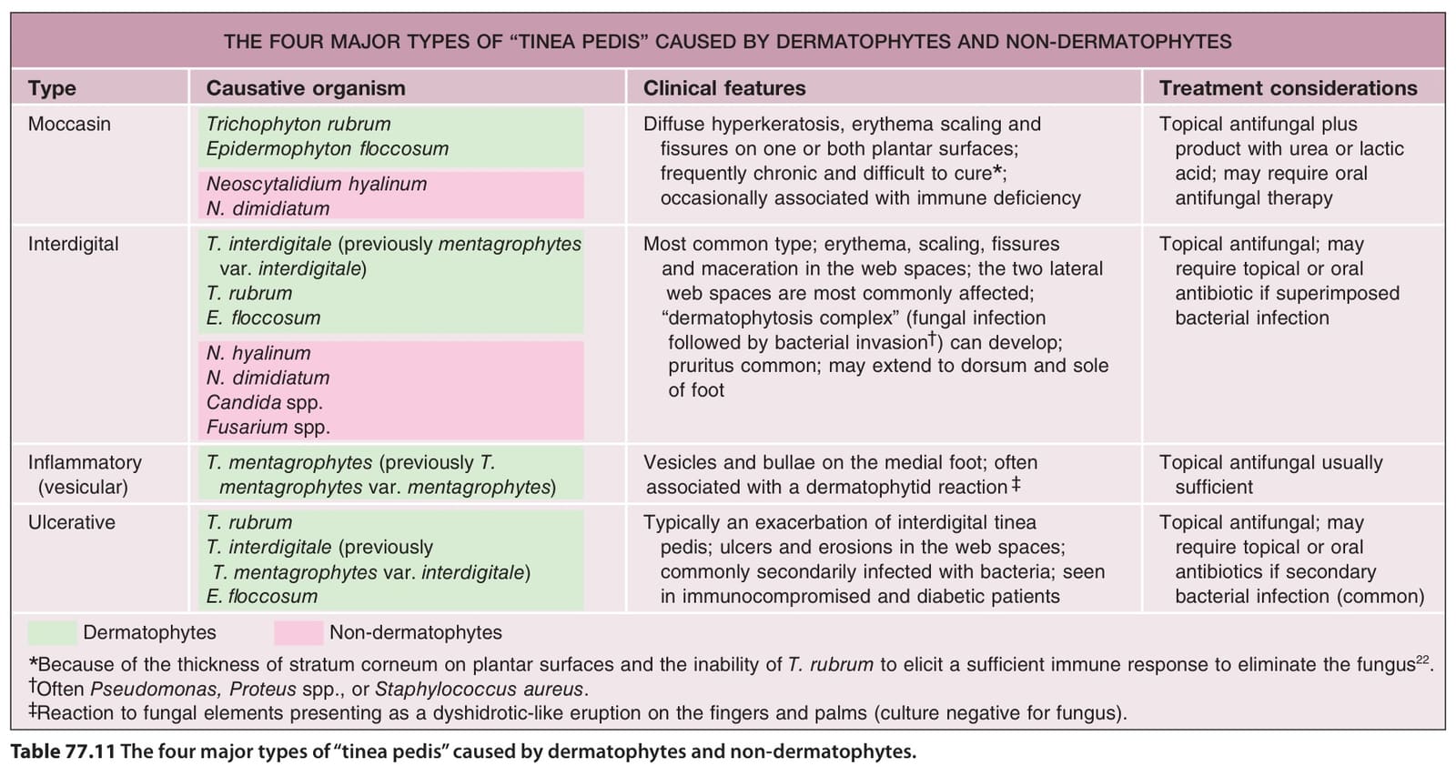
Table 77.11 The four major types of “tinea pedis” caused by dermatophytes and non-dermatophytes.
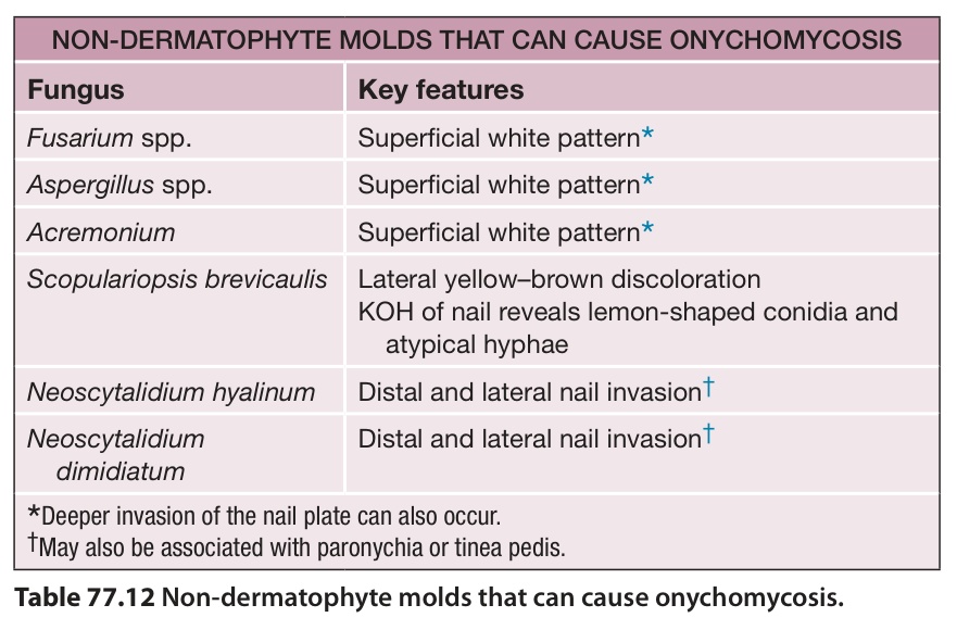
Table 77.12 Non-dermatophyte molds that can cause onychomycosis.
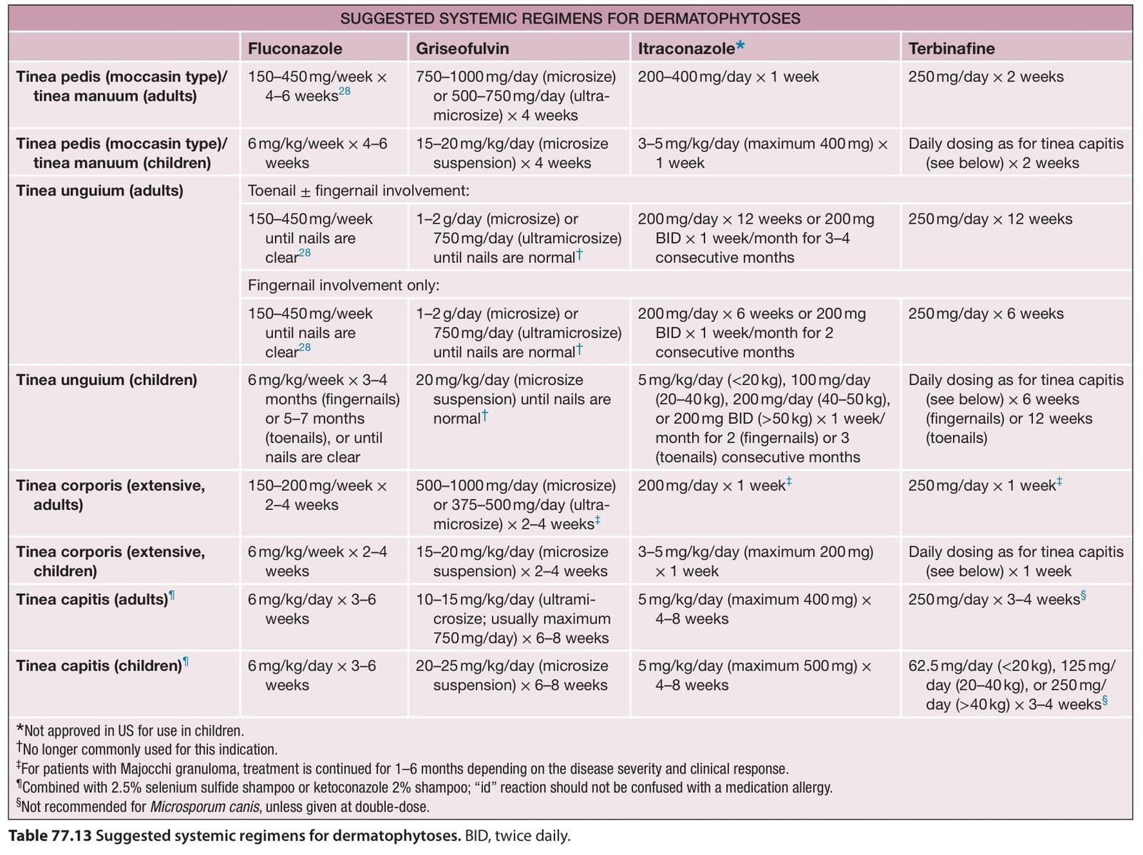
Table 77.13 Suggested systemic regimens for dermatophytoses. BID, twice daily.
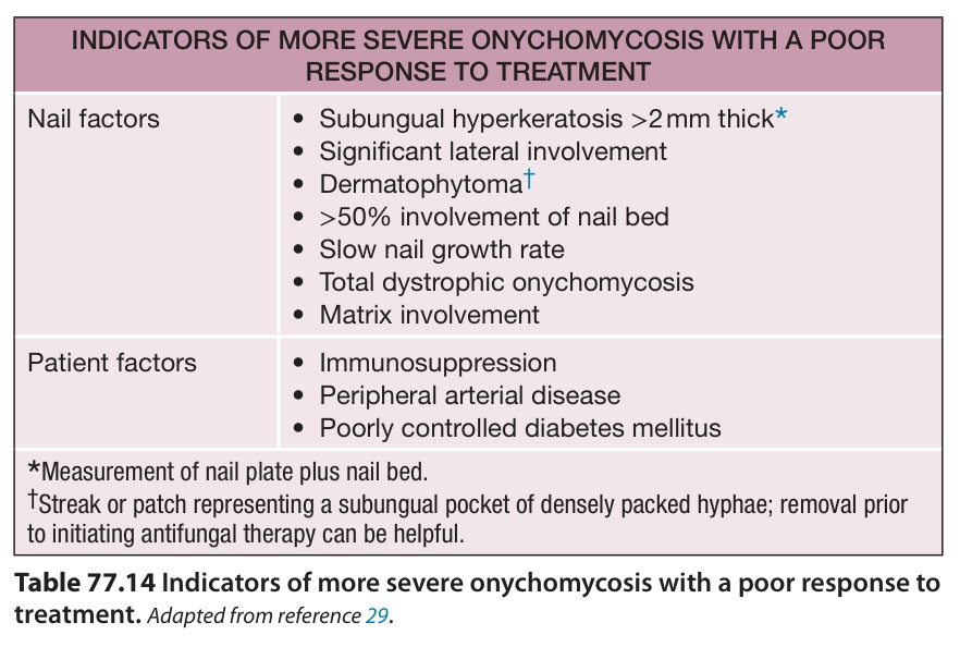
Table 77.14 Indicators of more severe onychomycosis with a poor response to treatment.Adapted from reference .
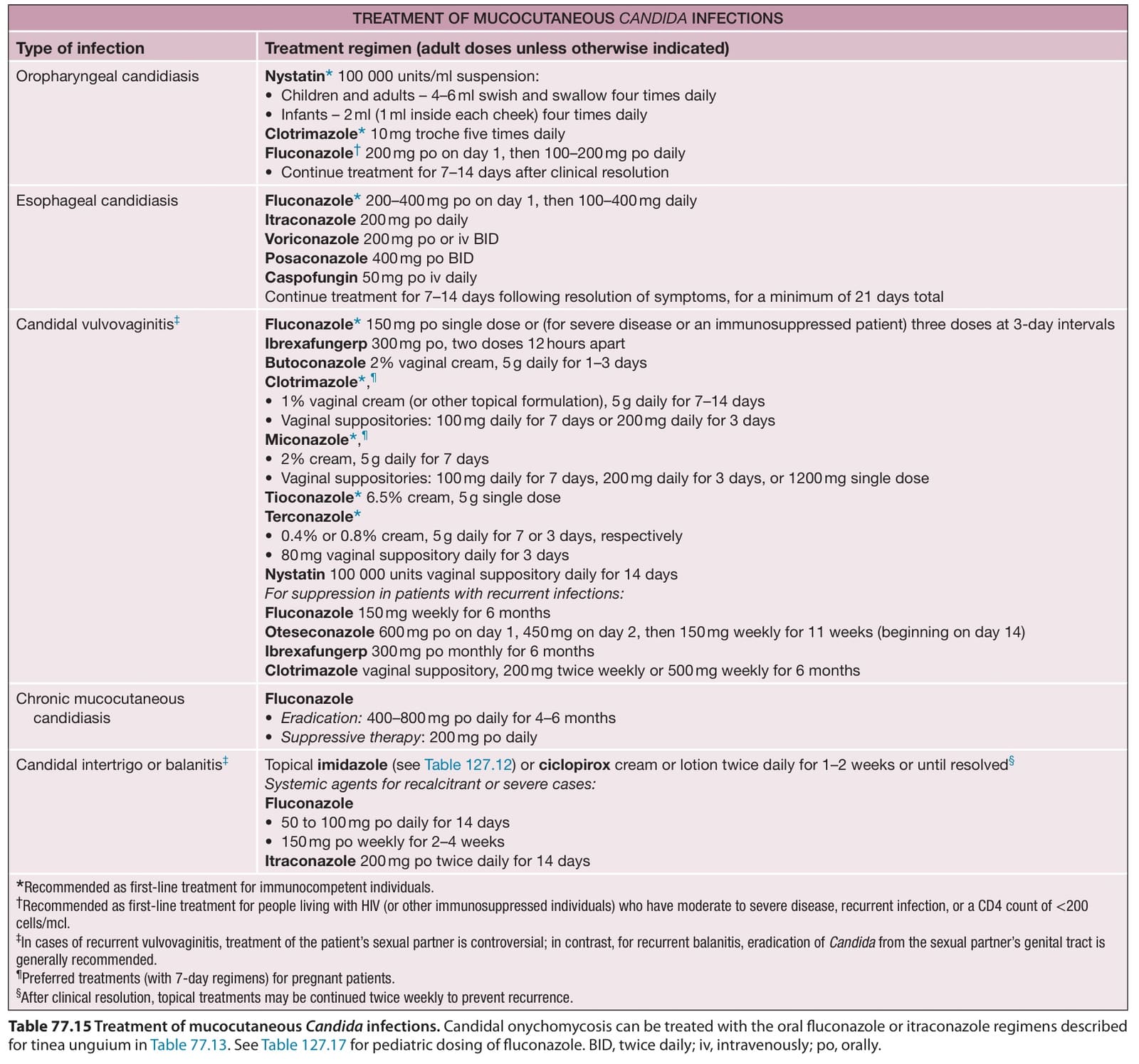
Table 77.15 Treatment of mucocutaneous Candida infections. Candidal onychomycosis can be treated with the oral fluconazole or itraconazole regimens described for tinea unguium in Table 77.13. See Table 127.17 for pediatric dosing of fluconazole. BID, twice daily; iv, intravenously; po, orally.