SUBCUTANEOUS MYCOSES
The “subcutaneous” mycoses are due to a large and diverse group of organisms that cause disease when implanted or otherwise introduced into the dermis or subcutis. Chromoblastomycosis, mycetoma, sporotrichosis, and lobomycosis are discussed in detail in this section; the
first three have been designated as neglected tropical diseases (NTDs) by the World Health Organization (WHO).
Another chronic subcutaneous fungal infection is basidiobolomycosis, which is caused by Basidiobolus ranarum. Although this organism is an environmental saprophyte that is found worldwide, the associated infection occurs most commonly in children living in tropical and subtropical climates. The most common portal of entry is the skin, typically after arthropod bites or minor trauma. Clinically, the disease manifests as a solitary, painless, indurated subcutaneous nodule or swelling of the thigh or buttock. The classic treatment is saturated solution of potassium iodide (SSKI; see Ch. 100), although successful results have been reported with oral azole antifungals and trimethoprim– sulfamethoxazole. Surgical excision is not recommended. A related organism, Conidiobolus coronatus, causes an infection that has similar clinical features but typically affects the face, especially the nasal region.
Although not a true fungal disease, protothecosis is treated with antifungal agents. Prototheca (usually P. wickerhamii) is a genus of achlorophyllic algae that can be introduced into the skin via trauma, often in the setting of exposure to contaminated water. Common clinical presentations include solitary cutaneous plaques (sometimes eczematous), nodules or ulcers, as well as olecranon bursitis. The extremities are the most frequent sites of involvement. Although immunocompetent patients typically have chronic, stable disease, patients who are immunocompromised can have widespread involvement, including algemia. Protothecosis is difficult to treat; surgical excision and systemic antifungals (e.g. amphotericin B) are more effective in immunocompetent hosts.
Skin and soft tissue infections with Pythium insidiosum, an aquatic oomycete, may present as progressive necrotic plaques in previously healthy children and adults as well as immunocompromised individuals. Pythiosis typically develops on the lower extremities or in the periocular area following exposure to stagnant water. It most often occurs in Thailand and India but has been reported worldwide, including in the southern US. These organisms resemble filamentous fungi but are more closely related to diatoms and algae. Treatment is difficult and includes surgical debridement together with antifungal and antibacterial agents.
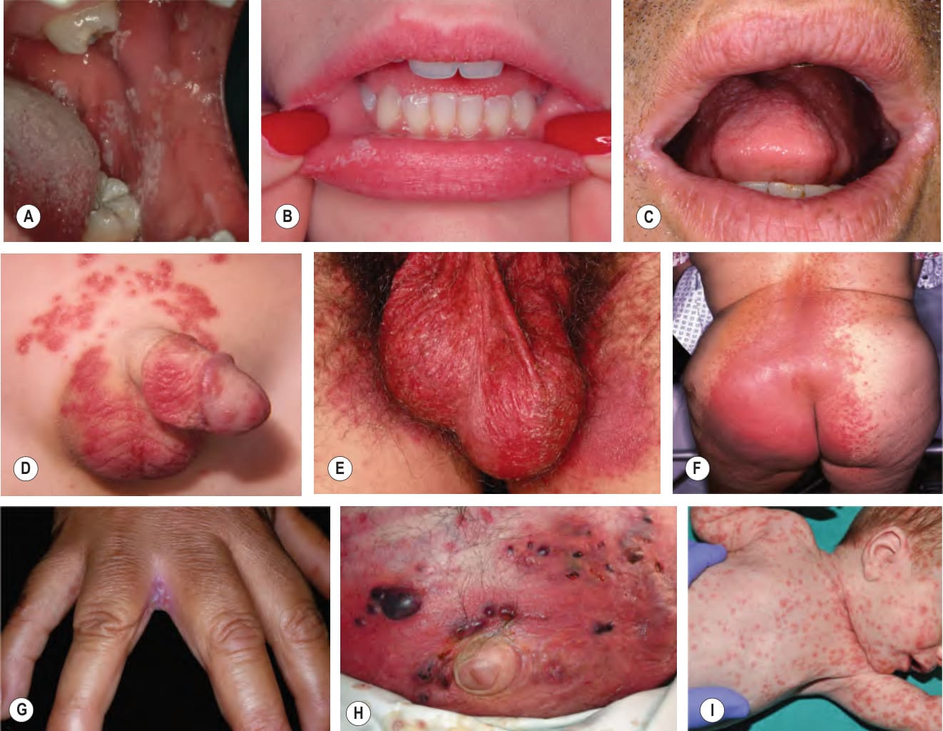
Fig. 77.22 Mucocutaneous candi- diasis.A Thrush with “cottage cheese”- like exudate on the buccal mucosa in a man with AIDS. B Thrush and candidal cheilitis. C Angular cheilitis (perlèche). D Candidiasis of the suprapubic area and penis in a young boy. Note the collarettes of scale on the coalescing, brightly erythematous papules. E Candidiasis of the scrotum and medial thighs with beefy red erythema, scale, and satellite papules. F This infection occurred in a hospitalized patient with diabetes mellitus who was receiving broad-spectrum antibiotics. Note the multiple satellite lesions. G Erosive interdigital candidiasis (erosio interdigitalis blastomycetica) in the classic location between the third and fourth fingers. H Dermal candidiasis in an immunosuppressed patient. I Early-onset cutaneous candidiasis in a term neonate. Widespread pink papules, some of which have collarettes of white scale, admixed with small pustules. This typically results from intrauterine acquisition and may be present at birth or appear during the first six days of life. B, D Courtesy Louis A. Fragola, Jr, MD; C, Courtesy Kalman Watsky, MD; E, G, Courtesy Eugene Mirrer, MD; I, Courtesy Antonio Torrelo, MD.
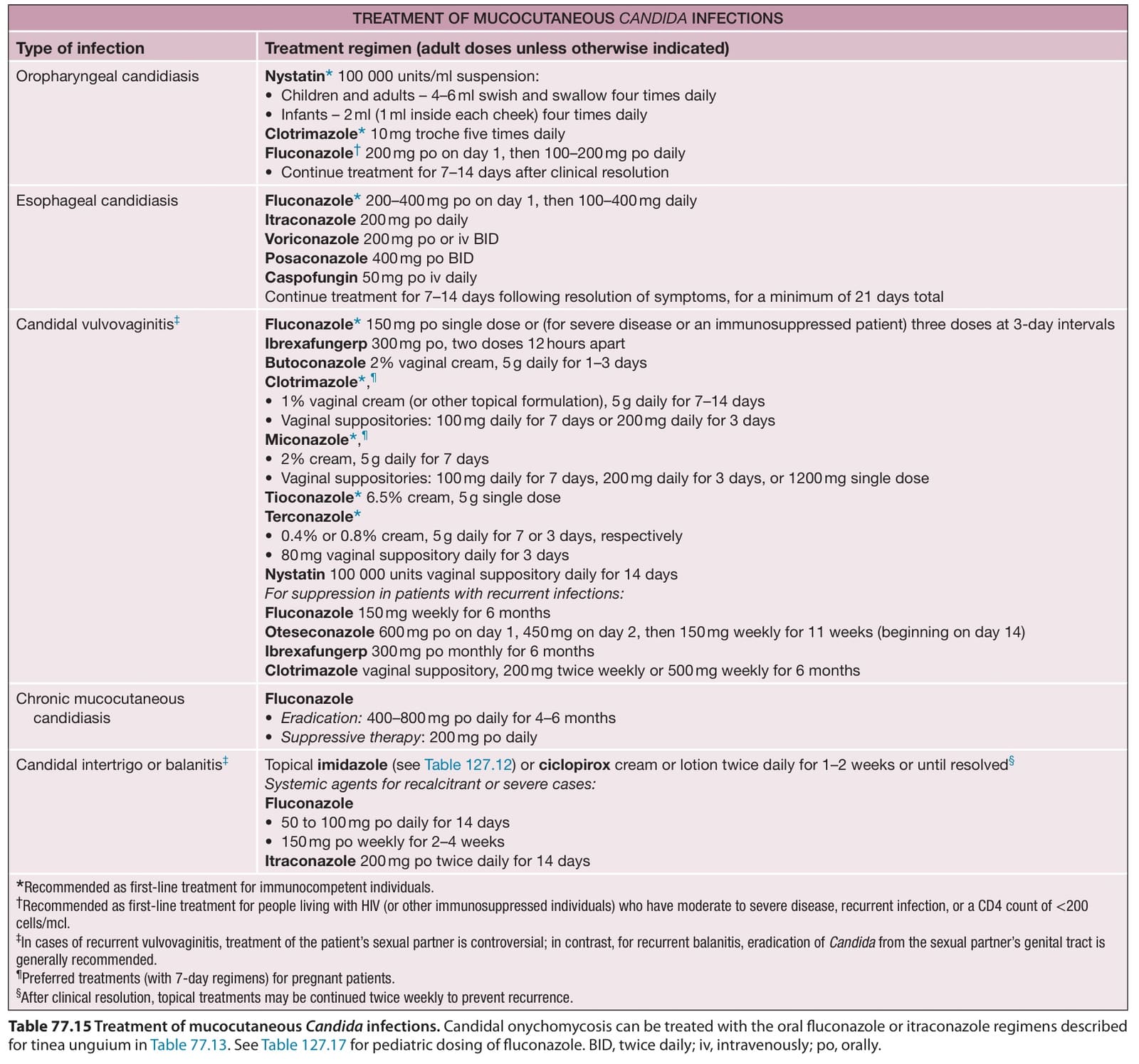
Table 77.15 Treatment of mucocutaneous Candida infections. Candidal onychomycosis can be treated with the oral fluconazole or itraconazole regimens described for tinea unguium in Table 77.13. See Table 127.17 for pediatric dosing of fluconazole. BID, twice daily; iv, intravenously; po, orally.