Chromoblastomycosis
Synonyms: Chromomycosis Cladosporiosis Verrucous dermatitis Phaeosporotrichosis Pedroso disease Fonseca disease
Introduction
Chromoblastomycosis presents as a “verrucous dermatosis” and is caused by several genera of dematiaceous (pigmented) fungi. Typically chronic in nature, an expanding verrucous plaque on the lower, or occasionally upper, extremity is the classic presentation. Two fungi, Fonsecaea pedrosoi and Cladophialophora carrionii, are responsible for most cases; less common causes include Fonsecaea compacta, Fonsecaea monophora, Phialophora verrucosa, and Rhinocladiella aquaspersa. The clinical presentation and colonial morphologies of each of these fungi are very similar, and differentiation is based on microscopic and conidial characteristics.
History
Pedroso first recognized chromoblastomycosis in 1911. Four years later, Medlar and Lane reported the first case in the US (in Boston). There has been confusion over the proper naming of this condition. At one point, chromoblastomycosis was thought to be closely related to blastomycosis; however, cellular division does not occur via budding (“blasto-”) but rather via internal septation, hence the preference for the term “chromomycosis”. Nonetheless, some authors have argued that the latter is also confusing, because it has the same meaning as phaeohyphomycosis (see below), and so they prefer the term “chromoblastomycosis”.
Epidemiology and pathogenesis
Chromoblastomycosis is an NTD that is most commonly found in tropical and subtropical climates and occasionally in temperate zones such as the US, Europe, and Canada. Farmers, miners, and others working in rural areas are at increased risk. Men 20–60 years of age are most often affected, probably due to increased occupational exposure, which accounts for up to 90% of cases.
The fungi responsible for causing chromoblastomycosis are found in the soil and in decaying plants and wood. The disease is typically contracted from trauma to the lower extremities, including from not wearing shoes. As a result, the organism is introduced into the dermis or subcutis via implantation.
Clinical features
The disease usually presents as a papule or nodule on the leg, which progresses to form a verrucous or granulomatous plaque (Fig. 77.24). The lesion may appear annular as the central portion resolves with scarring. Several lesions may coalesce to form a multinodular mass, or multiple lesions may exist as discrete islands scattered within unaffected skin. It is thought that autoinoculation from scratching may be responsible for the spread of infection. Typically, only one extremity is affected. A subcutaneous nodule or mass is occasionally the presenting lesion. There are usually no constitutional symptoms.
Pathology
Pseudoepitheliomatous hyperplasia, intraepidermal abscesses, and suppurative and granulomatous inflammation within the dermis are the typical histologic findings (Fig. 77.24C). Pathognomonic round, pigmented “Medlar bodies” or sclerotic bodies, which are 6 to 12 microns in diameter and said to resemble “copper pennies”, are present in the dermis, both extracellularly and within giant cells. Organisms can also be detected via PCR of tissue samples.
Differential diagnosis
Although characteristic reddish brown to black dots may be evident using dermoscopy, a biopsy is indicated to exclude other infectious diseases characterized by granulomatous lesions associated with scarring, such as cutaneous tuberculosis, tertiary syphilis, blastomycosis, and leishmaniasis. Mycetoma is another implantation mycosis that commonly affects the lower extremity, but edema, draining sinuses, and grains point to this diagnosis. If a biopsy is not feasible, a KOH examination of scrapings from a pigmented portion of the lesion can be performed. The presence of Medlar bodies is diagnostic (excluding other entities such as blastomycosis), but hyphae may also be seen. Culture growth at 25–30°C is slow and the different genera produce similar colonial morphologies. The major differences between genera are the microscopic features, in particular the type of conidia produced in culture. The three major types are:
●Cladophialophora-like (long branching side chains with shield cells at branch points)
●Phialophora-like (conidia resemble overflowing buds in a vase)
●Rhinocladiella-like (overall configuration similar to a mascara brush).
Treatment
Therapeutic options are limited. Some authors have advocated heat therapy based on evidence that the causative organisms will not grow at high temperatures. For small lesions, surgical excision can be attempted together with systemic antifungal treatment. 5-Flucytosine combined with either intravenous amphotericin B or an oral triazole has been reported to be efficacious. Itraconazole alone (200–400 mg/day) administered for at least 6 months may have cure rates of up to 80%–90%. Successful treatment with voriconazole or posaconazole has also been described. In a small series, oral terbinafine (500 mg/day) given for at least 7 months was effective. Other treatment considerations include cryosurgery and the addition of antibiotics if the lesion is secondarily infected.
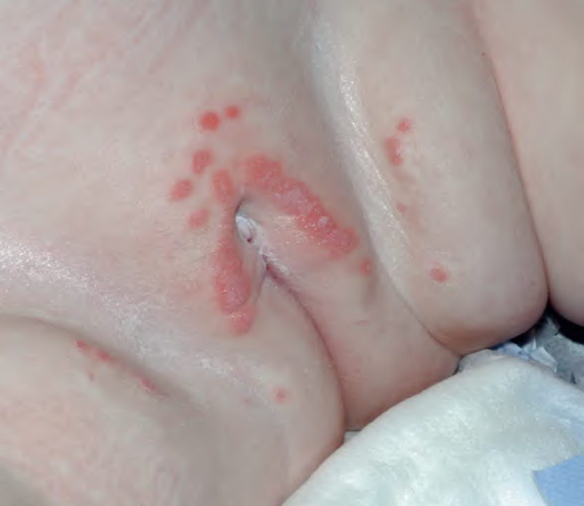
Fig. 77.23 Granuloma gluteale infantum. Coalescing moist, pink papules on the vulva and suprapubic area of an infant. Courtesy Julie V. Schaffer, MD.
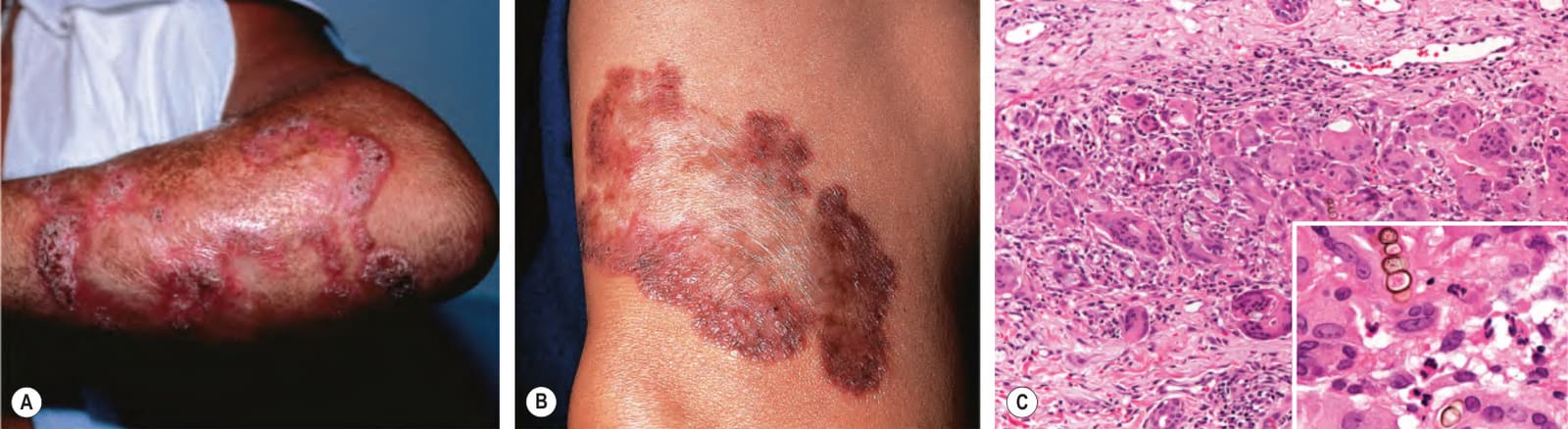
Fig. 77.24 Chromoblastomycosis. Annular and figurate plaques due to central clearing and scarring with a verrucous surface on the arm (A) and a more granulomatous appearance on the leg (B). C Brown-colored sclerotic body (inset) within a mixed granulomatous and neutrophilic dermal infiltrate. C, Courtesy C. Massone, MD.