Mycetoma
Synonyms: Madura foot Maduromycosis
Introduction
Mycetoma, often referred to as “Madura foot”, is a Greek term for “fungal tumor”. Due to implantation, it is a granulomatous infection of dermal and subcutaneous tissue that may extend to muscle or even bone. Mycetoma is differentiated from other mycoses by its characteristic draining sinuses containing grains (sclerotia, sulfur granules) and local edema. Two subtypes exist: (1) actinomycotic mycetoma – caused by filamentous aerobic and anaerobic organisms, e.g. Nocardia brasiliensis, Actinomadura madurae; and (2) eumycotic mycetoma – caused by true fungi. Botryomycosis caused by true bacteria (e.g. Staphylococcus aureus, Pseudomonas spp.) is also associated with grains, as is actinomycosis (see Ch. 74).
History and epidemiology
Dr. John Gill provided the first formal description of mycetoma in 1842, in Madura (India), hence the name “Madura foot”. Mycetoma, now classified as an NTD, is endemic to tropical and subtropical climates. Actinomycotic mycetoma is more prevalent in Mexico, while eumycotic
mycetoma is more common in other areas including Africa. The typical patient is a man between 20 and 50 years of age.
Pathogenesis
In most cases, causative organisms are acquired from the soil via direct inoculation into the skin, leading to a dermal and subcutaneous fungal infection. Deeper invasion to muscle or bone can subsequently occur. This process may occur rapidly or over many years. Sometimes, multiple sites of involvement appear in the vicinity of the original lesion, likely due to multiple injuries rather than contiguous spread of disease. Lack of protective footwear, malnutrition, and exposed cuts and abrasions are risk factors for infection.
Clinical features
The foot is the most common site of infection, followed by the hand, trunk, and scalp. Involvement is typically unilateral and begins as a painless papule. Once the subcutaneous tissues are infected, there is swelling and eventual formation of purulent draining sinuses (Fig. 77.25A). The drainage contains the characteristic grains, which represent compact masses of fungal colonies and vary in size from minute to half a centimeter in diameter. Invasion of deeper tissues then ensues, and sometimes cavities are formed within involved bone. Surprisingly, this disease is generally asymptomatic. All subtypes can have a similar clinical presentation.
Pathology
Pseudoepitheliomatous hyperplasia is a common histologic finding that accompanies suppurative and granulomatous inflammation of the dermis and subcutis as well as fibrosis. The characteristic grains represent tightly packed colonies of organisms (Fig. 77.25B). Estimation of the diameter of the filaments comprising the grain as compared to the size of the nuclei of surrounding inflammatory cells allows a distinction between eumycotic (thicker filaments) and actinomycotic (thin, fine filaments) mycetomas. Special stains, e.g. methenamine silver, Brown–Brenn, can aid in distinguishing the subtype (see Ch. 0).
Differential diagnosis
To establish the diagnosis of mycetoma, both the history and clinical appearance are important considerations followed by laboratory evaluation. In addition to noting the color of the grains (Table 77.16), KOH examination, Gram stain, and culture (bacterial and fungal) of exudate from a sinus tract and/or tissue should be performed. Molecular techniques such as PCR-based assays can also be utilized to identify organisms. Mycetoma has to be differentiated from botryomycosis and actinomycosis, which are also associated with grain formation (see Ch. 74).
Treatment
For the best outcome, eumycotic mycetoma must be diagnosed early and surgically excised (including a large margin of normal surrounding tissue) before the underlying bone becomes involved. Systemic antifungal therapy is administered before and after excision. Depending upon the particular fungus, itraconazole, voriconazole, posaconazole, and terbinafine have been used to treat eumycotic mycetomas. The medical treatment of actinomycotic mycetoma typically consists of streptomycin or amikacin plus either trimethoprim–sulfamethoxazole or dapsone; therapy is continued for months to years. Other useful drugs include moxifloxacin and imipenem.
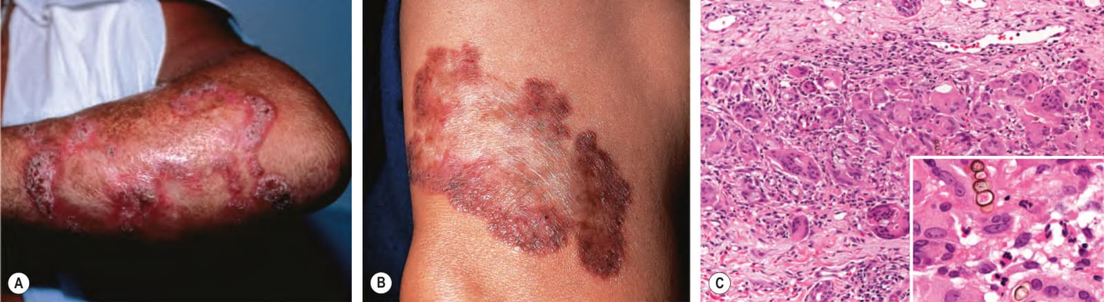
Fig. 77.24 Chromoblastomycosis. Annular and figurate plaques due to central clearing and scarring with a verrucous surface on the arm (A) and a more granulomatous appearance on the leg (B). C Brown-colored sclerotic body (inset) within a mixed granulomatous and neutrophilic dermal infiltrate. C, Courtesy C. Massone, MD.
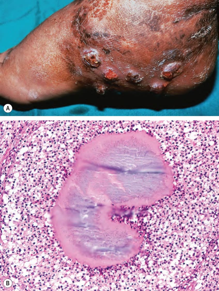
Fig. 77.25 Mycetoma (Madura foot).A Note the soft tissue swelling of the foot as well as multiple nodules with pustular discharge. B Histology of a grain, which represents tightly packed colonies of fungal organisms. B, Courtesy Lorenzo Cerroni, MD.
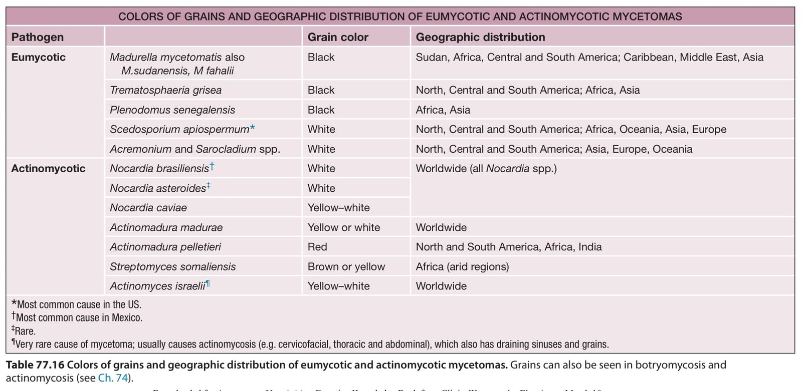
Table 77.16 Colors of grains and geographic distribution of eumycotic and actinomycotic mycetomas. Grains can also be seen in botryomycosis and actinomycosis (see Ch. 74).