Sporotrichosis
Synonym: Rose gardener’s disease
Introduction
Sporotrichosis is a subcutaneous mycosis caused by dimorphic organisms of the genus Sporothrix (e.g. S. schenckii, S. braziliensis, S. globose, S. mexicana). A gardener being pricked by a contaminated rose thorn is a classic vignette for the acquisition of sporotrichosis. Although various clinical presentations are possible, the most common one is a lymphocutaneous or “sporotrichoid” pattern, which refers to the primary inoculation site plus spread of the infection along the lymphatics in a nodular pattern.
Epidemiology
Sporothrix is present in the soil throughout the world. However, sporotrichosis is endemic to Mexico, Central America, and South America as well as other areas such as South Africa. In these locations, exposure to the organism typically occurs in individuals who work outdoors. In temperate climates, such as in the US and Canada, the disease most often develops in gardeners, especially those growing orchids and roses. All age groups can be affected, but it is more common in adults. In the past several decades, the incidence of sporotrichosis in the Rio de Janeiro region of Brazil has increased >100-fold due to transmission of S. brasiliensis by infected cats, which (in contrast to humans) harbor large numbers of organisms in cutaneous ulcers. This epidemic has spread to other areas of Brazil and adjacent countries such as Argentina, with recent reports outside of South America (e.g. in the United Kingdom).
Pathogenesis
Sporotrichosis is most commonly acquired from cutaneous inoculation, especially by vegetation such as thorns and wood. With the exception of S. brasiliensis from cats, animal-to-human transmission is rare. Multiple inoculations may occur simultaneously, not to be confused with spread of a single primary lesion. The presentation and course of sporotrichosis depends on the host immune response as well as the size and virulence of the inoculum. In naive hosts, involvement of regional lymphatics ensues. However, in some patients, especially those with a prior history of exposure to Sporothrix, lymphatic spread does not occur and a “fixed” ulcer or granulomatous plaque develops at the site of inoculation; the latter is often seen on the face, especially in children living in endemic areas.
Extensive cutaneous disease with or without systemic involvement is also possible, especially in an immunocompromised host. Cases of inhaled sporotrichosis have been reported with systemic and cutaneous dissemination, similar to disseminated histoplasmosis and other dimorphic fungal infections.
Clinical features
The initial presentation of sporotrichosis is a single papule at a site of injury, most commonly on the hand, appearing several weeks after inoculation. The lesion then becomes eroded or ulcerated with purulent drainage, but it is generally not painful. Additional lesions appear weeks later, typically developing as dermal and subcutaneous nodules and ulcers along the path of lymphatic drainage (often up the arm; Fig. 77.26). This is the well-known “sporotrichoid” pattern. The involved lymphatic vessels may become fibrosed. Lesions of “fixed” cutaneous sporotrichosis can have a granulomatous appearance, often with ulceration, while disseminated lesions typically present as subcutaneous nodules. Reactive conditions such as erythema multiforme and erythema nodusum have been reported in patients with sporotrichosis, especially that due to S. brasiliensis.
Pathology
Histologically, suppurative and granulomatous inflammation is found in the dermis and subcutis. The causative organisms are rarely evident; staining with fluorescent-labeled antibodies may aid in recognition of the sparse cigar-shaped yeast forms. Asteroid bodies are often seen. When numerous fungi are present (e.g. in an immunocompromised host), budding yeast and cigar-shaped organisms can often be visualized with PAS (Fig. 77.26D) or silver stains.
Differential diagnosis
When there is a sporotrichoid pattern, the major entity to be excluded is an atypical mycobacterial infection, in particular with M. marinum; less common causes are outlined in Table 77.17. As organisms are not typically seen by KOH or histopathologic examination, culture (of pus or tissue) is often required to definitively diagnose sporotrichosis; a nested PCR assay can also be used to detect organisms in clinical samples. Sporothrix spp. are dimorphic fungi (see below), and whether a hyphal or yeast form develops depends on the temperature. Growth is rapid at 25°C, revealing a glabrous white to brown mold that becomes firm, wrinkled, and more darkly pigmented over time. Microscopically, delicate conidia are clustered at the ends of conidiophores (specialized
hyphae); single, thick-walled pigmented conidia can also arise from hyphae. At 37°C, on enriched glucose-containing media, there are slowgrowing, whitish, pasty, yeast-like colonies. On microscopic examination of the latter, cigar-shaped budding yeasts are seen. Full speciation depends on molecular studies. The differential diagnosis of fixed plaque sporotrichosis and disseminated lesions is broad and includes other granulomatous disorders, both infectious and inflammatory.
Treatment
Topical therapy is not effective. Oral saturated solution of potassium iodide (SSKI) has been used with success. Although neither fungistatic nor fungicidal, SSKI is thought to affect the host’s immune reaction to the organism. Its cost is low, but it has a bitter taste as well as potential side effects including iododerma, gastrointestinal upset, and thyroid suppression (see Ch. 100). Based upon multicenter, non-randomized trials and case series, the Mycoses Study Group of the Infectious Diseases Society of America has recommended itraconazole (100–200 mg/day) for 3–6 months as the treatment of choice for lymphocutaneous or fixed cutaneous sporotrichosis. The drug is generally safe and well tolerated, and the relapse rate is low. Amphotericin B may be indicated in severe or disseminated disease.
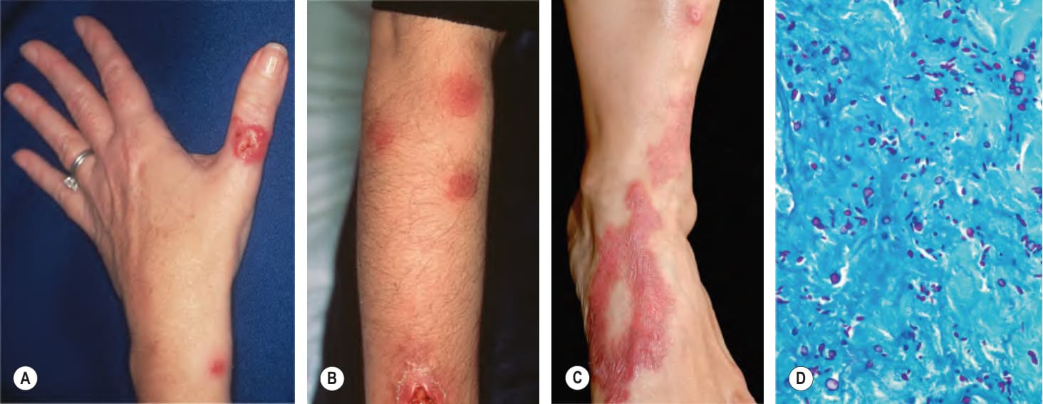
Fig. 77.26 Lymphocutaneous (sporotrichoid) pattern.A An eroded nodule on the thumb representing the primary lesion with a secondary lesion along the lymphatics due to sporotrichosis. B Ulcerated nodule on the extensor forearm with multiple more proximal nodules due to nocardiosis in a patient with lymphoma who was receiving systemic corticosteroids. C Granulomatous plaques with evidence of upward spread along the lymphatics. D Multiple yeast forms of Sporothrix schenckii in the dermis; note that many of the organisms have the characteristic cigar shape. B, Courtesy Jean L. Bolognia, MD; C, Courtesy Luis Requena, MD; D, Courtesy Ronald P. Rapini, MD.

Table 77.17 Infectious causes of a lymphocutaneous (“sporotrichoid”)