Lobomycosis
Synonyms: Keloidal blastomycosis Lacaziosis Lobo’s disease
Lobomycosis is a chronic fungal infection characterized by cutaneous nodules that resemble keloids. The causative organism, Lacazia loboi
pattern. There are also non-infectious causes such as lymphoma, Langerhans cell histiocytosis, and in-transit metastases. In addition, perineural spread of leprosy can mimic a lymphocutaneous pattern.
(previously known as Loboa loboi), is closely related to Paracoccidioides brasiliensis and has never been cultured in vitro. Lobomycosis occurs in Central and South America and has been linked to contact with dolphins and a marine environment as well as soil and vegetation in rural areas. Infection typically occurs after minor trauma and most often affects men.
Lobomycosis presents as firm, keloid-like nodules that may be pruritic or asymptomatic. It favors the distal extremities, ears (especially the helices), and face but can also affect the trunk (Fig. 77.27A,B). The lesions enlarge slowly over years, forming broad, multinodular plaques that may have a smooth surface or (occasionally) a verrucous appearance and ulceration. Histologic evaluation shows highly characteristic chains of thick-walled, yeast-like cells (known as “brass knuckles”) with intercellular bridges (Fig. 77.27C); PAS or silver stains highlight these organisms, which can also be identified in scrapings from the surface of skin lesions. Additional findings include a dermal granulomatous infiltrate and either atrophy or pseudoepitheliomatous hyperplasia of the epidermis. Treatment with surgical excision or cryosurgery is employed if feasible, since antifungal medications are typically ineffective. However, posaconazole or clofazimine plus itraconazole may be of benefit for patients with widespread disease.
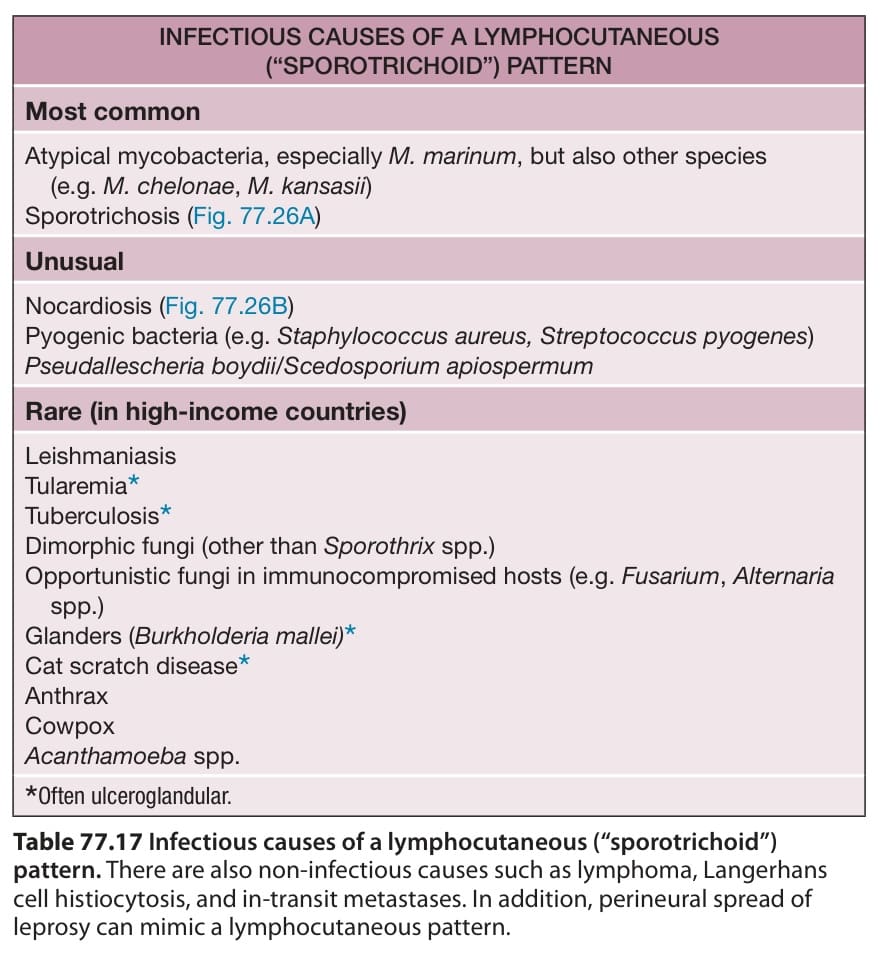
Table 77.17 Infectious causes of a lymphocutaneous (“sporotrichoid”)