SYSTEMIC MYCOSES
This section reviews systemic infections caused by opportunistic as well as true fungal pathogens. The true pathogens are generally dimorphic and cause infection in hosts with normal immune status, while opportunistic pathogens, which are generally less virulent, take advantage of the immunocompromised state.
True Pathogens
Introduction
The systemic mycoses due to true pathogens include histoplasmosis, blastomycosis, coccidioidomycosis, and paracoccidioidomycosis. They are caused by dimorphic fungi, meaning the organisms can exist in two forms – as molds (with septate hyphae and conidia) in nature and as other forms (usually yeasts) in living tissue and (with the exception of Coccidioides spp.) at 37°C. Each pathogen has certain geographic preferences (Fig. 77.28; see below). Acquisition of disease is typically through inhalation of the causative fungi, leading to pulmonary symptoms and pneumonitis. The vast majority of infections resolve spontaneously, and patients are left with strong, specific immunity. Disseminated histoplasmosis and coccidioidomycosis may occur in people living with HIV, as can disseminated infections with S. schenckii (dimorphic) and Talaromyces (Penicillium) marneffei (talaromycosis; dimorphic and opportunistic). Emergomyces spp. are dimorphic fungal pathogens that were initially described in South Africa (E. africanus; formerly Emmonsia pasteuriana) and subsequently reported in Europe, Asia, and the Americas (E. canadensis, E. europaeus, R. orientalis, E. pasteurianus). Disseminated infection in immunocompromised patients (e.g. those with HIV infection) can present with multiple crusted papules/nodules or ulcers, with parasitized macrophages similar to histoplasmosis and penicilliosis evident histologically (Table 77.18).
Histoplasmosis
Synonyms: Darling’s disease Cave disease Ohio valley disease Reticuloendotheliosis
Multinodular pinkish-brown plaques with a keloid-like appearance on the helix and back. C Spherical organisms with doubly refractile walls are seen within the dermis. Chain formation is commonly observed (inset; methenamine silver stain). A, B, Courtesy Regina Carneiro, MD and Caroline Brandao, MD; C, Courtesy C. Massone, MD.
Histoplasmosis is caused by the dimorphic fungus Histoplasma capsulatum var. capsulatum.
H. capsulatum is found in the soil in warm, moist climates, particularly the Mississippi, Ohio, and St Lawrence river valleys in the US and Canada. Between 80% and 90% of persons from endemic areas may exhibit positive histoplasmin skin testing. Birds, fowl, and bats are significant reservoirs for histoplasmosis. Because the feces of these animals contain the organisms, the areas in which they live can harbor infectious fungal spores. Caves, schoolyards, construction sites, unoccupied buildings, and chicken coops tend to be high-risk areas. H. capsulatum var. capsulatum is also endemic along river basins in some areas of Africa, India, Southeast Asia, and Australia.
Histoplasmosis is caused either by inhalation of H. capsulatum or, rarely, by direct cutaneous inoculation of the fungus. Immunocompetent hosts can acquire the disease, but immunocompromised hosts have a higher risk of dissemination. Clinical presentations include acute and chronic pulmonary histoplasmosis, disseminated histoplasmosis, and primary cutaneous histoplasmosis. In disseminated histoplasmosis, the most common sites of involvement (after the lung) are the spleen, lymph nodes, bone marrow, and liver; calcifications within the lymph nodes, lungs, and spleen can serve as evidence of prior infection. Cutaneous manifestations most commonly result from disseminated disease. Severe disseminated histoplasmosis has been reported in individuals with impaired interferon-γ responses due to mutations in IFNGR1 or STAT1, which encode interferon-γ receptor 1 and signal transducer and activator of transcription 1, respectively (see Ch. 60).
The skin lesions of histoplasmosis are nonspecific, making the diagnosis virtually impossible with physical examination alone. When chronic disseminated histoplasmosis occurs in immunocompetent hosts, the most common mucocutaneous finding is oral ulcers but occasionally nodules and vegetative plaques are also seen. Disseminated histoplasmosis in immunocompromised hosts (including people living with HIV) can present with mucocutaneous erosions or ulcers as well as multiple erythematous papules or nodules with scale or crust (Fig. 77.29A,B). In African histoplasmosis, a variant caused by H. capsulatum var. duboisii, most patients present with mucocutaneous, subcutaneous, and bone lesions.
Microscopic examination of biopsy specimens demonstrates characteristic intracellular yeast forms surrounded by a rim of clearing (Fig. 77.29C). Histiocytes and giant cells are the host cells in histoplasmosis, which is in the differential diagnosis of “parasitized macrophages” (see Table 77.18). For easier identification of the fungi, tissue may be stained with PAS or methenamine silver (Fig. 77.29D).
Because cutaneous histoplasmosis has a wide spectrum of clinical presentations, the diagnosis usually rests on histologic examination and culture of involved skin tissue. To evaluate for disseminated disease, bone marrow (sensitivity 70%–90%) and blood should be cultured at 25°C and 37°C, with plates held for up to a month. At 25°C, characteristic tuberculate macroconidia develop. Additional diagnostic tests include cultures of sputum and body fluids, polysaccharide antigen testing of blood and urine (useful in detecting disseminated disease), histoplasmin skin testing (indicated only in non-endemic areas), and serologic assays to measure antibody responses (complement fixation, immunodiffusion). Highly sensitive and specific PCR-based assays can be used to identify histoplasmosis in blood and tissue. Diseases in the differential diagnosis include other dimorphic fungal infections (e.g. paracoccidioidomycosis), mucocutaneous tuberculosis, major aphthae, and oral squamous cell carcinoma. For primary cutaneous histoplasmosis, entities in Table 77.17 should be considered.
In primary, self-limited, asymptomatic histoplasmosis, treatment may not be necessary. However, in symptomatic or disseminated disease, systemic antifungal therapy is required. Amphotericin B, given intra-venously and titrated up to 1 mg/kg/day, is currently the most effective therapy and should be used initially for severe disease, followed by itraconazole. In immunocompetent hosts with mild to moderate, stable disease, itraconazole (200–400 mg/day) is the treatment of choice. HIV-infected patients with disseminated histoplasmosis require lifelong maintenance therapy with itraconazole after initial treatment with amphotericin B (if required for severe disease). Successful treatment with voriconazole, posaconazole, and isavuconazole have also been described, but ketoconazole is no longer routinely used.
Blastomycosis
Synonyms: North American blastomycosis Gilchrist’s disease
Blastomyces dermatitidis is a dimorphic fungus and the most common causative agent of blastomycosis. Additional species that have more recently been associated with blastomycosis include B. gilchristii, B. helicus (formerly Emmonsia helica), B. emzantsi, and B. percursus.
Blastomycosis due to B. dermatitidis is endemic to North America, particularly the Mississippi and Ohio river valleys, Great Lakes region, and southeastern states; within this region, especially in the northern US and Canada, blastomycosis is also occasionally caused by B. gilchristii. While all ages and sexes can be affected, adult men are most likely to develop systemic infection, and children are more likely to develop acute pulmonary blastomycosis rather than chronic or cutaneous disease. The soil is an important source of infection, making those who have occupations with frequent outdoor exposure at higher risk than the remainder of the population. Disseminated disease due to B. helicus in immunocompromised hosts has been reported in the Western US and Canada. Blastomycosis also rarely occurs in other areas of the world, including Africa, India, and the Middle East; recent investigations suggest that B. emzantsi and B. percusus underlie most disease in these regions.
The lungs are typically the first site of infection, via inhalation of organisms. In contrast to infections with other dimorphic fungi, secondary cutaneous dissemination is a common occurrence and may even be the first sign of disease. Cutaneous manifestations are sometimes seen in the absence of overt pulmonary disease. Primary cutaneous blastomycosis is uncommon and results from direct inoculation of the skin from trauma, such as in the laboratory.
The most common cutaneous findings are papulopustules and well-demarcated verrucous plaques with scale-crust and pustules within their borders (Fig. 77.30A,B). Central ulceration can occur, and more advanced disease may have an appearance similar to pyoderma gangrenosum. The number of lesions can vary from one to several, and they tend to occur on exposed skin. Healing begins centrally and is followed by cribriform scarring. Mucous membrane involvement is occasionally observed.
Pulmonary infection is subclinical in up to 50% of patients with inhalation blastomycosis. Bone involvement may occur and presents as osteomyelitis with rare extension to muscle; genitourinary disease is uncommon.
Histologic examination of skin lesions demonstrates pseudoepitheliomatous hyperplasia, suppurative and granulomatous inflammation, and round yeast forms with characteristic broad-based budding and thick, double-contoured walls. This budding pattern helps to differentiate blastomycosis from other fungal infections. Methenamine silver and PAS staining allow better visualization of the fungi within giant cells and neutrophilic abscesses (Fig. 77.30D). Similar budding yeast forms can be seen in sputum samples.
Cutaneous blastomycosis must be differentiated from other cutaneous infections (e.g. verrucae, folliculitis, nocardiosis, tuberculosis, chromoblastomycosis, those due to other dimorphic fungi), inflammatory dermatoses (e.g. pyoderma gangrenosum, halogenoderma, sarcoidosis) and neoplasms (e.g. squamous cell carcinoma). Lesions of blastomycosis-like pyoderma, an exaggerated vegetative response to a prolonged primary or secondary bacterial infection, can also resemble cutaneous blastomycosis. Current or past residence in an area in which blastomycosis is endemic is a helpful clue.
Identification of characteristic broad-based budding yeast forms in pus by KOH or calcofluor examination is diagnostic of blastomycosis. Either skin tissue or pus should be cultured at both 37°C and 25°C. The former temperature allows identification of the budding yeast form, while the characteristic colonial morphology with white spikes (coremia) is observed at 25°C. Other tests such as blastomycin skin testing and complement fixation testing are not usually helpful, but PCR-based assays are available.
Blastomycosis, like histoplasmosis, requires systemic treatment with amphotericin B (given at the same doses) when severe or progressive. In mild to moderate non-CNS disease, itraconazole (200–400 mg/day) is the treatment of choice. Fluconazole (400–800 mg/day), voriconazole (especially following amphotericin B for CNS disease), posaconazole, and isavuconazole are alternative medications; ketoconazole is no longer routinely used. Occasionally, surgical treatment is indicated.
Coccidioidomycosis
Synonyms: Valley fever Desert rheumatism San Joaquin valley fever California disease
Coccidioidomycosis is caused by two dimorphic and highly virulent pathogens that are closely related, Coccidioides immitis and C. posadasii.
Coccidioidomycosis occurs predominantly in the summer and fall in the southwestern US, northern Mexico, and Central and South America; C. immitis is found primarily in California and C. posadasii elsewhere within these regions. The arthroconidia of Coccidioides spp. are inhaled via dust particles. Primary infection of the respiratory tract shows no sex or age preference, but disseminated disease is more common in Mexicans (5×), African-Americans (25×) and Filipinos (175×). Immunosuppressed states (e.g. HIV infection) and pregnancy also predispose to dissemination; White women are most likely to have more limited disease.
C. immitis and C. posadasii are thought to be the most virulent of all fungi. Infectious arthroconidia are inhaled and cause pulmonary infection in the vast majority of people living in endemic areas. The infection is asymptomatic in ~60% of affected individuals, while the remainder develop flu-like symptoms several weeks after exposure. Dissemination to the skin and other organs can occur and depends on host factors (see above). Of note, similar to histoplasmosis, severe disseminated disease has been associated with an impaired interferon-γ response due to mutations in IFNGR1 or STAT1 (see Ch. 60). Rarely, primary cutaneous coccidioidomycosis results from direct inoculation of the organism into the skin.
Symptomatic primary inhalation coccidioidomycosis typically presents as a flu-like syndrome with fatigue, anorexia, fever, cough, and pleuritic chest pain. The cutaneous manifestations can occur alone or in combination with the above symptoms and are grouped into four patterns:
●papules, pustules, plaques, abscesses, and sinus tracts favoring the face (Fig. 77.31A,B)
●ulcerations
●toxic erythema presenting as a diffuse macular eruption early in the disease course and sometimes mimicking contact dermatitis
●hypersensitivity reactions such as erythema multiforme and erythema nodosum. In people living with HIV, the papules may resemble molluscum contagiosum. Bone and meningeal involvement can also occur, presenting as osteomyelitis and meningitis, respectively.
Biopsy specimens of specific lesions (types 1 and 2 above) reveal characteristic endospore-containing spherules that measure 30–60 microns when mature (Fig. 77.31C). PAS and methenamine silver stains are helpful in identifying immature spherules. A suppurative granulomatous reaction with histiocytes, lymphocytes, and giant cells typically surrounds the spherules and released endospores.
Coccidioidomycosis can be diagnosed via direct examination (with KOH or calcofluor) and culture of infected tissue, pus, or body fluids. Histologically, larger sporangia (up to 300 microns in diameter) containing numerous endospores can also be seen in mucosal polyps due to Rhinosporidium seeberi. When cultured at 25°C or 37°C, Coccidioides spp. proliferate quickly (within days) and yield white to tan-brown colonies with varying morphology. Microscopically, septate hyphae that form thick-walled, barrel-shaped arthroconidia are seen at both temperatures. These arthroconidia alternate with thin-walled, vacant cells, which rupture and release the infectious arthroconidia when mature (Fig. 77.32). The infectious hyphal forms produced in culture should be examined with extreme caution, as the examiner is at risk for contracting the disease.
Serologic testing and exoantigen testing are useful in making the diagnosis of coccidioidomycosis. PCR-based assays are also available and in situ hybridization has been described.
As in the other dimorphic fungal infections, systemic antifungal therapy is required for severe or disseminated disease. Amphotericin B is the most effective medication, and the intravenous dose for severe disease is 1.0–1.5 mg/kg per day (higher dose for patients with AIDS). The chronic nature of disseminated disease requires prolonged therapy with itraconazole (400 mg/day) or, in the case of meningitis, fluconazole. Voriconazole, posaconazole, and isavuconazole, which have in vitro activity against coccidioidomycosis equal or superior to that of itraconazole, have been used successfully (alone or in combination with liposomal amphotericin B) to treat disseminated disease. Ketoconazole is no longer routinely used. Successful use of targeted immunomodulation with dupilumab and interferon-γ in addition to antifungal therapy in a child with refractory disseminated coccidioidomycosis associated with increased interleukin-4 and reduced interferon-γ has been reported.
Paracoccidioidomycosis
Synonyms: South American blastomycosis Brazilian blastomycosis
Paracoccidioidomycosis is caused by the dimorphic fungus Paracoccidioides brasiliensis. It is endemic to the Central and South American countries of Brazil, Argentina, Venezuela, Ecuador, and Colombia.
P. brasiliensis is a dimorphic saprophyte that is found in the soil. In most cases, infection occurs via inhalation of the conidia from the environment. Pulmonary disease ensues and can be followed by dissemination to the skin, mucous membranes, gastrointestinal tract, spleen, adrenal glands, and lymph nodes.
Paracoccidioidomycosis has several different clinical presentations with varying prognoses. In many patients, the primary pulmonary disease is subclinical to mild. When progressive disseminated disease occurs, mucocutaneous involvement is commonly seen. Lesions are often painful and ulcerative or verrucous; they are typically found on the face and in the nasal and oral mucosae (Fig. 77.33A,B). The ulcerations are referred to as “moriform stomatitis”. Lymphatic spread may occur, resulting in cervical lymphadenopathy.
Another clinical presentation is that of primary mucocutaneous paracoccidioidomycosis. This frequently has an intraoral and perioral distribution due to trauma from chewing contaminated sticks and leaves. Granulomatous lesions are typically seen. Primary cutaneous paracoccidioidomycosis can also occur via direct inoculation of the skin, resulting in the development of a verrucous papule or plaque or cutaneous ulceration.
The histologic features of paracoccidioidomycosis resemble those of blastomycosis (see above). Pseudoepitheliomatous hyperplasia is accompanied by suppurative and granulomatous inflammation. Yeast forms are found within and outside of giant cells. Large, thick-walled organisms with multiple narrow-based buds may be seen, leading to an outline that resembles a “mariner’s wheel” (Fig. 77.33C).
The differential diagnosis includes mucocutaneous leishmaniasis, granulomatosis with polyangiitis, NK cell lymphoma, and syphilis (endemic and epidemic) (see Table 45.3). Microscopic examination and culture of the organism are essential for diagnosing paracoccidioidomycosis. KOH or calcofluor examination of an appropriate specimen (sputum, dermal scraping, or pus) may reveal round to ovoid cells with narrow-based buds, which are also seen when the organism is cultured at 37°C.
There are several effective treatment options for paracoccidioidomycosis, including amphotericin B, systemic azoles, and sulfonamides. Prolonged therapy is generally required. In disseminated disease, systemic therapy with amphotericin B or an azole antifungal is indicated. Itraconazole is a preferred treatment because it provides reasonable efficacy and sideeffect profile, as well as low potential for recurrent disease. Treatment with voriconazole or isavuconazole can also be effective. Attempts to modulate the immune system via peptides derived from the P. brasiliensis 43 kDa glycoprotein have been described.
Opportunistic Pathogens
Introduction
Opportunistic mycotic infections may occur in individuals with primary or secondary immunodeficiencies. Worldwide, disseminated candidiasis and aspergillosis are the two most common systemic mycoses in neutropenic patients. Less commonly, mucormycosis, phaeohyphomycosis (due to pigmented fungi; see below), and hyalohyphomycosis (due to non-pigmented [hyaline] fungi; most often fusariosis) are seen (Fig. 77.34). Disseminated histoplasmosis, coccidioidomycosis, cryptococcosis, and talaromycosis are the systemic mycoses seen most commonly in the setting of HIV infection. Opportunistic pathogens are less virulent than true pathogens and impart no specific protective immunity to those infected.
History
Aspergillosis was among the first mycoses recognized, with the initial report of a human case by Sluyter in 1847. In 1855, Kurchenmeister isolated Mucor from the lung and described zygomycosis. Benham distinguished cryptococcosis from blastomycosis in the 1930s. Fusarium emerged as an opportunistic pathogen in neutropenic oncology patients in the 1990s.
Ulcerated plaques on the palate (A) and verrucous granulomatous red– brown plaques with crusting involving the perioral region and upper lip (B). C Dermal histiocytic infiltrate and yeast forms with multiple small, narrow-based buds. The organisms are highlighted with a methenamine silver stain (inset). A, B, Courtesy Marcia Ramos-e-Silva, MD, PhD; C, Courtesy C. Massone, MD.
Epidemiology
With the exception of several dimorphic fungi, opportunistic fungal infections occur worldwide and have no geographic predilection. The causative pathogens are commonly found in the environment. For example, Aspergillus spp., Fusarium spp., and Mucormycetes are soil saprophytes found in decaying vegetation. Cryptococcus neoformans can be isolated from avian (especially pigeon) droppings. T. marneffei is endemic to Southeast Asia and China, where the reservoir is thought to be in bamboo rats, but imported infections have been reported in the US. Lastly, Candida spp. commonly colonize the skin and gastrointestinal tract.
Overall, the incidence of opportunistic mycoses increased in conjunction with the greater use of immunosuppressive therapy and the HIV pandemic. In fact, some opportunistic mycoses are considered to be AIDS-defining diseases (see Ch. 78). Notably, the incidence of disseminated candidiasis and aspergillosis has fallen with the use of prophylactic voriconazole in high-risk oncology patients (e.g. neutropenic patients with hematologic malignancies, hematopoietic stem cell transplant
recipients). However, voriconazole lacks activity against Mucormycetes, which are covered by newer antifungals (e.g. posaconazole, isavuconazole).
Pathogenesis
Aspergillosis is an opportunistic mycosis that is caused by fungi in the genus Aspergillus, most often A. fumigatus and A. flavus. Burns, sites of trauma, surgical wounds, intravenous catheters (Fig. 77.35A), and macerated skin underlying occlusive dressings typically serve as portals of entry in primary cutaneous aspergillosis. The risk of dissemination is significant in immunocompromised individuals. In secondary cutaneous aspergillosis (a more common form), the conidia are inhaled, producing a primary lung infection that is followed by dissemination to the skin. Immunosuppression – particularly neutropenia plus chronic use of corticosteroids and sometimes late-stage AIDS – is a major risk factor for the development of invasive or disseminated aspergillosis.
Fungi in the subphylum Mucoromycotina, class Mucormycetes (also known as Mucoromycetes; formerly Zygomycetes), and order Mucorales cause mucormycosis, which was previously called zygomycosis. The genera most commonly responsible are Rhizopus, Mucor, Rhizomucor, and Lichtheimia (formerly Absidia). Risk factors include poorly controlled diabetes mellitus as well as other forms of immunosuppression, especially when associated with neutropenia. Primary infections can be rhino-orbitocerebral, pulmonary, or gastrointestinal as well as cutaneous via direct inoculation of the skin in settings similar to those of primary cutaneous aspergillosis; hematogenous dissemination may occur in immunocompromised hosts. Skin lesions that arise from contiguous spread (e.g. from the sinuses) or hematogenous dissemination are referred to as secondary cutaneous mucormycosis. Aspergillosis and mucormycosis are both characterized by necrotic skin lesions due to the angioinvasive nature of these organisms. Rapidly progressive mucormycosis with a mortality rate of up to ~30% has been described in association with SARS-CoV-2 infection, most often rhino-orbital(-cerebral) disease in patients with diabetes mellitus who recently received systemic corticosteroids.
Cryptococcus neoformans represents the predominant pathogen in cryptococcosis, which is known for producing meningitis in immunocompromised patients. In contrast, symptomatic infections with the C. gattii complex more commonly develop in otherwise healthy individuals and have led to outbreaks of life-threatening pneumonia in the Pacific Northwest. Both of these organisms, which are present in the soil and bird droppings (pigeons for C. neoformans), result in a primary pulmonary infection via inhalation. Subsequent dissemination to the CNS, bone, and skin may occur, especially with C. neoformans infections in people living with HIV and other immunocompromised hosts. Resultant skin disease is referred to as secondary cutaneous cryptococcosis. Primary cutaneous cryptococcosis can also occur, but the diagnosis is made after a systemic evaluation has been performed. The use of antiretroviral therapy (ART) in people living with HIV has led to a dramatic decrease in the incidence of cryptococcosis.
Yeasts belonging to the genus Candida are responsible for candidiasis, which can present as a mucocutaneous condition (see above) or, in immunocompromised hosts, a disseminated infection that often originates from the gastrointestinal tract. The ability of Candida to
exist in the form of pseudohyphae in tissue and blastoconidia in the bloodstream allows for adaptation and evasion of host defenses. Neutropenia, especially in patients who have received prolonged courses of corticosteroids, sets the stage for disseminated candidiasis; patients are usually also receiving broad-spectrum antibiotics. C. albicans is the most common species, but C. tropicalis is often implicated in fungemia in patients with leukemia. C. auris has been detected in ~50 countries, with increased frequency since the SARS-CoV-2 pandemic and a tendency to colonize the skin, medical equipment, and other surfaces. This organism presents a global health threat due to multidrug resistance, high transmissibility, and challenges in laboratory identification, with a mortality rate of ~20%–60% for invasive infection.
Hyalohyphomycosis includes infections with Fusarium, Talaromyces, and Paecilomyces spp. (see Fig. 77.34). These molds are found in the soil and throughout the environment and can cause disseminated infections in immunocompromised hosts. Disseminated infections with Fusarium, an angioinvasive organism, most often affect neutropenic oncology patients and may originate from a periungual focus (e.g.
paronychia, onychomycosis). Talaromyces marneffei is a dimorphic fungus, and people living with HIV are particularly at risk.
Clinical features
All of the opportunistic mycoses can present with several different types of cutaneous lesions (see Fig. 77.34). Skin findings range from firm papules (Fig. 77.35B) and purpuric or necrotic papulonodules (Fig. 77.35C) to hemorrhagic bullae (Fig. 77.35D,E) and ulcers. Subcutaneous nodules and cellulitis, often with areas of necrosis, can also be seen (Fig. 77.35F,G,H,K).
In disseminated aspergillosis, the CNS, kidneys, and heart (in addition to the lungs) are often involved in severe widespread disease. The prognosis is poor but improves when the patient is no longer neutropenic or corticosteroids are discontinued. Secondary cutaneous mucormycosis due to an underlying sinus infection may initially present with subtle unilateral facial swelling and mild erythema, while advanced disease is characterized by large areas of necrosis covered by a thick hemorrhagic crust.
Secondary cutaneous cryptococcosis is associated with a poor overall prognosis, with a mortality rate of up to 80% if left untreated. Findings range from papulonodules to cellulitis and panniculitis to necrotic ulcers and abscesses (Fig. 77.35H,K); they may also resemble lesions of molluscum contagiosum, herpes viral infections, or basal cell carcinoma (Fig. 77.35I,J). Approximately 15% of patients with systemic cryptococcosis have skin lesions, and no predictable body distribution has been identified.
Cutaneous lesions of disseminated candidiasis frequently present as firm pink papules or nodules (see Fig. 77.35B) on the trunk and extremities. The lesions often have a pale center but may be purpuric in patients with thrombocytopenia. Other presentations include ecthyma gangrenosum-like lesions (hemorrhagic bulla followed by a necrotic eschar; see Fig. 77.22H), pustules, abscesses, and purpura (in thrombocytopenic patients). While C. albicans is the most commonly associated species, C. tropicalis is more likely to produce cutaneous lesions. Systemic sites of involvement include the liver, spleen, muscle, kidney, retina, and heart valves. A sepsis-like syndrome may occur, with tachycardia, hypotension, dyspnea, and high fevers.
Like histoplasmosis, T. marneffei typically infects the liver, spleen, and lymph nodes. The skin may be involved with umbilicated papules that resemble molluscum contagiosum, necrotic nodules, and acneiform lesions. The most common sites are the face (particularly the forehead), arms, and trunk (Fig. 77.35L). When mucosal surfaces are involved, ulcers and papules are seen; prominent lymphadenopathy is often present.
Skin lesions develop in ~75% of patients with disseminated fusariosis, typically presenting as targetoid erythematous, ecthyma gangrenosumlike, or purpuric nodular lesions in a widespread, scattered distribution (see Fig. 77.35C). Affected individuals often have high fevers and severe myalgias.
Pathology
Histologic examination of lesions of cutaneous aspergillosis demonstrates 45° dichotomous branching of hyaline, septate hyphae that are best demonstrated with methenamine silver or PAS staining (Fig. 77.36A). Suppurative and granulomatous inflammation and/or necrosis may be observed.
In mucormycosis, there are highly characteristic broad, non-septate hyphae that have 90° branching and have been likened to ribbons (Fig. 77.36B). PAS staining may be helpful. Suppuration and necrosis, as well as blood vessel invasion by the fungi, are often seen.
Histologic findings in cryptococcosis are described as either gelatinous with numerous organisms and little inflammation, or granulomatous with fewer organisms, more inflammation, and little necrosis. While a PAS stain highlights the central yeast forms (Fig. 77.36C), mucicarmine or Alcian blue is used to visualize the characteristic capsule. India ink preparations, e.g. of CSF or dermal scrapings, also demonstrate the presence of the capsule. The wider the capsule and the more severe the immunocompromised state, the less the inflammation. Of note, cryptococcoid Sweet syndrome can mimic cryptococcosis both clinically and histologically, with cytoplasmic vacuolization of inflammatory cells resembling the capsule of Cryptococcus organisms and acellular basophilic bodies mimicking the yeast itself.
In disseminated candidiasis, budding yeast and pseudohyphae are seen in the dermis, rather than in the stratum corneum as in mucocutaneous candidiasis.
T. marneffei has microscopic characteristics similar to histoplasmosis. The small organisms are numerous and found within macrophages and giant cells (see Table 77.18). The histologic findings of fusariosis are similar to those of aspergillosis, with acute-branching, septate hyphae, angioinvasion, necrosis, and a variable granulomatous infiltrate.
Differential diagnosis
Microscopic examination of dermal scrapings (using KOH or calcofluor), histologic analysis, and culture of skin tissue are used to diagnose opportunistic fungal infections. The former studies are important considering that many of these organisms can also represent culture contaminants. The clinical differential diagnosis ranges from folliculitis and molluscum contagiosum to cellulitis and ecthyma gangrenosum due to a disseminated bacterial infection. When there are necrotic eschars, the possibility of herpes zoster or vasculitis may be raised (see Fig. 77.34).
Macroscopically, cultures of Aspergillus flavus and A. fumigatus have been likened to “south sea islands” because the central portion is khaki
green (flavus) or blue–green (fumigatus) with a rim of white to yellow color. Microscopically, spherical “heads” with conidia forming on the exterior are seen. In zygomycoses, culture reveals fast-growing “woolly” colonies in most instances. Microscopically, rhizoids, round sporangia, and sporangiospores (asexual spores formed within sporangia) are seen (see Fig. 77.34). Culture of Cryptococcus neoformans yields a cream-colored colony that is similar in appearance to Candida colonies. On birdseed agar, the C. neoformans colony becomes brown. Because T. marneffei is a dimorphic fungus, at 25°C the mold form is seen with a diffusible red pigment, and at 37°C the colony appears as a yeast.
Antibodies against the capsule can be detected in cryptococcosis, and high titers herald a poor prognosis. Serum assays for components of the fungal cell wall, such as 1,3 β-D-glucan (detects a variety of fungi, including Aspergillus and Fusarium) and galactomannan (relatively specific for Aspergillus but somewhat less sensitive), have been used to screen high-risk patients for early evidence of an invasive fungal infection. PCR-based assays have also become available for detection of many opportunistic fungi (especially Aspergillus).
Treatment
Prevention of opportunistic mycoses in high-risk patients can be aided by reduced exposure to environments such as construction sites and the use of ventilation systems. Prophylaxis with systemic antifungals (e.g. fluconazole, posaconazole, voriconazole, isavuconazole) is commonly administered to patients receiving myelosuppressive chemotherapy and to hematopoietic stem cell transplant recipients, both before and during periods of neutropenia. Even in the absence of neutropenia, individuals on chronic high-dose systemic corticosteroids (e.g. with chronic GVHD) are at significant risk for opportunistic fungal infections and represent another group now receiving prophylaxis. In the past, a febrile neutropenic patient unresponsive to systemic broad-spectrum antibiotics was empirically begun on amphotericin B. Nowadays, that patient might receive voriconazole, posaconazole, isavuconazole, or one of the echinocandins (e.g. caspofungin; Table 77.19; see Ch. 127). Of note, voriconazole is a triazole that has activity against Aspergillus and Fusarium as well as Candida (but not Mucormycetes); it causes photosensitivity associated with an increased risk of squamous cell carcinoma and melanoma and, like itraconazole, drug–drug interactions due to inhibition of cytochrome P450. Posaconazole and isavuconazole, triazoles with a similar range of activity (except for less Fusarium coverage with isavuconazole) that also cover Mucormycetes, are used for both treatment and prophylaxis of opportunistic fungal infections.
Once an opportunistic mycosis is diagnosed, prompt treatment is essential. Primary cutaneous infections with opportunistic molds, if localized, can be surgically excised, followed by administration of oral antifungals. Disseminated infections carry a poor prognosis and are frequently fatal, particularly when treatment is delayed or ineffective. Amphotericin B (including lipid formulations) had been the standard treatment for several decades. However, voriconazole has proven superior for invasive fungal infections due to Aspergillus, and, in the case of disseminated candidiasis, caspofungin can also be administered (see Ch. 127). For systemic infections due to other opportunistic fungi, amphotericin B is sometimes still employed, but voriconazole has activity against Fusarium, while posaconazole has activity against both Fusarium and Mucormycetes and isavuconazole is approved for the treatment of invasive mucormycosis.
In addition to amphotericin B and several triazoles, terbinafine may have activity against T. marneffei, which has a high mortality rate if inadequately treated. Unfortunately, recurrence is likely unless treatment is continued indefinitely.
Phaeohyphomycosis
Phaeohyphomycosis refers to infections with a group of dematiaceous fungi (i.e. pigmented due to melanin in the cell wall) that produce distinct brown to black hyphae in tissue rather than Medlar bodies or grains (see chromoblastomycosis and eumycotic mycetoma above). Although systemic disease can occur, particularly spread to the CNS from a primary lung source, this section will focus on the other three forms of phaeohyphomycosis – superficial, cutaneous, and subcutaneous.
Ajello and colleagues coined the term “phaeohyphomycosis” in 1974. Initially, the list of causative pathogens was limited, but it is now extensive and continues to lengthen.
Because phaeohyphomycosis encompasses infections caused by many fungi, the epidemiology is complex. All age groups and both sexes may be affected. There may be a slightly higher incidence in men due to increased environmental and occupational exposure. The culprit fungi in phaeohyphomycosis are found in plants and soil, making individuals with certain occupations, activities, and cultural habits (such as not wearing protective footwear) at higher risk of infection.
Superficial phaeohyphomycosis includes tinea nigra and black piedra (see above), while cutaneous phaeohyphomycosis includes infections of the hands, feet, and nails that mimic tinea infections in the same locations (e.g. N. hyalinum, N. dimidiatum; see Table 77.11). Trauma is typically the cause of subcutaneous phaeohyphomycosis. Both immunocompetent and immunocompromised hosts may acquire this disease. The immune status of the patient has a significant effect on the severity and the likelihood for disseminated disease; Cladophialophora bantiana is most commonly associated with CNS disease. Secondary cutaneous phaeohyphomycosis occurs when dematiaceous fungi spread from other sites to the skin.
The clinical presentation of cutaneous phaeohyphomycosis is similar to that of dermatophyte infections of the hands, feet, or nails. Subcutaneous phaeohyphomycosis most commonly presents as a nodule or plaque in regions of the body that are prone to trauma (Fig. 77.37A). The course is usually chronic, with slowly progressive disease. Clinical manifestations of disseminated disease include nonspecific skin lesions (papules, plaques, nodules) and signs of cerebral involvement.
A Purpuric plaque with central necrosis and surrounding erythema in a patient with subcutaneous phaeohyphomycosis due to Exerohilum rostratum. B PAS-positive hyphae with focal pigmentation within a phaeohyphomycotic cyst. B, Courtesy Luis Requena, MD.
Histologic examination of cutaneous and subcutaneous phaeohyphomycosis reveals pigmented hyphae in the stratum corneum and dermis/subcutis, respectively (Fig. 77.37B). In the latter, there are cyst-like abscesses that can have a fibrous capsule, hence the term
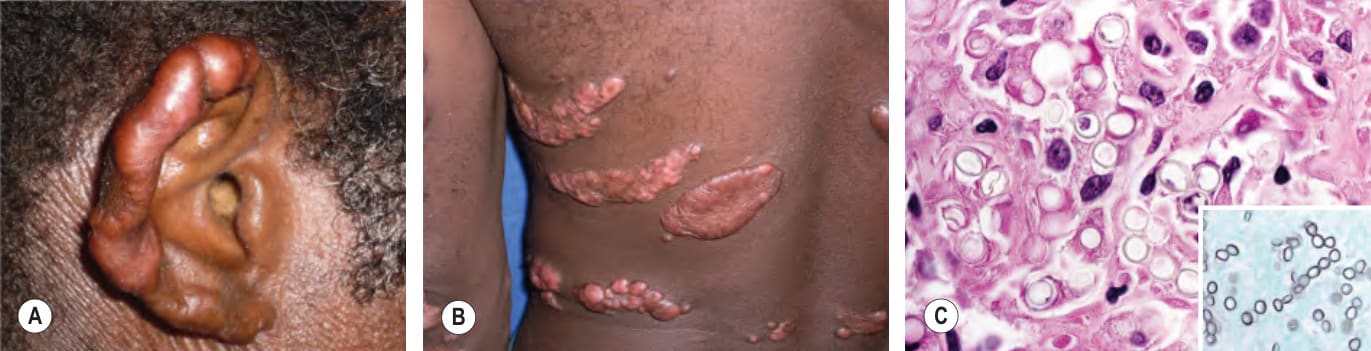
Fig. 77.27 Lobomycosis.A, B
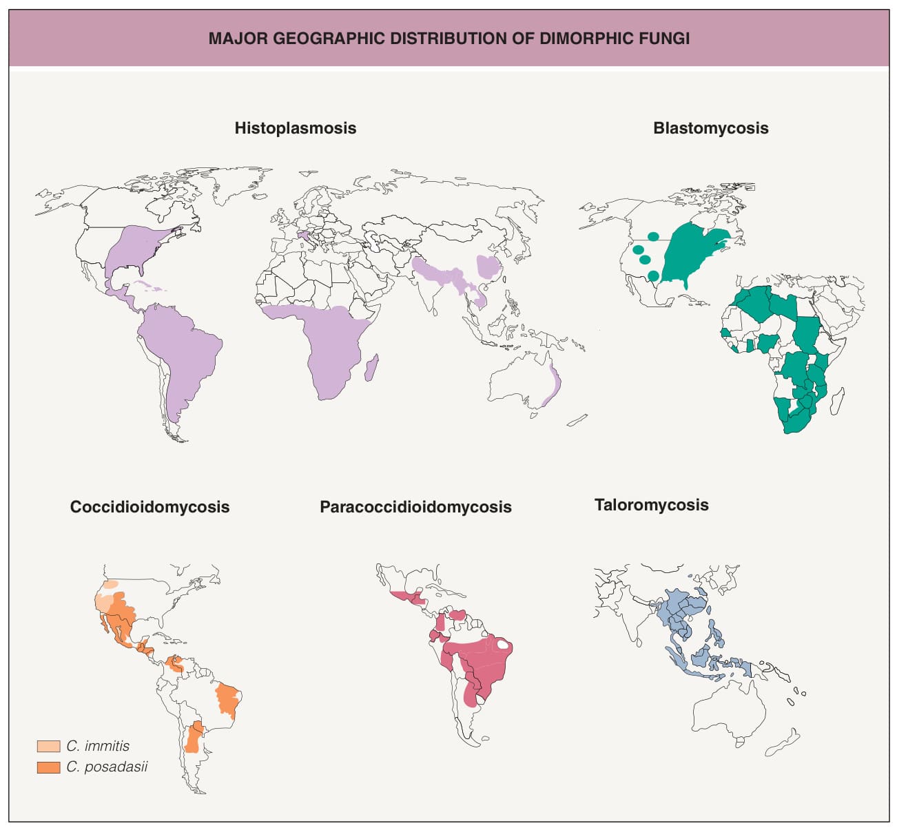
Fig. 77.28 Major geographic distribution of dimorphic fungi. Histoplasmosis has a worldwide distribution, with cases reported outside of these areas, and blastomycosis has also been reported in India and the Middle East. In addition, emergomycosis due to several dimorphic fungal species (Emergomyces africanus, E. canadensis, E. europaeus, R. orientalis, E. pasteurianus) has been reported in Africa, Europe, Asia, and the Americas.
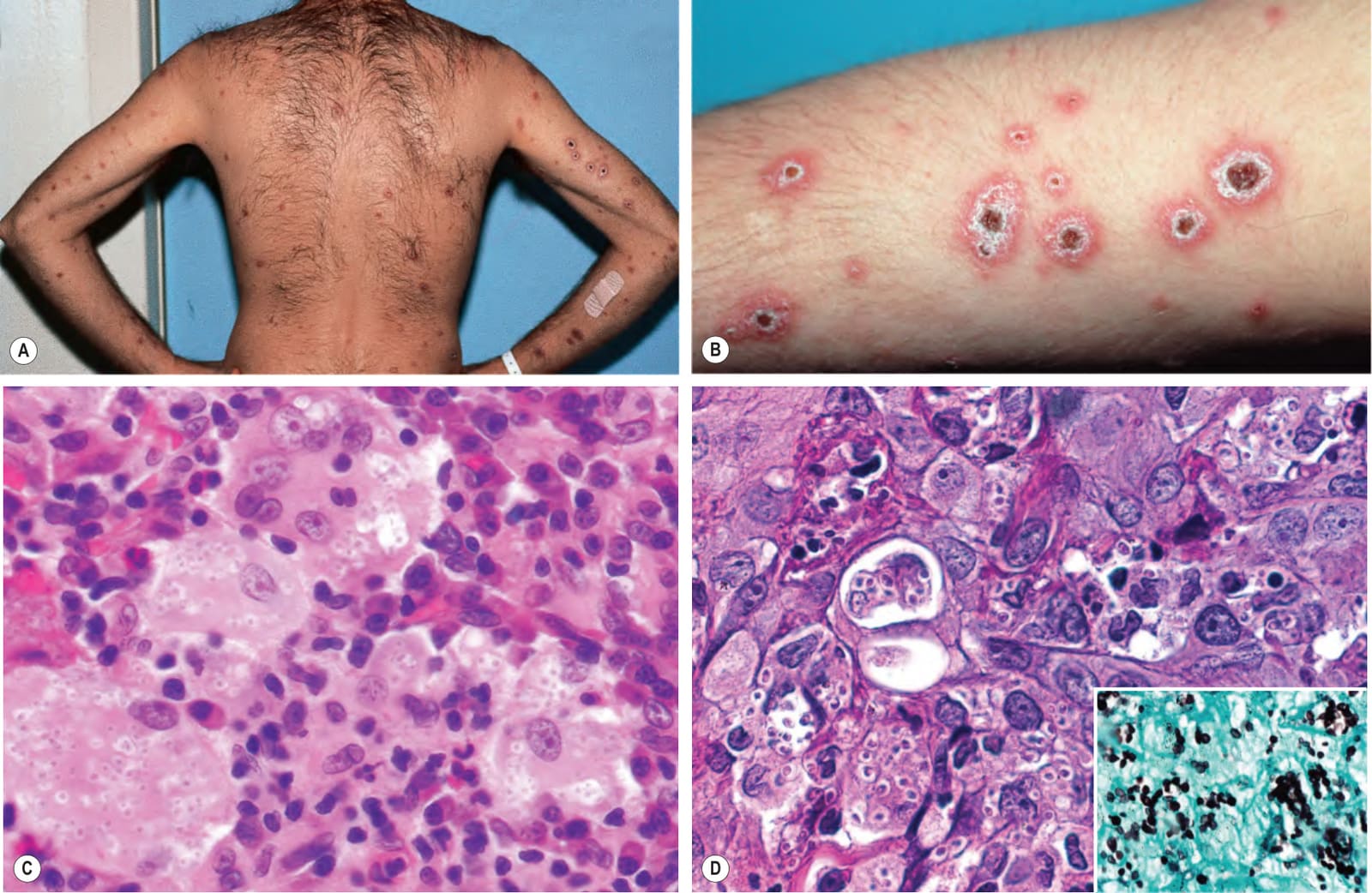
Fig. 77.29 Histoplasmosis.A, B Disseminated papules and nodules with scale-crust in a patient with AIDS. C Numerous yeasts within giant cells as well as dermal macrophages. D The organisms are highlighted with a methenamine silver stain (inset). C, Courtesy Jennifer McNiff, MD; D, Courtesy C. Massone, MD.
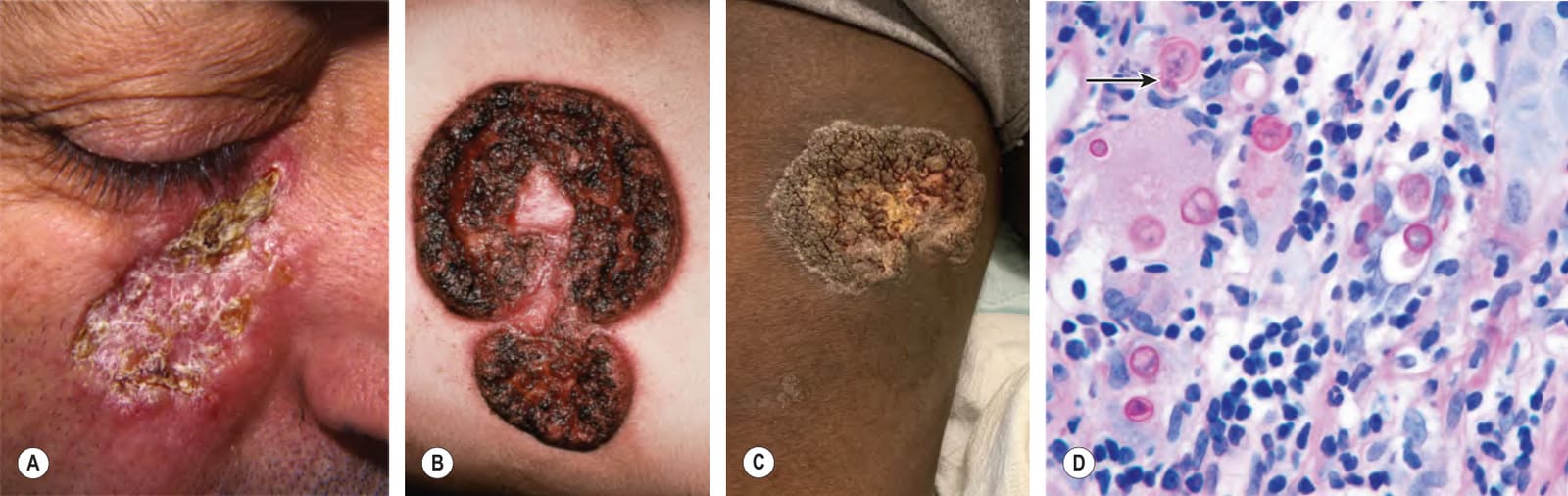
Fig. 77.30 Blastomycosis.A Facial plaque with scale-crust and a border with a granulomatous appearance. B Well-demarcated plaques with erosions, central scarring, and black crusting. C Verrucous plaque with focal crusting on the medial thigh. D Budding yeast forms in the dermis, several of which are within a giant cell (PAS stain). Note the single, broad-based budding (arrow). A, Courtesy Louis A. Fragola, Jr, MD; B, Courtesy Paul Lucky, MD; C, Courtesy Elizabeth Ergen, MD; D, Courtesy Mary Stone, MD.
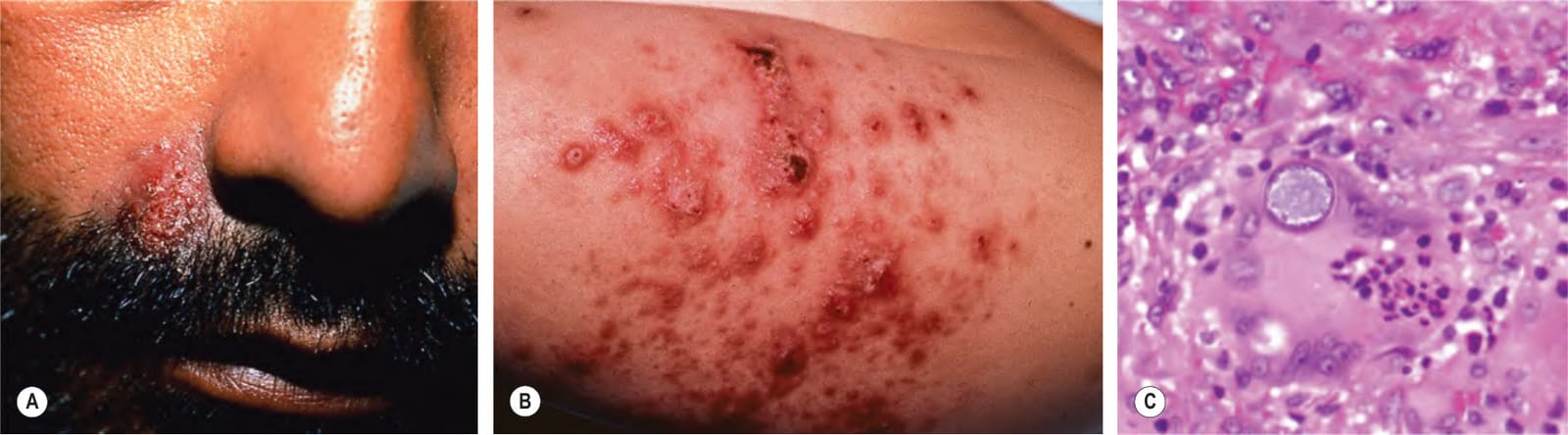
Fig. 77.31 Coccidioidomycosis. Moist erythematous plaque on the face (A) and multiple papules and suppurative nodules on the arm (B) in two patients living in the southwestern US. C An endospore-containing spherule within a giant cell. C, Courtesy Jennifer McNiff, MD.
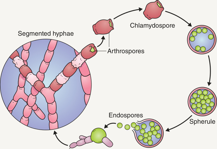
Fig. 77.32 Life cycle of Coccidioides immitis and C. posadasii.
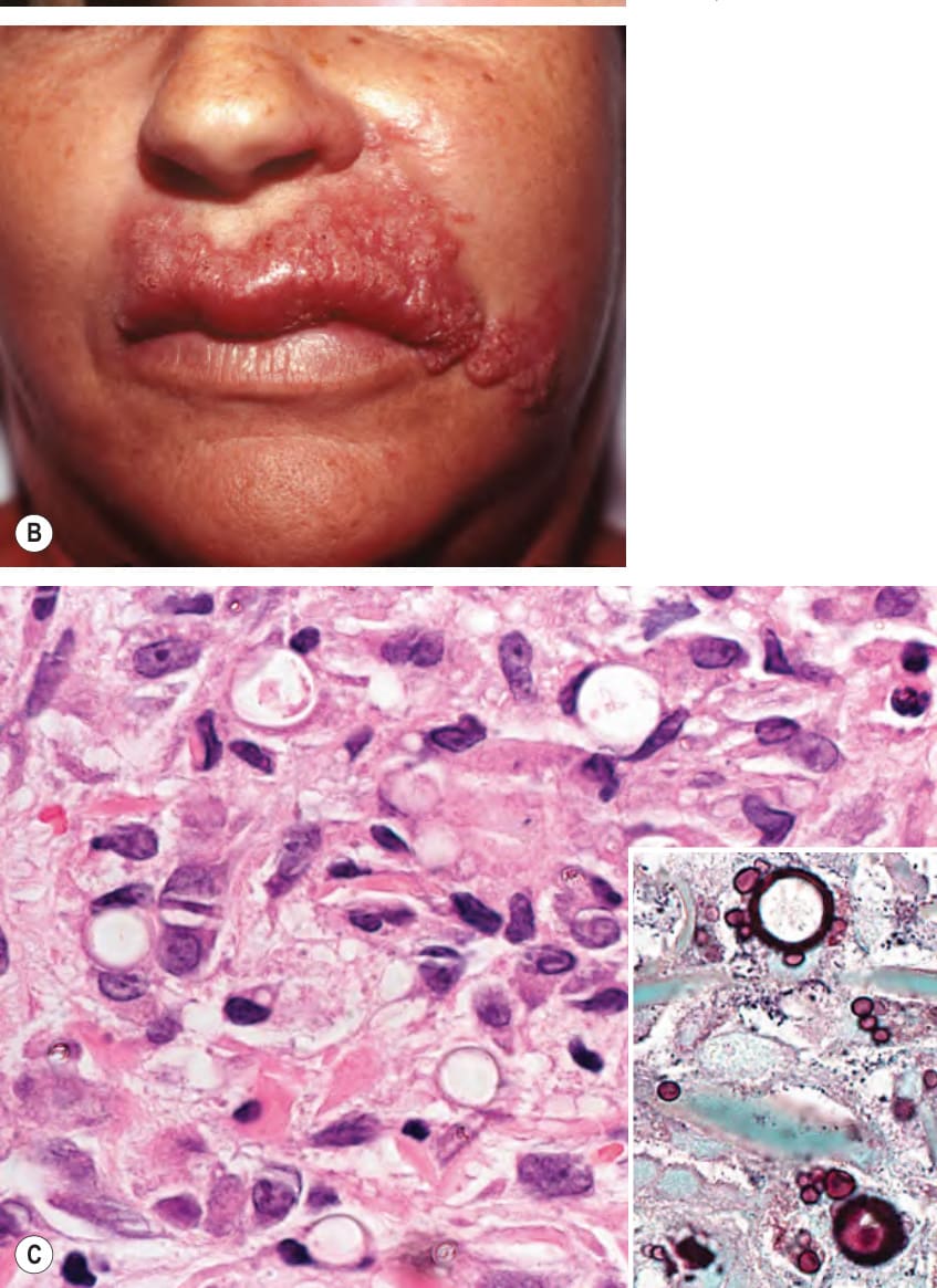
Fig. 77.33 Paracoccidioidomycosis.
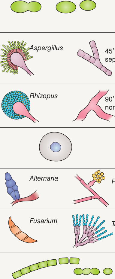
Fig. 77.34 Cutaneous features of common opportunistic mycoses. While the lesions of opportunistic fungal infections seem nonspecific, certain presentations may be more likely associated with a particular species.
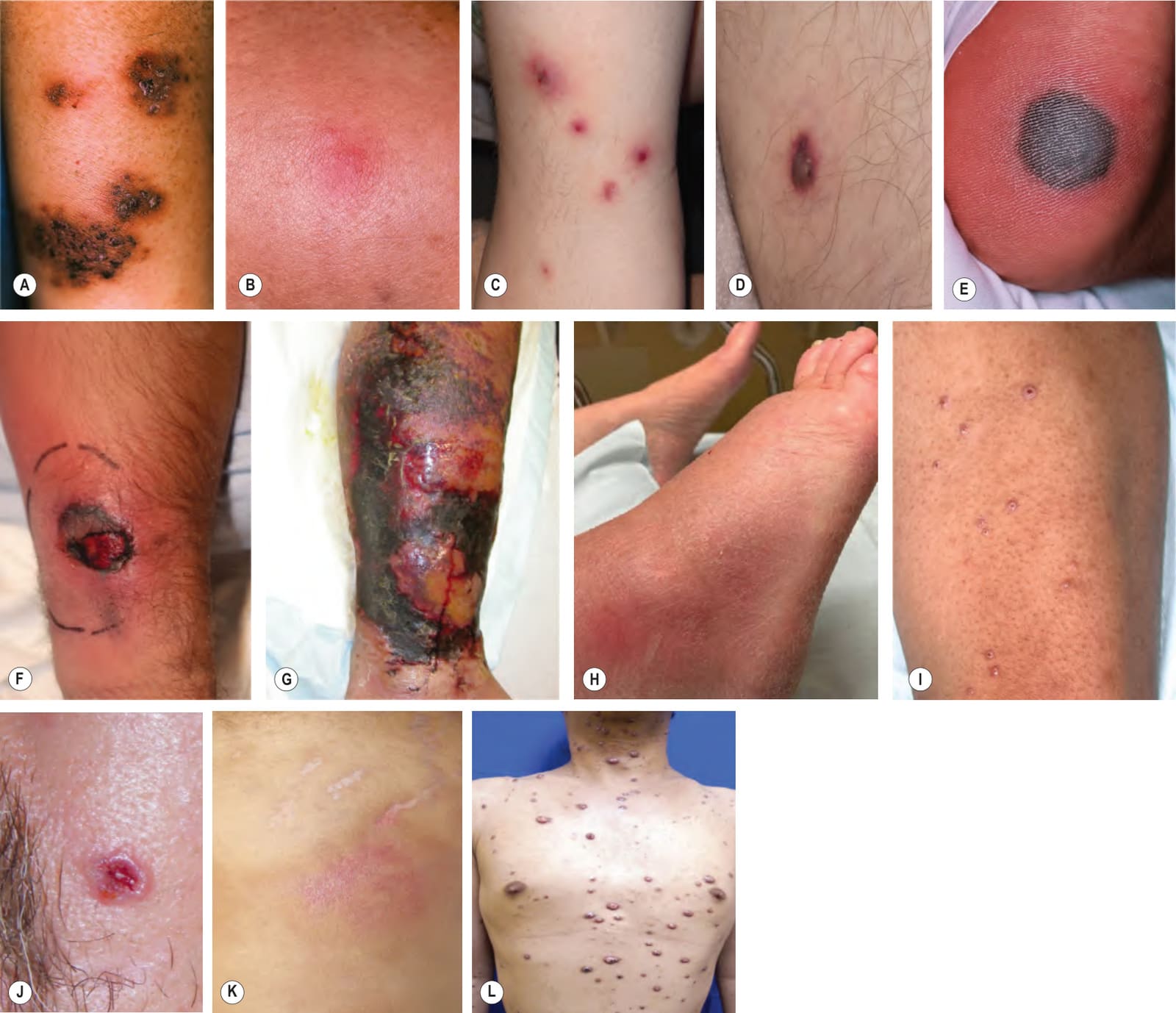
Fig. 77.35 Clinical findings of opportunistic fungal infections in immunocompromised hosts. Primary cutaneous aspergillosis characterized by hyperpigmented plaques with brown–black scale-crusts at the site of intravenous catheters on the arm (A); firm pink papulonodule due to disseminated candidiasis (B); erythematous papulonodules with central purpura, vesiculation and/or crusting in a leukemic patient with disseminated fusariosis (C); hemorrhagic vesicle representing a Fusarium solani septic embolus in a patient with acute lymphoblastic leukemia (D); necrotic hemorrhagic bulla due to embolus of Aspergillus flavus (E); cellulitis with large areas of necrosis due to Rhizopus (F) and cryptococcosis (G); cryptococcal cellulitis mimicking bacterial cellulitis (H); disseminated cryptococcosis presenting with crusted lesion(s) resembling molluscum contagiosum (I) and a basal cell carcinoma (J); cryptococcal cellulitis (K); numerous papules and nodules with central umbilication and crusting in disseminated Talaromyces (Penicillium) marneffei infection (L). D, Courtesy Edward Cowen, MD; H, Courtesy Catherine Baker, MD; L, Courtesy Evangeline Handog, MD and the Dermatology Department, Research Institute for Tropical Medicine.
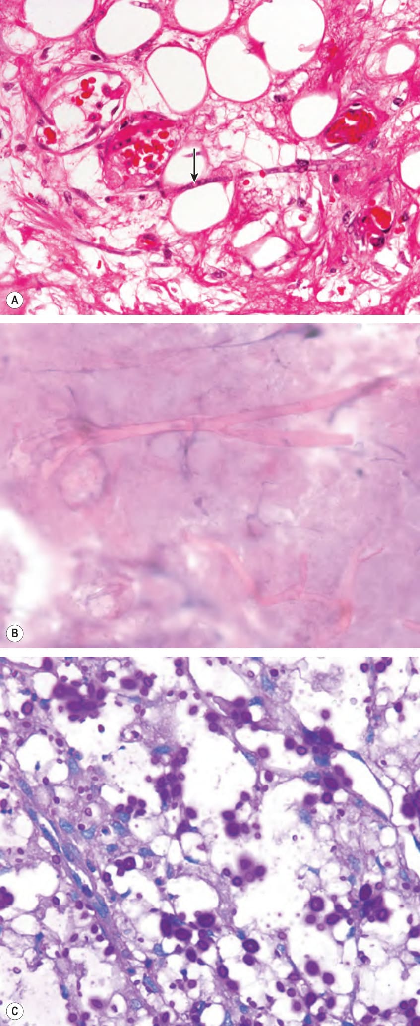
Fig. 77.36 Histologic findings of opportunistic fungal infections in immuno- compromised hosts.A Septate hyphae (arrow) within necrotic skin from a patient with disseminated aspergillosis. B Ribbon-like, non-septate Rhizopus hyphae in a “touch” preparation of dermal scrapings from the base of a necrotic bulla. C Numerous dermal yeast forms with gelatinous capsules in secondary cutaneous cryptococcosis (PAS stain). A, Courtesy Lorenzo Cerroni, MD; B, Courtesy Jean L. Bolognia, MD; C, Courtesy Athanasia Syrengelas, MD, PhD.
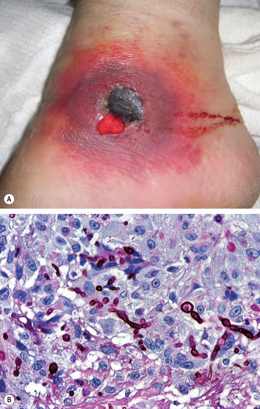
Fig. 77.37 Phaeohyphomycosis – clinical and histopathologic findings.
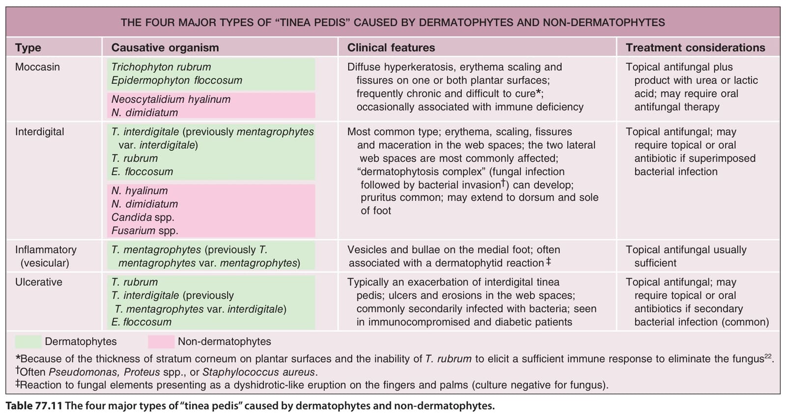
Table 77.11 The four major types of “tinea pedis” caused by dermatophytes and non-dermatophytes.
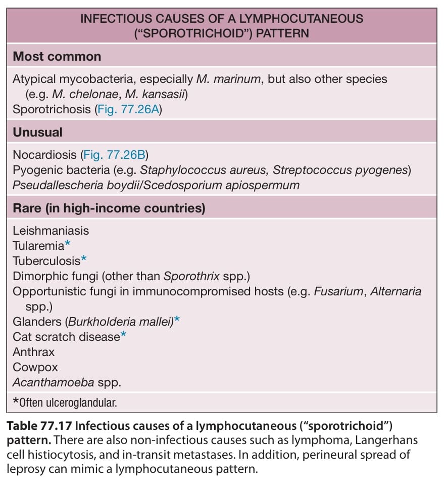
Table 77.17 Infectious causes of a lymphocutaneous (“sporotrichoid”)
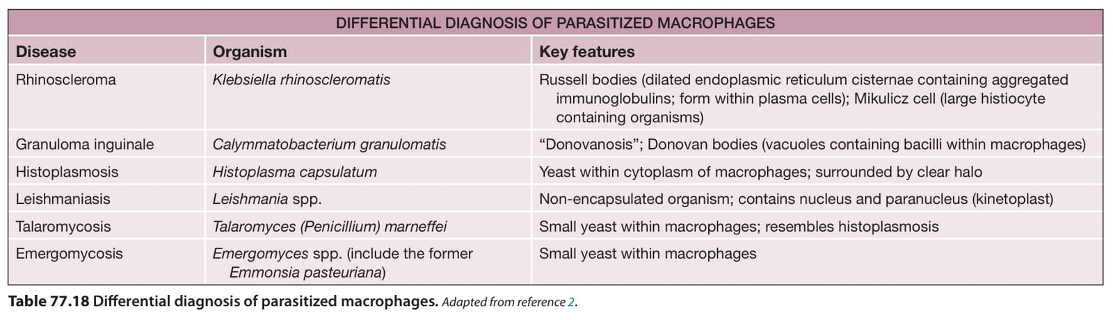
Table 77.18 Differential diagnosis of parasitized macrophages.Adapted from reference .
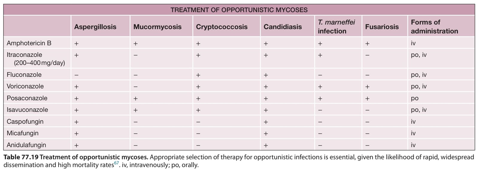
Table 77.19 Treatment of opportunistic mycoses. Appropriate selection of therapy for opportunistic infections is essential, given the likelihood of rapid, widespread dissemination and high mortality rates. iv, intravenously; po, orally.
“phaeomycotic cysts”. Occasionally, implanted material (e.g. a splinter) may also be observed.
For the subcutaneous and systemic forms, the differential diagnosis may include a foreign body granuloma, cutaneous leishmaniasis, chromoblastomycosis, and dimorphic fungal infections. KOH examination (superficial and cutaneous types), culture (all types), and histopathology (usually just subcutaneous and systemic types) allow the diagnosis to be made. Determination of genera and species requires fungal culture, but histopathology is useful in determining the extent of invasion and in differentiating causative pathogens from contaminants in culture specimens. Exophiala jeanselmei and E. dermatitidis are the organisms most commonly isolated from subcutaneous phaeohyphomycosis.
Subcutaneous disease is surgically excised, if possible; complete surgical excision is usually curative. Systemic disease due to dematiaceous fungi continues to be a therapeutic dilemma, as it is difficult to eliminate with oral antifungals alone. Nevertheless, itraconazole, given at a dose of 200 mg twice daily for a prolonged course (>12 months), has some efficacy for the non-surgical treatment of phaeohyphomycosis. Close follow-up is required and includes repeat cultures and histologic analyses of affected sites. Additional treatments for systemic disease include voriconazole, posaconazole, amphotericin B, and flucytosine.
In the past, Pneumocystis jirovecii (formerly referred to as Pneumocystis carinii) was classified as a protozoan. Currently, it is considered to be a fungus, based on genetic and biochemical analyses. This opportunistic infection occurs in individuals who are immunocompromised, including those with AIDS. The lungs are the most common site of infection (pneumonia), but, on occasion (<3% of patients), there is dissemination to other sites such as the liver, spleen, and lymph nodes.
Cutaneous involvement is rare and is characterized by erythematous or skin-colored, non-tender papules or nodules; there may be associated ulceration. The external ear (including the auditory canal) represents the most commonly reported site of involvement. Histologically, a diffuse infiltrate of foamy cells is seen within the dermis. Special stains (e.g. methenamine silver, Giemsa, Steiner) can be used to highlight the organisms, as can monoclonal antibodies; detection of P. jirovecii is also possible via PCR.
The most common treatment for pneumocystosis is trimethoprim– sulfamethoxazole (TMP-SMX). Additional therapies include parenteral pentamidine and oral atovaquone. Prophylaxis in the setting of immunosuppression usually consists of oral TMP-SMX or, less often, aerosolized pentamidine, oral dapsone, or oral atovaquone. Prophylaxis is important for dermatologic patients receiving systemic corticosteroids combined with immunosuppressive agents such as azathioprine and cyclophosphamide.
diseases. In: Elewski BE, ed. Cutaneous Fungal Infections. 2nd ed. Massachusetts: Blackwell Science; 1998.9. Gupta AK, Bluhm R, Summerbell R. Pityriasis versicolor.
the Pathogenic Actinomycetes. 3rd ed. Philadelphia: WB Saunders; 1988.3. Colombo AL, Padovan ACB, Chaves GM. Current