PATHOGENESIS
AIDS is caused by HIV-1 and much less often HIV-2. Both are enveloped, single-stranded RNA lentiviruses in the Retroviridae family. They are zoonotic and originated from simian immunodeficiency viruses (SIVs) that infect African primates: HIV-1 from chimpanzees and HIV-2 from sooty mangabeys. HIV-1 has four distinct lineages, each resulting from an independent cross-species transmission event; group M is responsible for ~99% of pandemic HIV disease, while groups N, O, and P are restricted to West Africa. HIV-2 is also largely restricted to West Africa, where its overall prevalence is declining. Compared with HIV-1, HIV-2 is less transmissible and virulent, resulting in lower viral loads and infrequent progression to AIDS. HIV exhibits substantial genetic variability due to the error-prone nature of its reverse transcriptase as well as very rapid virus replication in infected individuals.
During HIV-1 infection, the viral envelope proteins gp120 and gp41 initially interact with the CD4 molecule followed by a second inter-action with a chemokine receptor, usually CCR5 or CXCR4 (Fig. 78.2).
These receptors are found on activated CD4+ T cells, monocyte-macrophages, and dendritic cells. CXCR5 is the predominant fusion cofactor in most HIV transmissions; however, CD4-independent infection of renal cells and astrocytes gives rise to chronic renal disease and neurocognitive disorders. Following binding, changes in conformation induce fusion of the viral envelope with the plasma membrane, and the virus particle is thereby internalized. The RNA genome is transcribed by a reverse transcriptase enzyme that produces a DNA copy of the HIV RNA. This viral DNA copy is spliced into the host DNA through the action of an integrase enzyme. Transcription of viral DNA into RNA yields genomic material for new viral particles or it can be translated into viral proteins. Cleavage of the latter into structural components of the virus is accomplished by proteases. Intact viruses are then produced and host cells are destroyed.
Infection by the transmitted/founder virus is followed by a rapid increase in HIV replication (Fig. 78.3). After ~6 months, the viral load falls to a set point that is determined by the host’s innate natural killer (NK) cell response plus an adaptive immune response by CD8+ cells and neutralizing antibodies. The initial antibody response develops within 1–3 months after transmission and is strain-specific. This antibody response drives viral escape, and virus mutants become resistant to neutralization in 80% of patients. However, in 20% of patients, variants of the transmitted/founder virus evolve and induce antibodies with considerable neutralization breadth. CD8+ cytotoxic T cells directed against HIV begin to function within 10 days of exposure, correlating with the reduction in viremia during the acute retroviral syndrome; these cells also help to control the HIV throughout the disease course.
Initial infection results in a reduction in circulating CD4+ T cells, which is followed by a recovery to nearly normal levels and a subsequent slow fall of about 50 to 100 cells/mm per year (see Fig. 78.3). In contrast, the CD4+ T cells residing within the gastrointestinal tract are rapidly depleted early on. The progressive depletion of CD4+ T cells is the result of killing by direct infection, bystander effects of syncytia formation, chronic immune activation, lymphoid tissue damage, and senescence.
The marked chronic immune activation that characterizes HIV disease is thought to be related to low-grade HIV replication, immune responses
In a person living with HIV, AIDS is defined by a CD4+ cell count of <200/mm or <14% CD4+ T cells and/or the presence of an AIDS-defining condition (see Table 78.1). The natural history of HIV disease varies considerably, with the rate of decline in CD4+ T cells correlating with viral burden. In the absence of treatment, the median time for progression to AIDS is ~10 years. However, ~10%–15% of untreated patients, historically referred to as “long-term non-progressors”, live with HIV for ≥8 years with no symptoms and CD4+ T cell counts of >500 cells/mm; in addition, <0.5% of patients are “elite controllers” with no evidence of viremia even without ART. The ability of ART to suppress viral replication and enable reconstitution of CD4+ T cells has an immense impact on the disease course. Immune reactivation due to ART can result in a variety of inflammatory sequelae referred to as IRIS, ranging from unmasking or paradoxical “worsening” of infections or neoplasms to exacerbations of inflammatory disorders (see below).
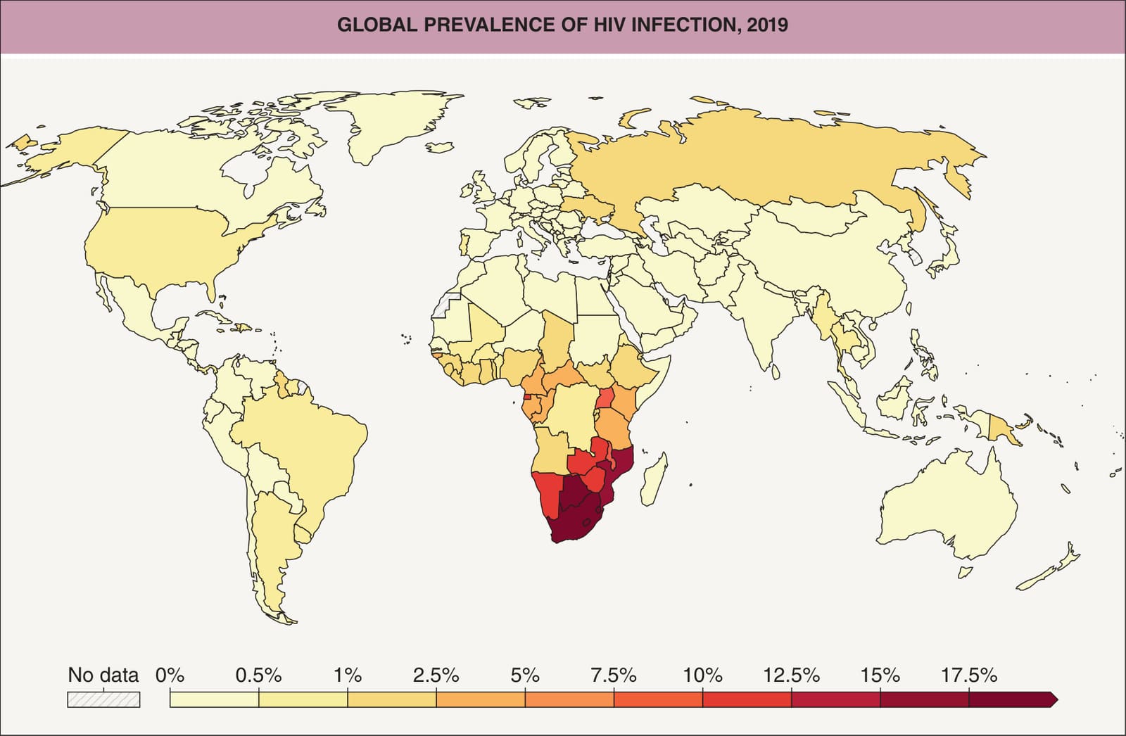
Fig. 78.1 Global prevalence of HIV infection, 2019. The worldwide estimate of the number of people living with HIV was 38.4 million in 2021, representing an overall prevalence of almost 0.5%. From the Institute for Health Metrics and Evaluation (IHME) 2019 Global Burden of Disease.
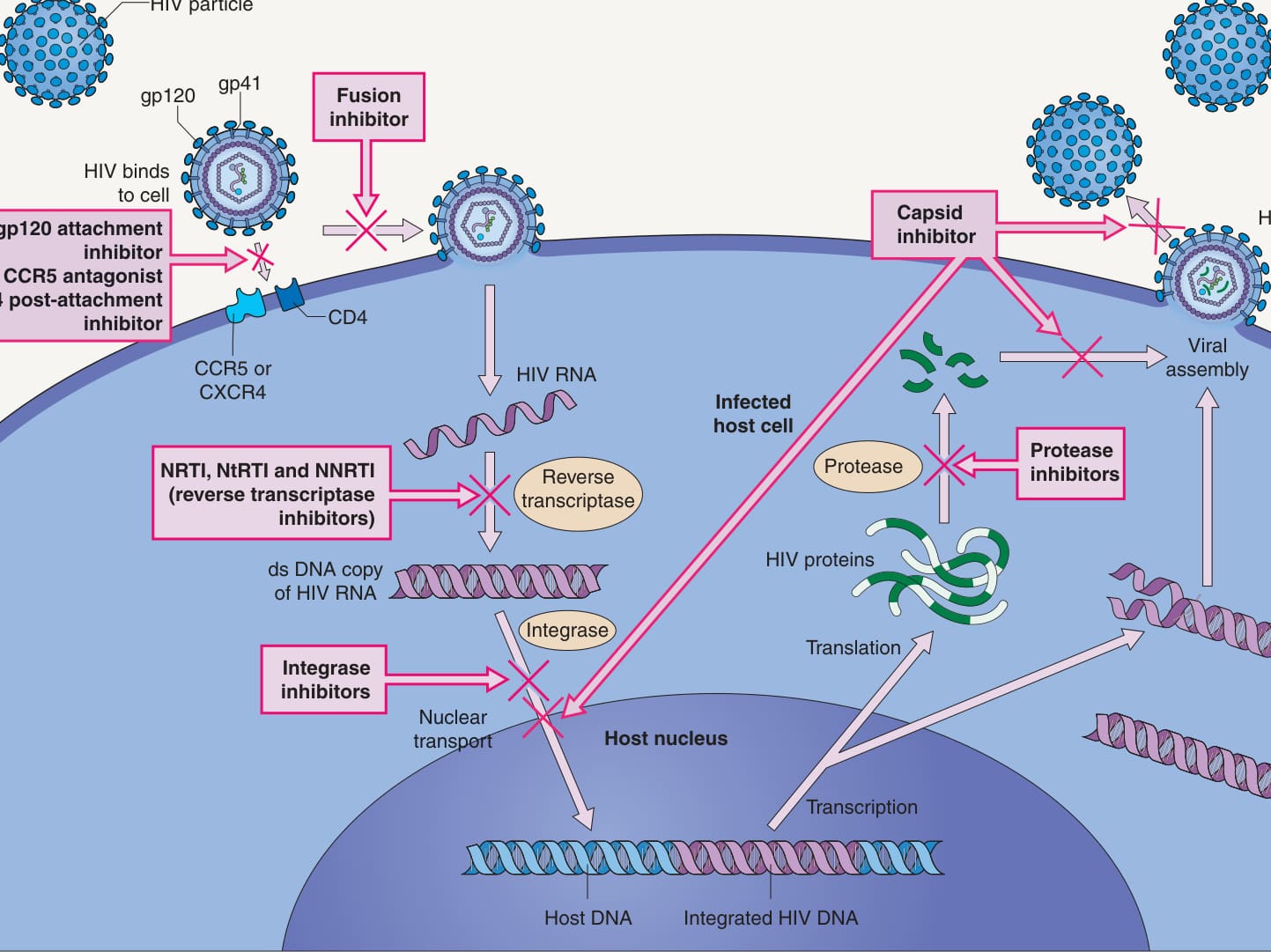
Fig. 78.2 Replication of HIV within CD4+ lymphocyte and target sites of antiretroviral drugs. CCR5, CC-chemokine receptor 5; NRTI, nucleoside reverse transcriptase inhibitor; NtRTI, nucleotide reverse transcriptase inhibitor; NNRTI, non-nucleoside reverse transcriptase inhibitor. Information on FDA-approved antiretroviral drugs is available at https://hivinfo.nih.gov/understanding-hiv/fact-sheets/fda-approved-hiv-medicines to HIV, reactivated infections, loss of mucosal integrity with consequent microbial translocation, and increased production of proinflammatory molecules. In patients receiving ART, increased levels of markers of inflammation have been associated with higher mortality rates and risk of cancer as well as cardiovascular, neurologic, and liver disease.
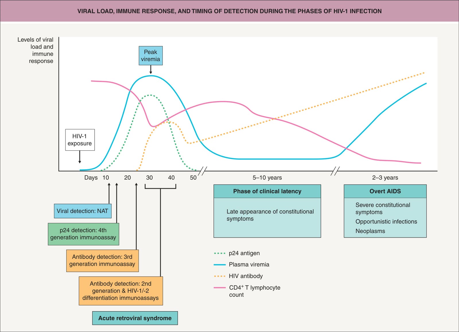
Fig. 78.3 Viral load, immune response, and timing of detection during the phases of HIV-1 infection. After HIV-1 exposure, initial virus replication and spread occur in the lymphoid organs, and systemic dissemination of HIV-1 is reflected by the peak of plasma viremia. A clinical syndrome of varying severity is associated with this phase of primary HIV-1 infection in up to 80% of HIV-1-infected persons. Downregulation of viremia during the transition from the primary to the early chronic phase coincides with the appearance of HIV-1-specific cytotoxic T lymphocytes and with the progressive resolution of the clinical syndrome. The long phase of clinical latency is associated with active virus replication, particularly in the lymphoid tissue. During the clinically latent period, CD4+ T lymphocyte counts slowly decrease, as does the HIV-1-specific immune response. When CD4+ T lymphocyte counts decrease below 200 cells/ml (i.e. when overt AIDS occurs), the clinical picture is characterized by severe constitutional symptoms and by the possible development of opportunistic infections and/or neoplasms. Adapted from Bart PA, Pantaleo G. The immunopathogenesis of HIV-1 infection. In: Cohen J, Powderly WG. Infectious Diseases. Edinburgh: Mosby, 2004:1236.
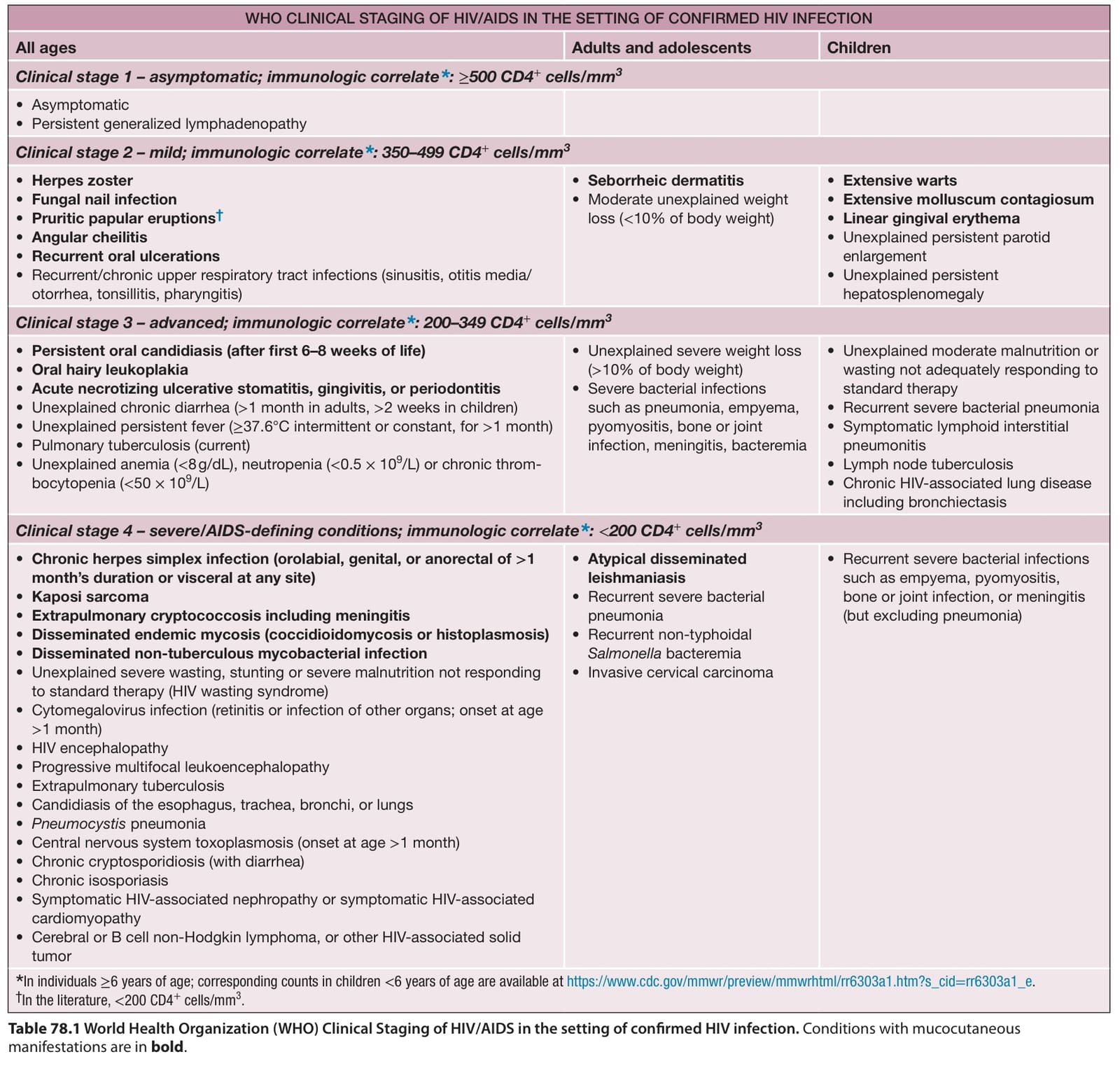
Table 78.1 World Health Organization (WHO) Clinical Staging of HIV/AIDS in the setting of confirmed HIV infection. Conditions with mucocutaneous manifestations are in bold.
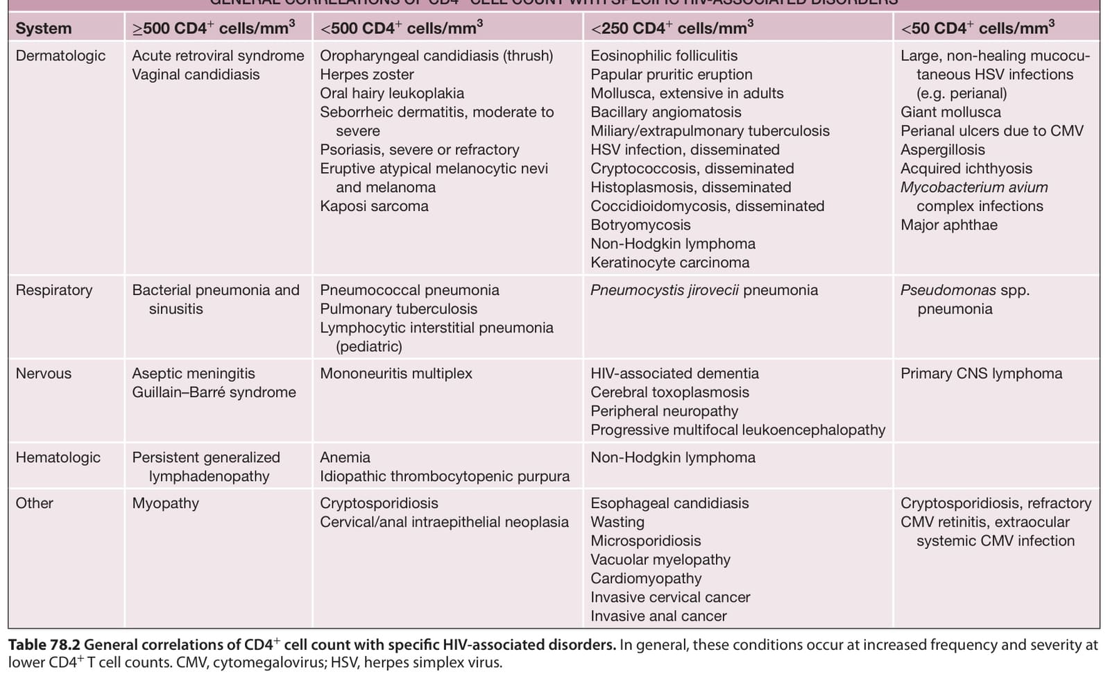
Table 78.2 General correlations of CD4+ cell count with specific HIV-associated disorders. In general, these conditions occur at increased frequency and severity at lower CD4+ T cell counts. CMV, cytomegalovirus; HSV, herpes simplex virus.