Viral Infections
Primary HIV exanthem (acute retroviral syndrome)
Primary HIV infection refers to the initial disease phase, when HIV-1 RNA, DNA, and/or p24 antigen are present in the plasma prior to the development of HIV-1 antibodies. Initially, up to 80% of patients develop an “acute retroviral syndrome” resembling influenza or infectious mononucleosis. Symptoms develop 2–6 weeks after HIV exposure and often include fever, headache, myalgia/arthralgia, pharyngitis, lymphadenopathy, and night sweats. A morbilliform exanthem also occurs in 40%–80% of patients; it lasts 4–5 days and is typically generalized, with the most prominent involvement on the face and trunk. Oral and genital ulcers are occasionally present. Symptoms are self-limited and resolve in a few days to weeks, with a median duration of 14 days. The initial phase of rapid HIV-1 replication causes a temporary fall in CD4+ cell counts, occasionally to an extent that gives rise to opportunistic infections.
Greater severity and longer duration of the acute retroviral syndrome is associated with a higher viral load set point and more rapid disease progression. Early initiation of ART during primary HIV infection may delay disease progression.
Herpes simplex virus (HSV) infection
Relatively immunocompetent people living with HIV usually have typical, self-limited HSV disease. As immunity wanes, the frequency of HSV recurrences increases and lesions take longer to heal, potentially evolving into chronic, extensive, deep, and painful ulcers that favor the perianal region, genitalia, and tongue (Fig. 78.4). Atypical presentations of HSV infection include folliculitis, verrucous plaques, and hypertrophic anogenital masses simulating neoplasia. Infection may become
disseminated or occur at unusual sites such as the non-keratinized (unattached) mucosa of the oropharynx (see Fig. 72.1) and the esophagus.
The ulcer edge should be scraped for HSV PCR (most sensitive), direct fluorescent antibody assay (DFA), and/or viral culture and, if negative, a skin biopsy performed; a Tzanck smear has low sensitivity. Higher doses and longer courses of antiviral medications are typically required, with continuation until all mucocutaneous lesions are healed. Resistance to acyclovir, usually due to reduced viral thymidine kinase activity, occurs more commonly in immunosuppressed (~5%) compared to immunocompetent (~1%) hosts. Alternative agents that do not rely on thymidine kinase include foscarnet, cidofovir, and imiquimod. Of note, HSV-2 infection increases the risk of HIV acquisition and promotes HIV-1 replication.
Varicella and herpes zoster
In people living with HIV, varicella tends to have new lesion formation over a longer period of time, a higher lesion count, and (particularly in adults) complications such as pneumonitis, hepatitis, and encephalitis. Lesions that persist as slowly healing ulcers have been reported.
People living with HIV have a 7- to 15-fold higher incidence of herpes zoster compared to the general population. Of children living with HIV who develop varicella, herpes zoster or a disseminated eruption reminiscent of varicella occurs in ~50% within 2 years. In addition to classic dermatomal eruptions, HIV-associated herpes zoster can present as chronic non-healing ulcers, hyperkeratotic verrucous plaques, and multidermatomal or disseminated skin lesions; potential complications include bacterial superinfection, systemic involvement, and multiple recurrences. The recombinant Shingrix® vaccine is recommended for the prevention of herpes zoster in immunocompromised adults, including those with HIV disease (see Ch. 80).
People living with HIV who do not have VZV immunity should receive varicella–zoster immune globulin within 10 days of exposure to VZV. Antiviral treatment of VZV infection in patients should be continued until clinical resolution occurs (see Ch. 80); acyclovir resistance is possible. Of note, patients treated with ART may experience paradoxical worsening of herpes zoster when their CD4+ T cell counts rise (see IRIS below).
Molluscum contagiosum
Molluscum contagiosum (MC) commonly occurs and tends to be more persistent in individuals with significantly reduced CD4+ T cell counts. In addition to the typical presentation of small umbilicated papules (Fig. 78.5), people living with HIV can develop larger (>1 cm), coalescent, verrucous, and widespread lesions. The clinical differential diagnosis may include basal cell carcinoma (BCC) and cutaneous lesions of cryptococcosis, histoplasmosis, or other dimorphic fungal infections. Dermoscopic evaluation, microscopic examination following curettage of lesional contents, or biopsy can help to confirm the diagnosis. In addition to curettage and other destructive modalities, topical and intravenous cidofovir have been used successfully for refractory MC in patients with HIV disease. MC may resolve spontaneously after initiation of ART but can also present as a manifestation of IRIS.
Human papillomavirus (HPV) infections
The spectrum of mucocutaneous lesions due to HPV infection ranges from common warts and condylomata acuminata to squamous intraepithelial neoplasia and carcinoma. The prevalence of HPV infection is higher in people living with HIV than in the general population and correlates with decreasing CD4+ T cell counts. People living with HIV have reduced HPV clearance, more treatment-resistant mucocutaneous lesions, and accelerated development of HPV-associated carcinoma. Skin lesions are often multiple, coalescent, and extensive (Fig. 78.6). There is evidence for synergistic interactions between HIV and HPV infections, suggesting that HPV vaccination may help control both infections at the population level in high prevalence settings.
Acquired epidermodysplasia verruciformis (AEDV) is characterized by hypopigmented to pink tinea versicolor-like lesions and numerous flat-topped papules that are due to HPV-5, -8, and other β HPV types. People living with HIV may develop squamous cell carcinomas (SCCs) in association with AEDV and other cutaneous warts, e.g. digital lesions due to high-risk HPV types, as well as anogenital warts.
Compared to the general population, women living with HIV have a 3-fold increase in the incidence of cervical intraepithelial neoplasia
(CIN) as well as higher rates of high-grade CIN. Anal HPV is present in >90% of MSM with HIV disease, and most have at least one high-risk HPV type. MSM with HIV disease also have an up to 50% prevalence of high-grade anal intraepithelial neoplasia (AIN) and a 30- to 50-fold higher risk of anal cancer than does the general population; a 5-fold increased risk of penile cancer has also been documented. Monitoring should include serial physical examinations of the anogenital area, colposcopy/proctoscopy, cervical/anal HPV determination and cytology, and histologic examination when indicated.
ART has not shown a demonstrable effect on the occurrence or clearance of cutaneous or anogenital warts. Enlargement or inflammation of pre-existing warts and an eruption of new warts can occur in the setting of IRIS. Some studies have shown a protective effect of ART on the development and progression of CIN and AIN. However, compared to the pre-ART era, incidence rates of cervical and anal cancers have not decreased. This is likely due to the longer life expectancy afforded by ART.
The choice of therapy for anogenital warts depends upon their size, number, and location. Options include destructive modalities such as cryotherapy, electrosurgery, laser therapy and surgical excision, in addition to patient-applied topical therapies including imiquimod and podophyllotoxin (see Ch. 79). Immunosuppressed patients require more treatment sessions to achieve clearance and have frequent recurrences. The threshold for biopsy should be low, and several sites may need to be sampled to exclude SCC. HPV vaccines are immunogenic and well-tolerated in people with HIV disease.
Oral hairy leukoplakia due to Epstein–Barr virus (EBV)
Oral hairy leukoplakia (OHL) results from Epstein–Barr virus (EBV) infection of the oral mucosa and typically presents as hyperkeratotic, corrugated white plaques with hair-like projections on the lateral aspect of the tongue (Fig. 78.7). It can occur at any stage of HIV disease but is more prevalent in men and those with CD4+ T cell counts below 200/ mm. It represents a predictor of rapid disease progression if ART is not initiated. OHL can also occur in patients with other forms of immunosuppression, such as organ transplant recipients. Although treatment is usually not necessary, topical or oral antiherpetic antiviral medications and topical podophyllin, tretinoin, or gentian violet may be of benefit; however, recurrence is common. OHL may also regress with ART.
Cytomegalovirus (CMV) infections
Reactivation of CMV is associated with CD4+ T cell counts that are <100/mm, and it remains a significant cause of morbidity and mortality in patients with AIDS. Retinitis, esophagitis, and colitis represent common clinical manifestations, and mucosal ulcers may occur in the setting of disseminated disease. Cutaneous presentations are uncommon and include ulcers favoring the anogenital area (Fig. 78.8), verrucous or hyperpigmented plaques, prurigo nodularis-like lesions,
purpuric papules, vesicles, and morbilliform eruptions. Histologically, characteristic cytomegalic intranuclear inclusion bodies are seen within endothelial cells in the dermis of lesional skin (see Fig. 78.8 inset and Fig. 80.23). Ulcerative lesions are often co-infected with HSV or VZV, and because CMV can also be detected in non-lesional skin, its pathogenic role has been questioned. Specific treatments include oral valganciclovir and intravenous ganciclovir, foscarnet, and cidofovir.
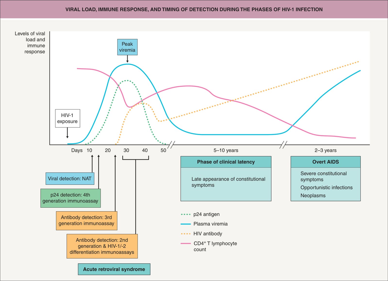
Fig. 78.3 Viral load, immune response, and timing of detection during the phases of HIV-1 infection. After HIV-1 exposure, initial virus replication and spread occur in the lymphoid organs, and systemic dissemination of HIV-1 is reflected by the peak of plasma viremia. A clinical syndrome of varying severity is associated with this phase of primary HIV-1 infection in up to 80% of HIV-1-infected persons. Downregulation of viremia during the transition from the primary to the early chronic phase coincides with the appearance of HIV-1-specific cytotoxic T lymphocytes and with the progressive resolution of the clinical syndrome. The long phase of clinical latency is associated with active virus replication, particularly in the lymphoid tissue. During the clinically latent period, CD4+ T lymphocyte counts slowly decrease, as does the HIV-1-specific immune response. When CD4+ T lymphocyte counts decrease below 200 cells/ml (i.e. when overt AIDS occurs), the clinical picture is characterized by severe constitutional symptoms and by the possible development of opportunistic infections and/or neoplasms. Adapted from Bart PA, Pantaleo G. The immunopathogenesis of HIV-1 infection. In: Cohen J, Powderly WG. Infectious Diseases. Edinburgh: Mosby, 2004:1236.
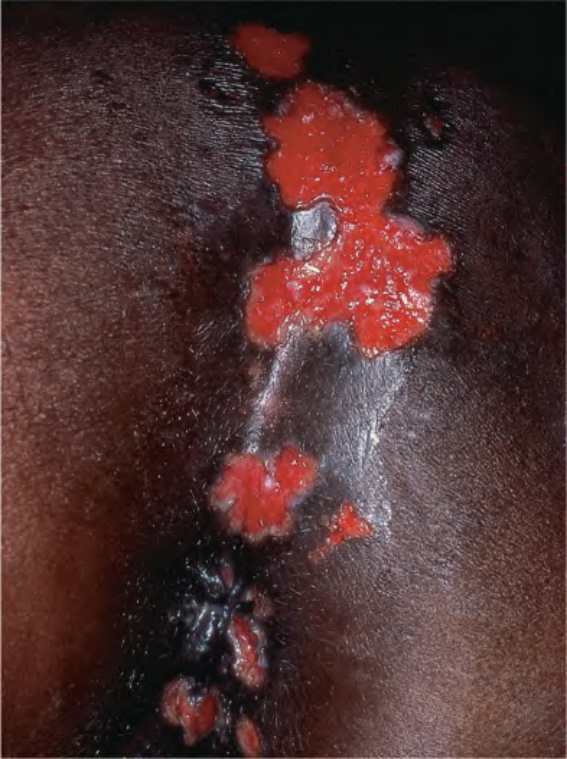
Fig. 78.4 Chronic ulcerative herpes simplex virus infection in a patient with HIV. Slowly enlarging ulcers on the buttocks and perianal area have a scalloped border.
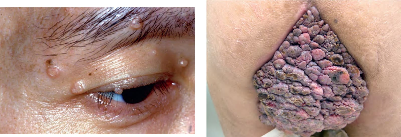
Fig. 78.5 Molluscum contagiosum. Multiple facial lesions are commonly seen in people living with HIV and less likely to resolve spontaneously. Disseminated fungal infections may present with molluscum contagiosum-like lesions and should be excluded.
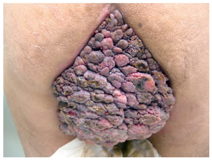
Fig. 78.6 Extensive perianal condylomata acuminata. This 65-year-old man was receiving antiretroviral therapy and had a CD4+ T cell count of 240/mm.
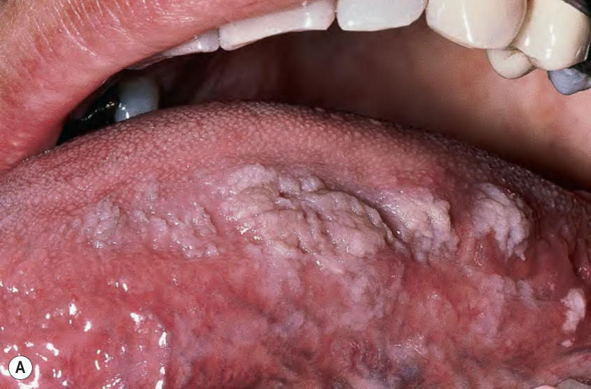
Fig. 78.7 Oral hairy leukoplakia.A Shaggy white keratotic plaques along the lateral aspect of the tongue. A corrugated pattern is often seen. B Histologic findings include irregular parakeratosis, acanthosis, and focal balloon cell change of the epithelium. The inset shows positive staining for Epstein–Barr virus-encoded RNA (EBER) in the nuclei of affected keratinocytes. A, Courtesy Charles Camisa, MD; B, Courtesy Lorenzo Cerroni, MD; Inset, Courtesy Luis Requena, MD.
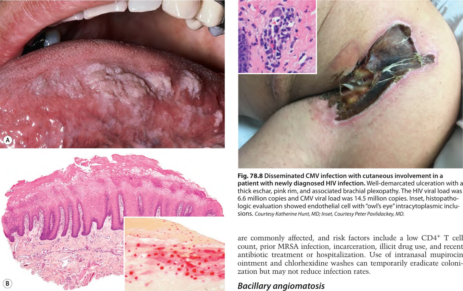
Fig. 78.8 Disseminated CMV infection with cutaneous involvement in a patient with newly diagnosed HIV infection. Well-demarcated ulceration with a thick eschar, pink rim, and associated brachial plexopathy. The HIV viral load was 6.6 million copies and CMV viral load was 14.5 million copies. Inset, histopathologic evaluation showed endothelial cell with “owl’s eye” intracytoplasmic inclusions. Courtesy Katherine Hunt, MD; Inset, Courtesy Peter Pavlidackey, MD.