Bacterial Infections
People living with HIV are at increased risk for the development of recurrent and potentially severe cutaneous bacterial infections, which can be localized or widespread and sometimes have unusual clinical appearances. In addition to diminished antibody- and cell-mediated immune responses, predisposing factors include skin barrier impairment from excoriations or other cutaneous infections, indwelling catheters, and malnutrition. In addition to the entities discussed below, cutaneous bacterial infections that can occur in association with HIV disease include necrotizing fasciitis, nocardiosis, malacoplakia, pseudomonal “hot tub” folliculitis, “malignant” otitis externa, and ecthyma gangrenosum.
Staphylococcus aureus is the most common bacterial pathogen in individuals with HIV disease. Cutaneous presentations include impetigo, folliculitis, furunculosis, wound infections, cellulitis, and rarely botryomycosis. Compared with the general population, people living with HIV have a higher prevalence of methicillin-resistant S. aureus (MRSA) colonization of the skin and a 6- to 18-fold higher incidence of MRSA skin/soft tissue infections; they also have more frequent recurrences. The lower extremities, buttocks, and scrotum
are commonly affected, and risk factors include a low CD4+ T cell count, prior MRSA infection, incarceration, illicit drug use, and recent antibiotic treatment or hospitalization. Use of intranasal mupirocin ointment and chlorhexidine washes can temporarily eradicate colonization but may not reduce infection rates.
Bacillary angiomatosis
Bacillary angiomatosis is a rare condition that is characterized by vascular proliferation and is due to the Gram-negative bacilli Bartonella henselae and B. quintana. It occurs in individuals with HIV disease who are severely immunocompromised, usually with CD4+ T cell counts <100/mm. Bacillary angiomatosis favors the skin and subcutaneous tissues, although virtually any internal organ can be affected. Patients typically present with a single to numerous, firm, red or violaceous papules and nodules; these lesions may be painful, ulcerate, or bleed profusely after trauma. Large subcutaneous nodules of bacillary angiomatosis may also ulcerate. Disseminated infection is associated with systemic symptoms such as fever, night sweats, and weight loss.
The differential diagnosis may include Kaposi sarcoma, pyogenic granulomas, and non-tuberculous mycobacterial infections. Histologically, a lobular proliferation of capillaries and venules with large, protuberant endothelial cells is seen, surrounded by a neutrophilic infiltrate. Lesional bacilli can be visualized with a Warthin–Starry stain or identified via PCR-based assays; serologic testing for anti-Bartonella antibodies may assist in diagnosis. Treatment consists of doxycycline or macrolides. The median time to complete response is ~1 month, and 3–4 months of therapy is recommended to prevent relapse.
Mycobacterial infections
A diverse clinical spectrum of tuberculous and non-tuberculous mycobacterial skin infections can develop in people living with HIV. Cutaneous findings include erythematous to violaceous papules and nodules as well as ulcers, acneiform eruptions, abscesses, and verrucous plaques (Fig. 78.9); a sporotrichoid (lymphocutaneous) distribution pattern is sometimes seen. The risk of infection increases with progressive immunosuppression, and the classic histologic finding of caseating granulomas may be absent in patients with markedly diminished cell-mediated immunity.
Syphilis
Rates of syphilis have risen over the past decade, with possible factors including riskier sexual behaviors (due in part to the introduction of PrEP) as well as potential impediment of innate and acquired immunity to Treponema pallidum by ART. In addition to classic papulosquamous lesions of secondary syphilis, people with HIV disease may
This 54-year-old man with HIV and a CD4+ T cell count of 350/ mm presented with a 4-month history of slowly enlarging, thick plaques on his arms and legs. The lesions resolved over 6 months with treatment including ciprofloxacin, clarithromycin, and ethambutol.
present with multiple primary chancres, concomitant lesions of primary and secondary syphilis, palmoplantar keratoderma, annular plaques, and lues maligna. The latter, which is also known as malignant or ulceronodular syphilis, is a severe form of secondary syphilis (see Ch. 82). A prodrome of fever, arthralgia/myalgia, headache, and photophobia precedes the appearance of papules, pustules, and necrotic nodules with ulceration and crusting (Fig. 78.10); lesions are symmetrically distributed, with frequent involvement of the palms and sometimes the oral mucosa. Individuals with HIV disease have a higher likelihood, earlier onset, and greater severity of neurosyphilis. The standard stagespecific treatment regimens for syphilis are recommended, but with longer monitoring.
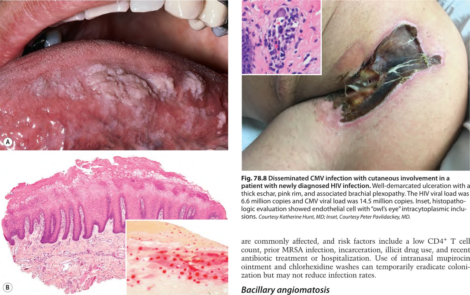
Fig. 78.8 Disseminated CMV infection with cutaneous involvement in a patient with newly diagnosed HIV infection. Well-demarcated ulceration with a thick eschar, pink rim, and associated brachial plexopathy. The HIV viral load was 6.6 million copies and CMV viral load was 14.5 million copies. Inset, histopathologic evaluation showed endothelial cell with “owl’s eye” intracytoplasmic inclusions. Courtesy Katherine Hunt, MD; Inset, Courtesy Peter Pavlidackey, MD.
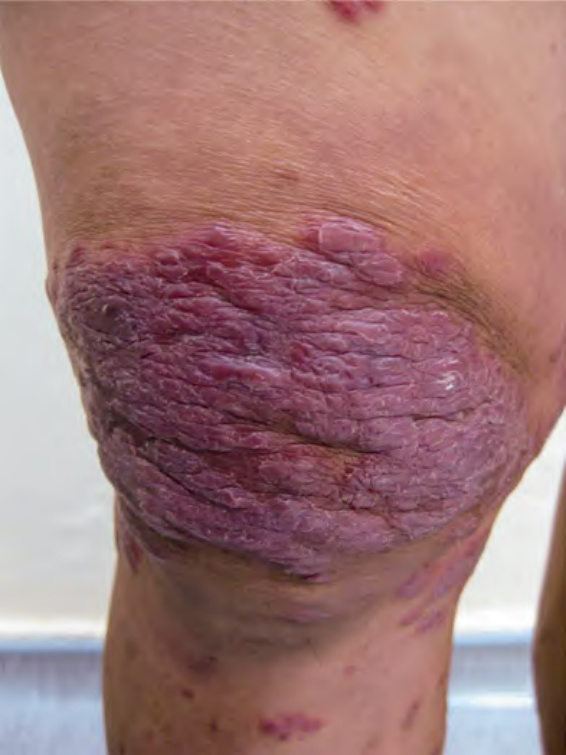
Fig. 78.9 Mycobacterium haemophilum infection.
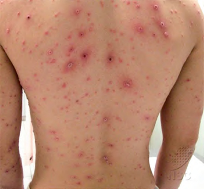
Fig. 78.10 Secondary syphilis presenting as lues maligna. This man with HIV had a fever, headache, arthralgias, and myalgias together with this widespread eruption of pustules and crusted papulonodules.