Parasitic Infections
Leishmaniasis
Leishmaniasis is caused by at least 20 species of intracellular protozoans of the genus Leishmania. It is typically transmitted by sandflies with only occasional spread via contaminated needles. HIV-associated leishmaniasis occurs in both endemic and non-endemic regions of the world, with endemic areas including portions of the Mediterranean basin, Middle East, South Asia, East Africa, and Central/South America (see Fig. 83.1). HIV enhances the severity of leishmaniasis by inducing a shift from a protective Th1-type immune response to a Th2-type reaction pattern. Cutaneous findings range from solitary ulcers in exposed areas to atypical diffuse induration of the skin, eroded erythematous plaques, extensive hyperpigmented macules and papules, and necrotic genital ulcers. Mucocutaneous leishmaniasis may result in disfiguring ulcerations of the lips, palate, and nose. The diagnosis of leishmaniasis can be established via microscopy, culture, or PCR of lesional tissue.
Leishmaniasis in people living with HIV has a higher rate of therapeutic failure, relapse, and associated mortality. Amphotericin B is recommended as first-line therapy, and lipid-based formulations are preferred due to their reduced systemic toxicity (particularly nephrotoxicity). Pentavalent antimonials do not represent first-line therapy in those co-infected with HIV and Leishmania due to a high incidence of side effects (e.g. pancreatitis, arrhythmias), treatment failure, and mortality. Oral miltefosine is well-tolerated but less effective in the setting of HIV infection. Early initiation of ART is recommended, as it contributes to higher survival rates and lower relapse rates.
Strongyloidiasis
Strongyloides stercoralis is a soil-transmitted intestinal helminth with a worldwide distribution, although it most commonly leads to infection in
HIV infection. Crusted and keratotic plaque on the elbow that is teeming with mites.
tropical and subtropical regions. Autoinfection due to penetration of the intestinal mucosa can cause hyperinfection and disseminated strongyloidiasis, both of which are more common in patients with impaired cellmediated immunity (see Ch. 83). People living with HIV have twice the risk of S. stercoralis infection compared to the general population, but not necessarily higher rates of hyperinfection or disseminated strongyloidiasis.
Cutaneous manifestations of strongyloidiasis include urticaria and larva currens – urticarial serpiginous tracks from larval penetration and migration through the dermis, often beginning in the perianal area. Disseminated strongyloidiasis produces rapidly progressing petechial and purpuric eruptions on the abdomen and proximal limbs (“thumbprint” purpura), and eosinophilia is common. Larvae are generally absent in skin biopsies of larva currens, but in disseminated disease they may be observed in the dermis in association with vascular damage. Ivermectin and albendazole have replaced thiabendazole as first-line treatment because of greater efficacy and tolerability. Mortality is high (up to 80%) with hyperinfection or disseminated disease.
Acanthamebiasis
Acanthamoeba are free-living amoebae present in the environment and secretions of healthy individuals. Infection occurs in people living with HIV who have CD4+ T cell counts <250/mm3;. The skin may be the initial portal of entry or a site of hematogenous dissemination. Cutaneous lesions include papules, pustules, ulcers, and indurated bluish or erythematous cellulitic plaques, which may or may not be painful. Trophozoites are scant and difficult to detect in routinely stained histologic sections. Optimal treatment is not well-defined and includes combinations of miltefosine with antifungal and antibacterial agents.
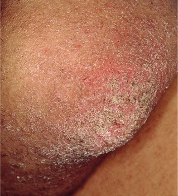
Fig. 78.12 Crusted scabies in the setting of