CONCLUSIONS
The number and variety of manifestations of HIV disease are greater in the skin than in any other organ. Cutaneous complications are a source of significant morbidity and may also be stigmatizing. Although ART has decreased the incidence of several HIV-associated skin conditions, other disorders such as IRIS, drug reactions, metabolic disturbances, HPV infections, and SCCs are still commonly observed. In fact, they may even be increasing in incidence due to prolonged survival of people living with HIV. By recognizing the spectrum of HIV-associated skin conditions and performing appropriate diagnostic tests, treatment can be administered in a timely fashion and outcomes optimized.
Additional figures available in our eBook (see inside front cover for access code).
-
Chelidze K, Thomas C, Chang AY, et al. HIV-related skin sites/default/files/media_asset/UNAIDS_FactSheet_en.pdf>.
-
UNAIDS. In Danger: UNAIDS Global AIDS Update 2022;
-
Kaushik SB, Lebwohl MG. Psoriasis: which therapy for disease in the era of antiretroviral therapy: recognition and management. Am J Clin Dermatol. 2019;20:423–442.14. Patil S, Majumdar B, Sarode SC, et al. Oropharyngeal
July 2022. <https://www.unaids.org/en/resources/ documents/2022/in-danger-global-aids-update>.
3. Rollenhagen C, Lathrop MJ, Macura SL, et al. Herpes which patient: focus on special populations and chronic infections. J Am Acad Dermatol. 2019;80:43–53.28. Menon K, Van Voorhees AS, Bebo BF Jr, et al. Psoriasis in candidosis in HIV-infected patients – an update. Front Microbiol. 2018;9:980.15. Daniel CR 3rd, Norton LA, Scher RK. The spectrum of nail simplex virus type-2 stimulates HIV-1 replication in cervical tissues: implications for HIV-1 transmission and efficacy of anti-HIV-1 microbicides. Mucosal Immunol. 2014;7:1165–1174.4. Vafai A, Berger M. Zoster in patients infected with HIV: a patients with HIV infection: from the medical board of the National Psoriasis Foundation. J Am Acad Dermatol. 2010;62:291–299.29. Adizie T, Moots RJ, Hodkinson B, et al. Inflammatory disease in patients with human immunodeficiency virus infection. J Am Acad Dermatol. 1992;27:93–97.16. Ramos-e-Silva M, Lima CMO, Schechtman RC, et al.
arthritis in HIV positive patients: a practical guide. BMC Infect Dis. 2016;16:100.30. Resnick SD, Murrell DF, Woosley JT. Pityriasis rubra pilaris,
Systemic mycoses in immunodepressed patients (AIDS). Clin Dermatol. 2012;30:616–627.17. Murakawa GJ, Harvell JD, Lubitz P, et al. Cutaneous review. Am J Med Sci. 2001;321:372–380.5. Coates SJ, Leslie KS. What’s new in HIV dermatology?
F1000Res 2019;8:F1000 Faculty Rev: 980. <https://doi. org/10.12688/f1000research.16182.1>
6. Looker KJ, Ronn MM, Brock PM, et al. Evidence of acne conglobata, and elongated follicular spines: an HIV-associated follicular syndrome? J Am Acad Dermatol. 1993;29:283.31. Eisman S. Pruritic papular eruption in HIV. Dermatol Clin.
aspergillosis and acquired immunodeficiency syndrome. Arch Dermatol. 2000;136:365–369.18. Alvar J, Aparicio P, Aseffa A, et al. The relationship synergistic relationships between HIV and human papillomavirus (HPV): systematic reviews and meta-analyses of longitudinal studies of HPV acquisition and clearance by HIV status, and of HIV acquisition by HPV status. J Int AIDS Soc. 2018;21:e25110.7. Poljak M, Sterbenc A, Lunar MM. Prevention of human
2006;24:449–457. vi.
32. Smith KJ, Skelton HG, DeRusso D, et al. Clinical and histo- between leishmaniasis and AIDS: the second 10 years. Clin Microbiol Rev. 2008;21:334–359.19. Jarvis JN, Lockwood DN. Clinical aspects of visceral pathologic features of hair loss in patients with HIV-1 infection. J Am Acad Dermatol. 1996;34:63–68.33. Woods EA, Foisy MM. Antiretroviral-related alopecia leishmaniasis in HIV infection. Curr Opin Infect Dis. 2013;26:1–9.20. Galarza C, Ramos W, Gutierrez EL, et al. Cutaneous papillomavirus (HPV)-related tumors in people living with human immunodeficiency virus (HIV). Expert Rev Anti Infect Ther. 2017;15:987–999.8. Tong WWY, Hillman RJ, Kelleher AD, et al. Anal intraepi- in HIV-infected patients. Ann Pharmacother. 2014;48:1187–1193.34. Prose NS, Abson KG, Scher RK. Disorders of the nails and acanthamebiasis infection in immunocompetent and immunocompromised patients. Int J Dermatol. 2009;48:1324–1329.21. Thomas C, Coates SJ, Engelman D, et al. Ectoparasites:
thelial neoplasia and squamous cell carcinoma in HIV-infected adults. HIV Med. 2014;15:65–76.9. Grulich AE, van Leeuwen MT, Falster MO, Vajdic CM.
hair associated with human immunodeficiency virus infection. Int J Dermatol. 1992;31:453–457.35. Cribier B, Mena ML, Rey D, et al. Nail changes in patients scabies. J Am Acad Dermatol. 2020;82:533–548.22. Aquilina C, Viraben R, Sire S. Ivermectin-responsive infected with human immunodeficiency virus. Arch Dermatol. 1998;134:1216–1220.36. Vega LE, Espinoza LR. Vasculitides in HIV infection. Curr
Demodex infestation during human immunodeficiency virus infection. A case report and literature review. Dermatol Basel Switz. 2002;205:394–397.23. Nair SP. Insect bite reaction and HIV infection. Indian
Incidence of cancers in people with HIV/AIDS compared with immunosuppressed transplant recipients: a metaanalysis. Lancet. 2007;370:59–67.10. Rigopoulos D, Paparizos V, Katsambas A. Cutaneous
Rheumatol Rep. 2020;22:60.37. Gregory N, DeLeo VA. Clinical manifestations of photo- sensitivity in patients with human immunodeficiency virus infection. Arch Dermatol. 1994;130:630–633.38. Mansourati FF, Stone VE, Mayer KH. Porphyria cutanea
J Dermatol Venereol Leprol. 2015;81:95.24. Motswaledi MH, Visser W. The spectrum of markers of HIV infection. Clin Dermatol. 2004;22:487–498.11. Sabbagh P, Riahi SM, Gamble HR, et al. The global
HIV-associated infective and inflammatory dermatoses in pigmented skin. Dermatol Clin. 2014;32:211–225.25. Mahlangeni GM, Tod BM, Jordaan HF, et al.
and regional prevalence, burden, and risk factors for methicillin-resistant Staphylococcus aureus colonization in HIV-infected people: a systematic review and metaanalysis. Am J Infect Control. 2019;47:323–333.12. Plettenberg A, Lorenzen T, Burtsche BT, et al. Bacillary tarda and HIV/AIDS: a review of pathogenesis, clinical manifestations and management. Int J STD AIDS. 1999;10:51–56.39. Wong SN, Khoo LSW. Chronic actinic dermatitis as the
Clinicopathological features of seborrheic-like dermatitis in HIV-infected adults: a single institutional descriptive cross-sectional study. Am J Dermatopathol. 2021;43:27–34.26. Alpalhao M, Borges-Costa J, Filipe P. Psoriasis in HIV angiomatosis in HIV-infected patients–an epidemiological and clinical study. Dermatol Basel Switz. 2000;201:326–331.
presenting feature of HIV infection in three Chinese males. Clin Exp Dermatol. 2003;28:265–268.40. Koethe JR, Lagathu C, Lake JE, et al. HIV and antiretro- viral therapy-related fat alterations. Nat Rev Dis Primers. 2020;6:48.
infection: an update. Int J STD AIDS. 2019;30:596–604.
In people with HIV infection, trimethoprim–sulfamethoxazole (TMP-SMX) is the most common cause of a cutaneous drug reaction. Often taken for prophylaxis or treatment of Pneumocystis jirovecii pneumonia or toxoplasmosis, intravenously administered TMP-SMX leads to an exanthematous eruption and fever (typically 8–12 days after initiating therapy) in 50%–60% of patients (Fig. 78.20). This occurrence rate is 10 times that observed in the general population. Additional side effects of TMP-SMX include fixed drug eruption, SJS, and TEN.
Close observation is often sufficient since most cutaneous drug eruptions spontaneously regress, especially the morbilliform type. However, if systemic signs such as fever develop or epidermal detachment is observed, the suspected offending agent(s) should be withdrawn immediately as this may herald a more serious, possibly life-threatening, complication. With some drugs such as zidovudine, sulfonamides, and dapsone, patients may be successfully desensitized after an adverse drug reaction. Drug rechallenge should be done under controlled circumstances. Rechallenge is contraindicated for abacavir and is not recommended for NNRTIs.
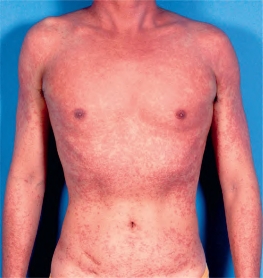
Fig. 78.20 Morbilliform drug eruption due to trimethoprim–sulfamethox- azole (TMP-SMX). This young man with HIV developed a widespread eruption of blanchable erythematous macules and papules 8 days after starting TMP-SMX. Note the coalescence on the upper trunk.